Abstract
Steatocystoma multiplex (SM) is a rare hamartomatous malformation of the pilosebaceous duct junction. Most cases of SM are sporadic, although less common autosomal dominant inherited forms have been reported. Steatocystoma multiplex suppurativa (SMS) is a much rarer inflammatory variant of SM, associated with severe inflammatory lesions resembling those of hidradenitis suppurativa. We describe herein a 28-year-old male with SMS who presented with extensive giant cysts on his neck, face, and scalp.
Key Words: Adnexa, Pilosebaceous duct, Steatocystoma multiplex, Hidradenitis suppurativa, Tumor
Introduction
Steatocystoma multiplex (SM) is a rare hamartomatous disorder of the pilosebaceous duct, manifesting clinically by multiple asymptomatic small dermal cysts [1]. Most of the cases of SM are sporadic and rarely inherited as an autosomal dominant trait, which is linked to a mutation in exon 1 of keratin 17 (KRT17) [2, 3]. SM starts in adolescence or early adulthood, but earlier and later onsets have been reported too, without gender or racial predilection [4, 5]. Clinically, SM manifests as skin-colored to yellowish, firm, cystic, or dome-shaped papules or nodules, with areas of involvement including the neck, chest, trunk, arms, axillae, and groin, but rarely the face and scalp [6, 7]. Inflammatory variants with lesions, resembling features of acne conglobata or hidradenitis suppurativa, have been reported in a few cases [8, 9, 10]. Giant SM – more than 20 mm – is extremely rare, and some cases of this giant form were recently reported as having an association with recurrent mutation in the KRT17 gene [7, 11, 12]. Here, we report a rare variant of SM with giant lesions on the neck, face, and scalp.
Case Presentation
A 28-year-old male patient presented with a history of progressive non-tender skin-colored nodules over the scalp, face, neck, trunk, and axillary regions for 10 years. The lesions were mostly asymptomatic, although at times became inflamed and ruptured spontaneously, discharging a small amount of yellowish oily material. Past clinical history was unremarkable. The patient's father and two of his siblings had similar lesions, yet theirs were smaller and asymptomatic.
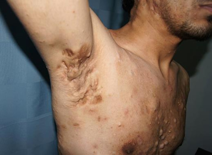
Physical examination revealed multiple, firm, skin-colored 0.5–1 cm subcutaneous nodules over the chest, abdomen, back, axillae, and extremities. The patient also exhibited multiple, giant, mobile, skin-colored subcutaneous nodules, approximately 4–6 cm in size, on the forehead, scalp, and neck (Fig. 1). There were fluctuant nodules and sinuses located in the axillary region (Fig. 2). Two excisional biopsies were carried out, revealing cysts lined by squamous epithelium with sebaceous glands (Fig. 3). A diagnosis of steatocystoma multiplex suppurativa (SMS) was made based on clinical and histopathological findings.
Fig. 1.
Multiple skin-colored papular and nodulocystic lesions varying in size (from 4 to 6 cm) over the forehead (a, b) and lateral cervical region (c).
Fig. 2.
Nodulocystic lesions, scarring, and sinuses in the right axilla.
Fig. 3.
Excisional biopsy reveals cysts lined by squamous epithelium with sebaceous glands (original magnification ×100, hematoxylin and eosin stain).
Discussion
First reported by Jamieson in 1873, SM is a rare sporadic, often inherited autosomal dominant trait, resulting from a mutation of keratin 17 on chromosome 17q21.2 [2, 3, 11, 13]. This mutation has been reported in pachyonychia congenita type 2, which involves SM as part of its clinical manifestation [14]. In our patient, although family history was positive, indicating the possibility of an autosomal dominant trait, no changes were found in the mucous membranes, hair, or nails.
SM consists of a spectrum of clinical morphology and anatomical distribution. Characteristically, it presents with multiple skin-colored dermal cysts on the trunk, axilla, groin, and proximal extremities [1]. In the literature, however, there are rare SM presentations reported for the breasts, face [15, 16], and scalp [17, 18], but in most of these atypical cases, the lesions were localized and limited to a single anatomical location.
Kim et al. [6] differentiated typical SM from SM limited to the scalp. They classified the typical form as mostly hereditary, appearing in childhood and early adulthood, with SM limited to the scalp as being sporadic and appearing in late adulthood. Lee et al. [17] reported sporadic cases of SM presentation limited to the scalp, with alopecia patches due to trichotillomania.
A case somewhat similar to ours involving face and scalp lesions of 10 years was recently reported by Vukicevic [19] in 2017. The patient, however, was a sexagenarian with a history of trauma without any family history, and was eventually treated with CO2 laser. Our case was referred for surgical excision.
While etiology of SM remains elusive, there have been reports indicating infection, trauma, and/or immunological insult as being responsible for it. Our patient did not demonstrate a history of trauma or infection. Clinical diagnosis of SM is confirmed by histology in order to exclude follicular infundibulum tumors, milia, epidermal inclusion cysts, and eruptive vellus hair cysts. The pathognomonic histopathological feature of SM is the presence of sebaceous lobules close to the cystic wall, made up of flattened, stratified squamous epithelium lacking a granular layer [1].
In 2016, Santana et al. [9] reported the first case from Brazil similar to ours, in which a young female presented with inherited SMS for 11 years, whereas Fekete and Fekete [8] reported a case of sporadic generalized SM with a partial suppurativa variant; this developed at some point in a young male patient, with a disease duration of 20 years. Concurrent presentation of SM with hidradenitis suppurativa is reported in the literature, although Hollmig and Menter [10] also reported a case similar to ours, having a strong familial link in a female and her sister, both with SM and hidradenitis suppurativa occurring simultaneously.
Various treatment modalities are used for SM, ranging from yttrium aluminum garnet and CO2 laser, intralesional steroid injections, tetracycline ointment, and oral isotretinoin or cryotherapy. However, surgical excision remains the mainstay in most cases [3, 5, 6, 11].
Our patient was a rare case of typical SMS affecting the scalp, neck, and face, and was referred to dermatological surgery for definitive treatment.
Statement of Ethics
Informed consent was obtained from the patient. The authors have no ethical conflicts to disclose.
Disclosure Statement
The authors have no conflicts of interest to declare. There were no funding sources for this work.
Author Contributions
L. Alotaibi: reviewed relevant literature, communicated with the patient for further information, participated in research introduction, case presentation and discussion, and final report writing. M. Alsaif: reviewed relevant literature, participated in research introduction, case presentation and discussion, and prepared the version for publication. A. Alhumidi: reviewed relevant literature, provided and highlighted the case for publication, participated in research introduction, case presentation and discussion, and final report writing. M. Turkmani: reviewed relevant literature, revised histopathology of the case, participated in research introduction, case presentation and discussion, and final report writing. F. Alsaif: reviewed relevant literature, participated in research introduction, case presentation and discussion, and revised the entire research process until the approval of final version for publication.
References
- 1.Cho S, Chang SE, Choi JH, Sung KJ, Moon KC, Koh JK. Clinical and histologic features of 64 cases of steatocystoma multiplex. J Dermatol. 2002 Mar;29((3)):152–6. doi: 10.1111/j.1346-8138.2002.tb00238.x. [DOI] [PubMed] [Google Scholar]
- 2.Antal AS, Kulichova D, Redler S, Betz RC, Ruzicka T. Steatocystoma multiplex: keratin 17 - the key player? Br J Dermatol. 2012 Dec;167((6)):1395–7. doi: 10.1111/j.1365-2133.2012.11073.x. [DOI] [PubMed] [Google Scholar]
- 3.Kamra HT, Gadgil PA, Ovhal AG, Narkhede RR. Steatocystoma multiplex-a rare genetic disorder: a case report and review of the literature. J Clin Diagn Res. 2013 Jan;7((1)):166–8. doi: 10.7860/JCDR/2012/4691.2698. [DOI] [PMC free article] [PubMed] [Google Scholar]
- 4.Park YM, Cho SH, Kang H. Congenital linear steatocystoma multiplex of the nose. Pediatr Dermatol. 2000 Mar-Apr;17((2)):136–8. doi: 10.1046/j.1525-1470.2000.01732.x. [DOI] [PubMed] [Google Scholar]
- 5.Rongioletti F, Cattarini G, Romanelli P. Late onset vulvar steatocystoma multiplex. Clin Exp Dermatol. 2002 Sep;27((6)):445–7. doi: 10.1046/j.1365-2230.2002.01027.x. [DOI] [PubMed] [Google Scholar]
- 6.Kim SJ, Park HJ, Oh ST, Lee JY, Cho BK. A case of steatocystoma multiplex limited to the scalp. Ann Dermatol. 2009 Feb;21((1)):106–9. doi: 10.5021/ad.2009.21.1.106. [DOI] [PMC free article] [PubMed] [Google Scholar]
- 7.Jeong SY, Kim JH, Seo SH, Son SW, Kim IH. Giant steatocystoma multiplex limited to the scalp. Clin Exp Dermatol. 2009 Oct;34((7)):e318–9. doi: 10.1111/j.1365-2230.2009.03274.x. [DOI] [PubMed] [Google Scholar]
- 8.Fekete GL, Fekete JE. Steatocystoma multiplex generalisata partially suppurativa—case report. Acta Dermatovenerol Croat. 2010;18((2)):114–9. [PubMed] [Google Scholar]
- 9.Santana CN, Pereira DD, Lisboa AP, Leal JM, Obadia DL, Silva RS. Steatocystoma multiplex suppurativa: case report of a rare condition. An Bras Dermatol. 2016 Sep-Oct;91((5 suppl 1)):51–3. doi: 10.1590/abd1806-4841.20164539. [DOI] [PMC free article] [PubMed] [Google Scholar]
- 10.Hollmig T, Menter A. Familial coincidence of hidradenitis suppurativa and steatocystoma multiplex. Clin Exp Dermatol. 2010 Jun;35((4)):e151–2. doi: 10.1111/j.1365-2230.2009.03742.x. [DOI] [PubMed] [Google Scholar]
- 11.Wang J, Li J, Li X, Lei D, Xiao W, Li Z, et al. A recurrent mutation in the KRT17 gene responsible for severe steatocystoma multiplex in a large Chinese family. Clin Exp Dermatol. 2018 Mar;43((2)):205–8. doi: 10.1111/ced.13311. [DOI] [PubMed] [Google Scholar]
- 12.Setoyama M, Mizoguchi S, Usuki K, Kanzaki T. Steatocystoma multiplex: a case with unusual clinical and histological manifestation. Am J Dermatopathol. 1997 Feb;19((1)):89–92. doi: 10.1097/00000372-199702000-00017. [DOI] [PubMed] [Google Scholar]
- 13.Smith FJ, Corden LD, Rugg EL, Ratnavel R, Leigh IM, Moss C, et al. Missense mutations in keratin 17 cause either pachyonychia congenita type 2 or a phenotype resembling steatocystoma multiplex. J Invest Dermatol. 1997 Feb;108((2)):220–3. doi: 10.1111/1523-1747.ep12335315. [DOI] [PubMed] [Google Scholar]
- 14.Ofaiche J, Duchatelet S, Fraitag S, Nassif A, Nougué J, Hovnanian A. Familial pachyonychia congenita with steatocystoma multiplex and multiple abscesses of the scalp due to the p.Asn92Ser mutation in keratin 17. Br J Dermatol. 2014 Dec;171((6)):1565–7. doi: 10.1111/bjd.13123. [DOI] [PubMed] [Google Scholar]
- 15.Uçmak D, Sula B, Meltem Akkurt Z, Fidan V, Firat U, Arica M. A rare case of facial steatocystoma multiplex. Acta Dermatovenerol Croat. 2013;21((3)):205–6. [PubMed] [Google Scholar]
- 16.Sardana K, Sharma RC, Jain A, Mahajan S. Facial steatocystoma multiplex associated with pilar cyst and bilateral preauricular sinus. J Dermatol. 2002 Mar;29((3)):157–9. doi: 10.1111/j.1346-8138.2002.tb00239.x. [DOI] [PubMed] [Google Scholar]
- 17.Lee D, Chun JS, Hong SK, Seo JK, Choi JH, Koh JK, et al. Steatocystoma multiplex confined to the scalp with concurrent alopecia. Ann Dermatol. 2011 Oct;23((2 Suppl 2)):S258–60. doi: 10.5021/ad.2011.23.S2.S258. [DOI] [PMC free article] [PubMed] [Google Scholar]
- 18.Pietrzak A, Bartosinska J, Filip AA, Rakowska A, Adamczyk M, Szumilo J, et al. Steatocystoma multiplex with hair shaft abnormalities. J Dermatol. 2015 May;42((5)):521–3. doi: 10.1111/1346-8138.12837. [DOI] [PubMed] [Google Scholar]
- 19.Vukicevic JP. Steatocystoma multiplex involving the face and scalp: a case report. Hong Kong J Dermat Venereol. 2017 Mar;25((1)):29–32. [Google Scholar]


