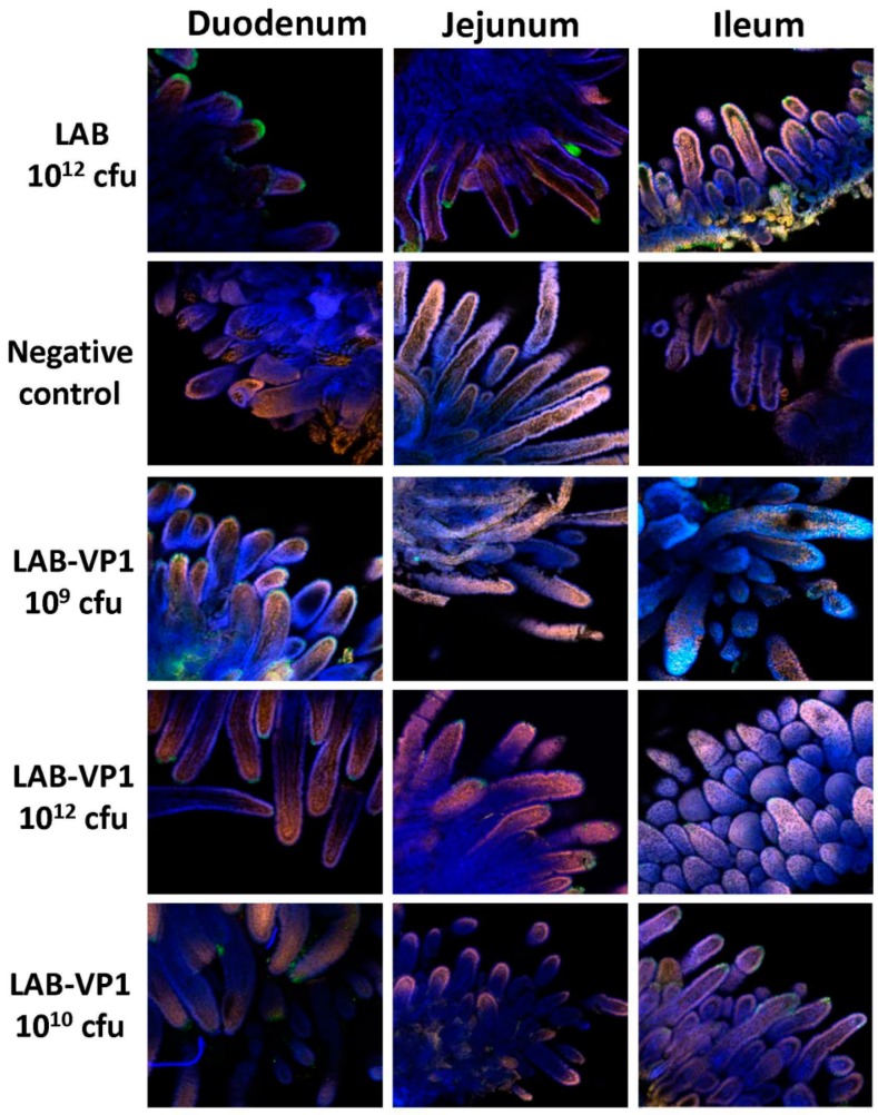
Figure 15.
Detection of HuNoV antigen-positive cells in the intestine by IFA. Fresh intestinal tissues collected from inoculated piglets at PID 5 were sectioned into small pieces, fixed, quenched, and permeabilized. The tissues were incubated with HuNoV VP1-specific polyclonal antibody, followed by incubation with goat anti-guinea pig IgG labeled with Alexa Fluor 488 antibody. Nuclei and actin were counterstained with SYTOX orange and phalloidin labeled with Alexa Fluor 633. The stained tissues were mounted onto slides and examined using an Olympus FV1000 confocal microscopy imaging system. Representative microscope images (10× magnifications) from each group were shown. The size of villus may be variable because of the individual difference of the piglets. A green signal represents HuNoV antigen, a blue signal is actin, and an orange signal is the nucleus.

