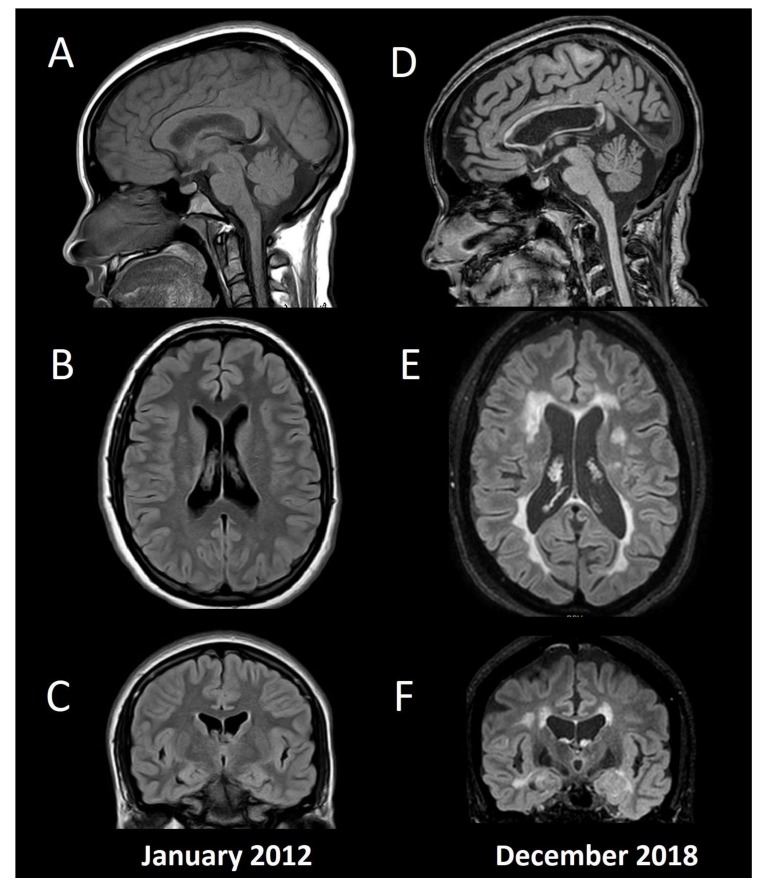
Figure 1.
Conventional MRI in a patient with multiple sclerosis and cognitive impairment. Baseline MRI (A: Sagittal T1, B: Axial FLAIR, C: Coronal FLAIR) from a 15-year-old female with fulminant MS (Marburg variant, EDSS 8.0). After initial aggressive treatment in 2012, including myeloablation with cyclophosphamide, the patient remained asymptomatic without disease modifying treatment, until a second supratentorial motor relapse in 2015, confirming her MS diagnosis and beginning fingolimod. Since then, no relapses or new T2/enhancing lesions have appeared, and she had an EDSS 1.0 by the time of the second MRI in 2018 (D: Sagittal T1, E: Axial FLAIR, F: Coronal FLAIR). Her Mini-Mental State Examination was 30 (normal), Beck Depression Inventory 4 (without depression), and her fatigue severity score was 4 (significative fatigue). She had below normal performance (≤1.5 standard deviation) in verbal and visual episodic memory, and in information processing speed tests, with the diagnosis of cognitive impairment according to the MACFIMS battery. Note the widespread brain volume loss including cortical grey matter, ventricular width, and corpus callosum atrophy. The patient gave her written informed consent to present this data.

