Abstract
Synovial sarcoma is a malignant tumor, it accounts for approximately 5–10% of the soft tissue sarcoma, and mainly develops in the para-articular regions in adolescents and young adults. We reported a case of synovial sarcoma extending into the vastus intermedius muscle beyond the intermuscular septum which is considered to be the greatest barrier to ensuring a safety margin in musculoskeletal surgery. Preoperative sequential MRI showed the intermuscular-septum line and direct tumoral invasion beyond the intermuscular septum. A histopathological examination showed the direct invasion to the intermuscular septum. Synovial sarcoma seldom shows such direct invasive pattern to barriers, much less the robust barrier like an intermuscular septum. To our knowledge, no previous study has reported such unique extension of a synovial sarcoma beyond the intermuscular septum. Surgeons should be aware of the potential vulnerability of the intermuscular septum.
INTRODUCTION
Synovial sarcoma is a malignant tumor, it accounts for approximately 5–10% of the soft tissue sarcoma, and mainly develops in the para-articular regions in adolescents and young adults [1]. The combination of surgery and chemotherapy has resulted in about 60% 5-year survival rate and 50% 10-year survival rate, mainly because of the metastasis in lung [2, 3]. In the patients with synovial sarcoma, wide-resection surgery is usually performed for local recurrent control, while neo/adjuvant chemotherapy is commonly performed to decrease the risk of metastasis recurrences [4]. We reported a case of synovial sarcoma extending into the vastus intermedius muscle beyond the intermuscular septum which is considered to be the greatest barrier to ensuring a safety margin in musculoskeletal surgery [5].
CASE REPORT
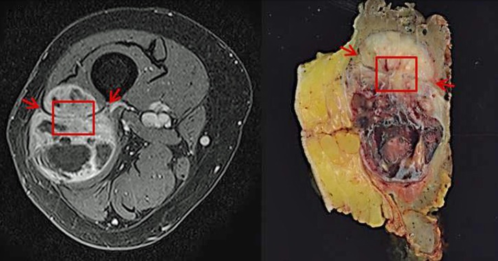
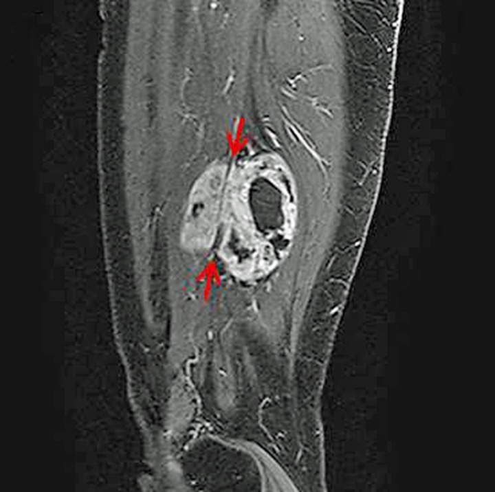
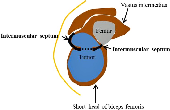
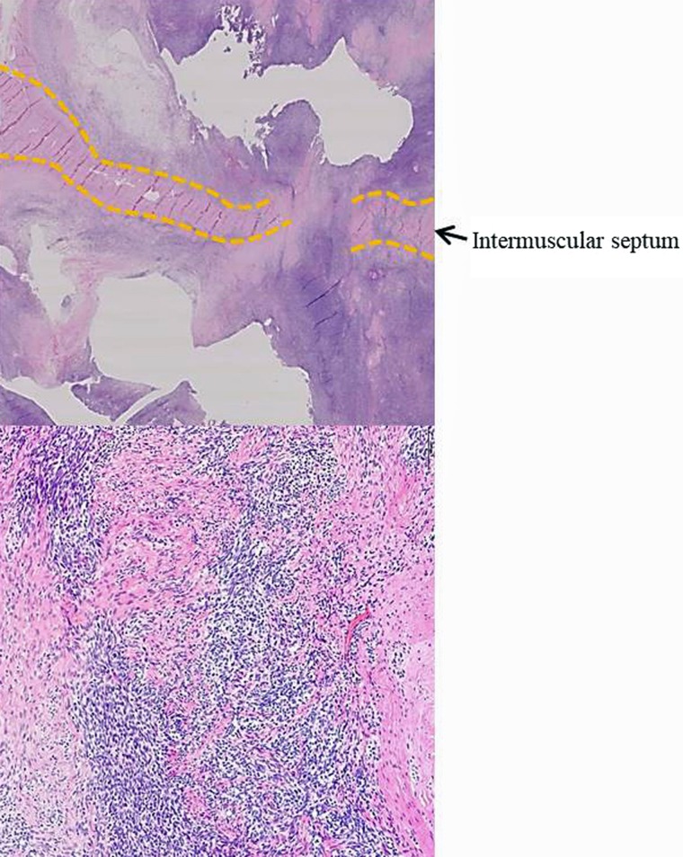
A 55-year-old female with a large mass was admitted to our hospital. She had no traumatic episode. Magnetic resonance imaging (MRI) revealed a well-defined and gadolinium-enhanced mass (5.5 × 4.9 cm) in her right thigh. MRI also revealed a mass extending into the vastus intermedius muscle beyond the intermuscular septum (red square and arrows in Figs 1 and 2, and original schema in Fig. 3). A needle biopsy from the mass confirmed the histological diagnosis of synovial sarcoma which was positive for SS-18 break apart by fluorescence in situ hybridization (FISH). After 2 cycles of neo-adjuvant chemotherapy, a wide resection of the tumor was performed. A histopathological examination (red square in Fig. 1) showed that the tumor had direct invasion into the intermuscular septum (Fig. 4). Five months after the surgery, the patient has shown no sign of recurrence.
Figure 1:
Preoperative sequential MRI clearly showed the intermuscular-septum line (red arrows) and direct tumoral invasion beyond the intermuscular septum (red square).
Figure 2:
Preoperative sequential MRI clearly showed the intermuscular-septum line (red arrows) and direct tumoral invasion beyond the intermuscular septum (red square).
Figure 3:
To facilitate the understanding of how the tumor extended beyond the intermuscular septum, we created the original schema, which revealed a mass extending into the vastus intermedius muscle beyond the intermuscular septum.
Figure 4:
A histopathological examination (red square in Fig. 1) showed that the tumor had direct invasion into the intermuscular septum.
DISCUSSION
The intermuscular septum is a robust fibrous connective tissue which divides muscular compartments of the thigh and it is considered to be the greatest barrier to ensuring a safety margin in musculoskeletal surgery and it is important to consider strength of the barriers to obtain a safe margin in sarcoma surgery [5]. Preoperative sequential MRI in this case clearly showed the intermuscular-septum line (red arrows in Figs 1 and 2) and direct tumoral invasion beyond the intermuscular septum. A histopathological investigation (red square in Fig. 1) showed the direct invasion to the intermuscular septum although synovial sarcoma should be an indolent tumor (Fig. 4). Although some soft tissue sarcoma such as myxofibrosarcoma sometimes show direct invasion to these barriers [6, 7], synovial sarcoma seldom shows such direct invasive pattern to barriers, much less the greatest barrier like an intermuscular septum. Moreover, in the patients with synovial sarcoma, wide-margin surgery with even though thin barriers resulted in good local control rates. Although synovial sarcoma usually cannot show such a local invasive pattern, a relatively indolent tumor growth might allow for such unfamiliar development of a tumor, even against a robust barrier. To our knowledge, no previous study has reported such unique extension of a synovial sarcoma beyond the intermuscular septum. We believe that these radiological and pathological findings have a huge impact to sarcoma surgeons. Surgeons should be aware of the potential vulnerability of the intermuscular septum. A careful preoperative examination is warranted to obtain a safe margin in sarcoma surgery.
ACKNOWLEDGMENTS
We thank members of our division for helpful discussions.
CONFLICT OF INTEREST STATEMENT
No conflicts of interest.
FUNDING
No sources of funding.
ETHICAL APPROVAL
The patients and/or their families were informed that data from medical records would be submitted for publication, and gave their consent. Ethical approval for this study was obtained from an ethic committee in our institution.
CONSENT
Written informed consent was obtained from the patients for their anonymized information to be published in this article.
GUARANTOR
Seiichi Matsumoto, MD. PhD, 3-8-31 Ariake, Koto-ward, Tokyo 135–8550, Japan. Tel: +81-3-3520-0111; Fax: +81-3-3520-0141; E-mail: smatsumoto@jfcr.or.jp
References
- 1. Fisher C. Synovial sarcoma. Ann Diagn Pathol 1998;2:401–21. [DOI] [PubMed] [Google Scholar]
- 2. Lewis JJ, Antonescu CR, Leung DH, Blumberg D, Healey JH, Woodruff JM, et al. Synovial sarcoma: a multivariate analysis of prognostic factors in 112 patients with primary localized tumors of the extremity. J Clin Oncol 2000;18:2087–94. [DOI] [PubMed] [Google Scholar]
- 3. Krieg AH, Hefti F, Speth BM, Jundt G, Guillou L, Exner UG, et al. Synovial sarcomas usually metastasize after >5 years: a multicenter retrospective analysis with minimum follow-up of 10 years for survivors. Ann Oncol 2011;22:458–67. [DOI] [PubMed] [Google Scholar]
- 4. Verweij J, Seynaeve C. The reason for confining the use of adjuvant chemotherapy in soft tissue sarcoma to the investigational setting. Semin Radiat Oncol 1999;9:352–9. [DOI] [PubMed] [Google Scholar]
- 5. Kawaguchi N, Ahmed AR, Matsumoto S, Manabe J, Matsushita Y. The concept of curative margin in surgery for bone and soft tissue sarcoma. Clin Orthop Relat Res 2004;419:165–72. [DOI] [PubMed] [Google Scholar]
- 6. Kikuta K, Kubota D, Yoshida A, Suzuki Y, Morioka H, Toyama Y, et al. An analysis of factors related to recurrence of myxofibrosarcoma. Jpn J Clin Oncol 2013;43:1093–1104. [DOI] [PubMed] [Google Scholar]
- 7. Kikuta K, Kubota D, Yoshida A, Morioka H, Toyama Y, Chumann H, et al. An analysis of factors related to the tail-like pattern of myxofibrosarcoma seen on MRI. Skeletal Radiol 2015;44:55–62. [DOI] [PubMed] [Google Scholar]


