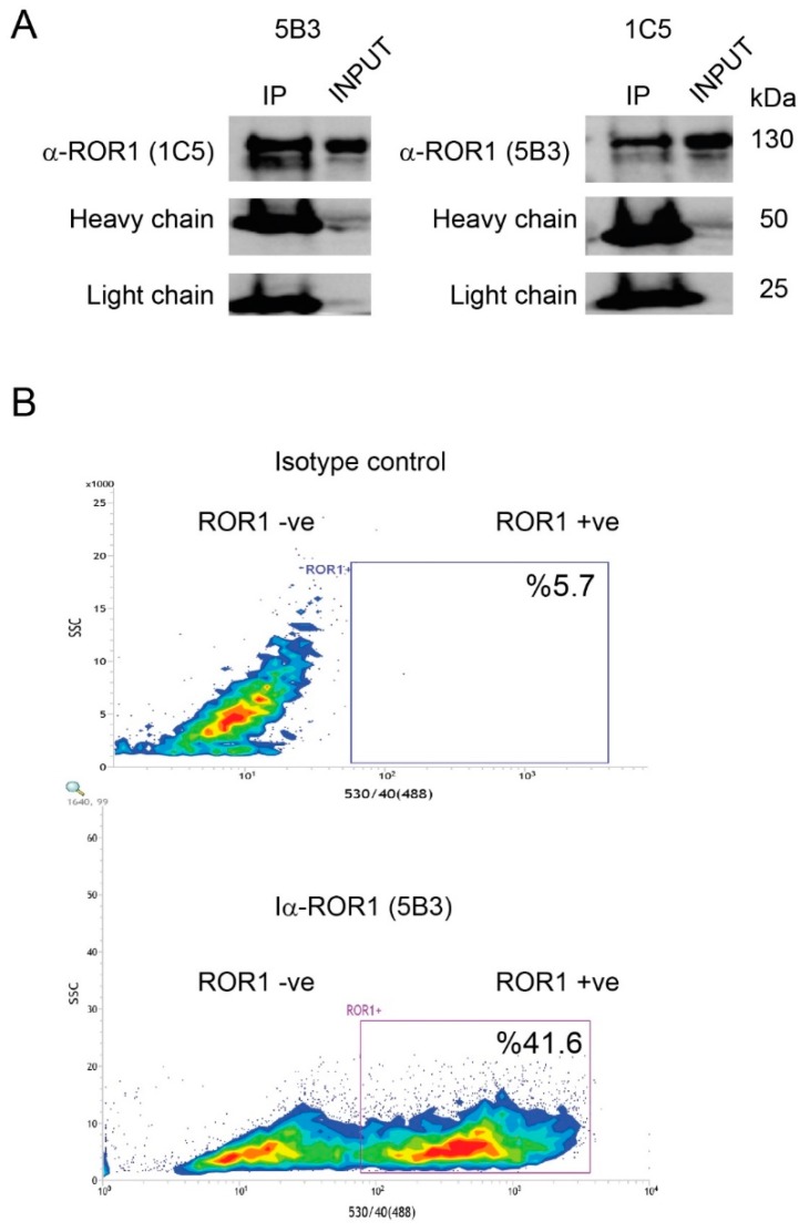
Figure A1.
ROR1 monoclonal antibodies detect native and denatured forms of ROR1. (A) ROR1 protein was immunoprecipitated by ROR1 monoclonal antibodies 1C5 and 5B3. The immunoprecipitate of 5B3 was probed with 1C5, and that of 1C5 was probed with 5B3 antibodies in a Western blot. Heavy and light chains of the antibodies were used as IP control. (B) ROR1 expression in PLC/PRF/5 cells was assessed in flow cytometry by using 5B3 antibody. The isotype staining and ROR1-specific detection are shown in upper and lower panels, respectively. Alexa488-anti-mouse IgG antibody was used as a secondary antibody.

