Abstract
Is an amygdala necessary to experience and perceive fear? Intriguing evidence comes from patient S.M. who lost her left and right amygdalae to disease. Initial testing suggested that S.M.’s most defining symptom was an inability to recognize fear in other people’s facial expressions. A fascinating paper by Adolphs and colleagues in 2005 examined one potential mechanism for this impairment: a failure to spontaneously attend to widened eyes, the most distinctive physical feature portrayed in symbolic fear expressions. This study helped invigorate debates about the brain basis of fear and paved the way for a more nuanced understanding of amygdala function.
Keywords: Fear, emotion, amygdala, prediction
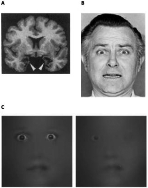
Since the 1800s, scientists have lesioned animal brains and observed the consequences. During this time, human patients with brain lesions have made similarly invaluable scientific contributions to understanding the brain basis of language, memory and emotion. A woman with rare bilateral lesions of the amygdala, known as S.M., is perhaps the most famous patient to shed light on nature of fear. S.M. suffers from Urbach-Wiethe disease (UWD), a rare genetic condition causing selective calcification of amygdala neurons (Figure 1A). Since S.M. was introduced to the scientific community in 1990, her abilities and deficits have been extensively documented in over 30 peer-reviewed journal articles (for a full listing, see Table 1.1 in [1]). Her most striking symptoms: an apparent inability to experience or perceive fear, confirming, for many scientists, the amygdala’s role in a neural system for fear. A closer examination of S.M.’s emotional life, however, suggests a more nuanced and interesting story.
Figure 1. Perceiving Fear Without An Amygala.
(A) Tl-weighted magnetic resonance (MR) image of S.M.’s brain in the coronal plane, showing amygdala lesions on the left and right (dark regions, denoted by arrows), from [4] with permission. S.M.’s lesions are largely confined to her amygdalae, and the fibers of passage therein (although additional damage was later detected in the anterior portion of her entorhinal cortex, the adjacent white matter, and a portion of her ventromedial prefrontal cortex; [1]). (B) An example of a posed, wide-eyed gasping face that symbolizes fear. (C) Facial information used to discriminate a wide-eyed gasping face from a smiling face in ten control subjects (left) and S.M (right) (from [10], used with permission).
The amygdala was first linked to fear in the 1930s when Heinrich Klüver and Paul C. Bucy removed the temporal lobes (including the amygdalae) of several rhesus monkeys and observed profound behavioral changes in the animals, including their willingness to approach snakes and other animals that they typically avoided before surgery. These initial studies launched an intense and long-lasting interest in the amygdala’s role in creating states of fear. In rats, mice, monkeys and other animal models, studying fear often means destroying or temporarily disabling amygdala neurons to learn how they organize behaviors such as escape from a dangerous situation, defense against a predator, and freezing in the face of uncertain threat. Such experiments aim to dissect the amygdala’s role in coordinating hypothalamic and brainstem nuclei to change autonomic nervous system activity, measured as increased skin conductance, heart rate and respiration, and in regulating neuromodulators like dopamine, all in the service of fleeing, fighting and freezing. And studying fear often means examining how amygdala neurons allow an animal to learn, for instance, that a neutral sound, such as a tone – referred to as the conditioned stimulus (CS) – comes to predict the presence of a mild threat, such as an electric shock – the unconditioned stimulus (US) – after the two have been repeatedly paired over time, a process that some scientists call “Pavlovian fear learning.” Such studies in non-human animal models contribute to an understanding of fear and the neural pathways that create it only by virtue of a complicated series of (often unstated) inferences [2]. By the early 1990s, scientists concluded that the amygdala was a necessary part of a central fear system. More recently, the amygdala has been called the “switchboard for fear” [3].
Do such conclusions fully and accurately reflect the amygdala’s role in human fear? And what about the ability to perceive fear in others, which seems so crucial to human behavior but is rarely examined in laboratory settings in the context of animal studies? S.M. offers a rare peek at an emotional life created by a human brain deprived of amygdala neurons. An early study of S.M., published in Nature, described her as unable to recognize facial expressions of fear [4]. In practice, when scientists conclude that a person is unable to “recognize a facial expression of fear,” they usually mean that the subject in question has difficulty applying the word “fear” to photographs of people posing wide-eyed, gasping faces that symbolize fear expression (Figure 1B). Such is the case for S.M. By contrast, S.M. has no difficulty explicitly labeling smiling faces as “happiness” and nose-wrinkled faces as “disgust.” And notably, S.M. understands the concept of being afraid – she understands the situations in which fear likely occurs and correctly describes how frightened people tend to behave. She also correctly identifies and uses synonyms for fear, such as “afraid,” “scared,” “worried,” “terrified,” and “alarmed.” Altogether, S.M. seemed to have a specific inability to perceive fear in other people’s faces, suggesting, at least initially, that the human amygdala may be indispensable for recognizing fear.
Some of the findings reported in initials studies of S.M. hinted at a more interesting puzzle, however. Photographs portraying other emotional expressions, such as scowling faces depicting anger and startled faces depicting surprise, also contain wider eyes. S.M.’s intensity ratings for these faces are similarly impaired compared to those of brain-damaged control subjects; only her ratings of faces without wide eyes – smiles depicting happiness and squinting, nose-wrinkled faces portraying disgust – are relatively normal. Further, and intriguingly, S.M. was unable to draw a wide-eyed gasping face when asked to draw the facial expression of fear, but she was able to draw other features depicting fear, such as cowering with hair standing on end [5]. These subtleties suggested that S.M.’s difficulties have less to do with perceiving fear per se, and are more specific to processing widened eyes in human faces.
The “it’s all in the eyes” hypothesis was reinforced by brain imaging experiments (in healthy individuals) published during the same time period. For example, the human amygdala plays an important role in monitoring human gaze [6] and is particularly responsive to widened eyes, specifically the amount of white sclera [7]. Perhaps the amygdala only appeared to be necessary for perceiving fear because fear perception is almost exclusively measured by asking test subjects to judge wide-eyed, gasping faces! Sure enough, a carefully designed brain imaging study published in 2003 showed that amygdala activity increases in response to wide-eyed, gasping faces when the gaze is directed forward towards the viewer, but not when the gaze is directed to the side [8].
This study reported another intriguing finding: wide-eyed scowling faces (portraying anger) elicit increased amygdala activity when gaze is averted from the viewer, but not when gaze is directed forward towards the viewer [8], suggesting that amygdala neurons are most responsive during ambiguity and uncertainty [9]. Humans are particularly skilled at teaching each other about salient features in the world by following each other’s gaze, called shared or joint attention. These ideas refined the working hypothesis: perhaps amygdala circuits direct spatial attention to human eyes particularly when it is important to learn about potential threats and rewards, i.e., anything salient that may impact the future physiological state of the body, a process called allostasis.
In 2005, Ralph Adolphs led a team of scientists to test the hypothesis that S.M. struggled to recognize a fearful facial expression precisely because – without her amygdalae – she did not spontaneously attend to the most distinctive physical feature in these portrayals: widened eyes [10]. S.M. and normal control subjects viewed a wide-eyed gasping face or a smiling face approximately 3,000 times. To examine how the subjects visually sampled information from the face images, the authors used a “bubbles” technique, pioneered by Frederic Gosselin & Philippe Schyns in 2001. Each time, most of the face was obscured, leaving only a small part visible through a Gaussian “bubble” at one of five spatial frequencies. After viewing part of a face through the small peep hole, S.M. and the control subjects then judged whether the revealed features belonged to a face portraying fear or happiness. As predicted, control subjects used information from high frequency bubbles over the eye regions of faces to identify wide-eyed gasping faces as portraying fear (vs. smiling faces portraying happiness). S.M., however, did not (Figure 1C). By contrast, when the authors examined the use of high spatial frequency information from the mouth region (where smiles appear), they observed no difference between control subjects and S.M. Additional follow up analyses confirmed that S.M. failed to make use of visual information from the eye region when judging emotions in a face. One of the most astounding observations was that S.M.’s impairment could be temporarily reversed by simply asking her to look at the eye region in faces. Based on these findings, the authors concluded that they had identified a mechanism to explain the amygdala’s role in fear recognition.
Nonetheless, curious findings emerged to suggest an even more nuanced interpretation of S.M.’s impairment: S.M.’s amygdala lesions, it seems, left her unable to spatially orient to human eyes in particular contexts. For example, S.M. spontaneously used high spatial frequency information from eyes when asked to judge the gender of the faces. Such findings are only suggestive, of course, but they evoke a hypothesis that is similar to one from brain imaging studies: amygdala neurons may be important for tracking another person’s gaze particularly in contexts that are ambiguous and uncertain. In such situations, gaze might provide the brain with clues to the allostatic value of sensory cues. Gaze might forecast an upcoming reward or threat, allowing the rest of the brain (by way of the amygdala) to learn the value of sensory cues so that it can better predict the body’s needs on future occasions (i.e., improving allostatic balance).
This hypothesis – that the amygdala acts as a context-sensitive sentinel for learning threat and reward – is consistent with emerging research on its role in the brain’s predictive architecture. A variety of computationally-formalized approaches to brain function (called predictive coding, active inference, belief propagation and the Bayesian brain hypothesis) are unified by the hypothesis that the brain creates an internal model of the world by “remembering” neural patterns from prior experiences, perceptions and actions, which then function as Bayesian filters for processing incoming information and guiding action. In this view, unexpected threats and rewards – called prediction errors– function as strong teaching signals for learning the value of sensory cues. The amygdala appears to be important for alerting the rest of the brain when to learn from these signals [11] and update its internal model to better predict salient (i.e., allostatically-relevant) events in the future. Supporting this hypothesis, optogenetic activation of neurons in the lateral nucleus of the amygdala respond preferentially to unexpected events [12], teaching an animal to better predict those events. Furthermore, action potentials in amygdala neurons, recorded when humans and monkeys viewed faces, are too slow to be reactions to bottom-up visual information sent from visual cortex [13], and instead may signal the rest of the brain to learn prediction errors that improve its ability to predict forward in time and space.
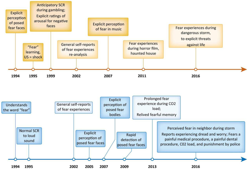
The overall constellation of findings (see Figure 2) suggests that S.M.’s deficits are not specific to some central fear system anchored in the amygdala. Instead, S.M. may have difficulty in recognizing wide-eyed gasping faces because she is less able to orient to human gaze when others find it useful and informative. This would leave S.M. at sea when required to predict threats and rewards in all but the most intense circumstances. For example, S.M. is unable to experience fear in a variety of situations, like watching horror movies, walking through a haunted house, and viewing live snakes and spiders. Yet she experiences intense fear when breathing air with high concentrations of carbon dioxide that leave her oxygen-hungry; she also spontaneously reports experiencing dread and worry in her everyday life. Similarly, S.M. is unable to learn that a neutral, visual cue predicted a mild threat like an electric shock, even after she had experienced their pairing many times (i.e., Pavlovian conditioning). But S.M. can indeed learn fear in everyday life, by mere association, when the threats are intense enough. For example, she has avoided seeking medical treatment several times because of pain she experienced on previous occasions.
Figure 2. An Abbreviated Summary of Published Observations About S.M.’s Impairments and Abilities.
Impairments are displayed on orange time line; abilities on blue time line. References can be found in [1]. Posed fear face = a posed, wide-eyed gasping face. US = unconditioned stimulus in Pavlovian conditioning. SCR = skin conductance response, which is a measure of sympathetic nervous system activity. Negative faces refers to posed portrayals of scowling (anger), wide-eyed gasping (fear), frowning (sadness) and nose-wrinkled (disgust) expressions. Re-analysis refers to data that were re-analyzed by a different group of scientists. C02 load = breathing air that has a higher concentration of carbon dioxide.
Finally, in addition to S.M.’s ability to experience fear in certain contexts, she is also able to perceive fear in many circumstances (again, see Figure 2). In the lab, S.M. has perceived fear in body postures that are posed to portray fear. In the context of her own life, S.M. has perceived her neighbor as fearful during a thunderstorm. And it’s important to note that in general, when it comes to facial expressions, widened eyes neither consistently [14] nor specifically express [15] fear. Whether S.M. can perceive fear in more naturalistic facial movements that occur in their everyday context and that may not always involve widened eyes remains an open question that awaits future research.
Acknowledgements
The paper was supported by grants from the U.S. Army Research Institute for the Behavioral and Social Sciences (W911NF-16-1-019), the National Cancer Institute (U01 CA193632) and the National Institute of Mental Health (R01 MH113234 and R01 MH109464), and the National Science Foundation (CMMI 1638234). The views, opinions, and/or findings contained in this paper are those of the authors and shall not be construed as an official U.S. Department of the Army position, policy, or decision, unless so designated by other documents.
Footnotes
Publisher's Disclaimer: This is a PDF file of an unedited manuscript that has been accepted for publication. As a service to our customers we are providing this early version of the manuscript. The manuscript will undergo copyediting, typesetting, and review of the resulting proof before it is published in its final citable form. Please note that during the production process errors may be discovered which could affect the content, and all legal disclaimers that apply to the journal pertain.
References
- 1.Feinstein JS, Adolphs R, and Tranel D, A tale of survival from the world of Patient S.M., in Living without an amygdala, Amaral DA and Adolphs R, Editors. 2016, Guilford: New York: p. 1–38. [Google Scholar]
- 2.Barrett LF and Satpute AB, Historical pitfalls and new directions in the neuroscience of emotion. Neurosci Lett, 2017. [DOI] [PMC free article] [PubMed] [Google Scholar]
- 3.Gross CT and Canteras NS, The many paths to fear. Nat Rev Neurosci, 2012. 13(9): p. 651–8. [DOI] [PubMed] [Google Scholar]
- 4.Adolphs R, et al. , Impaired recognition of emotion in facial expressions following bilateral damage to the human amygdala. Nature, 1994. 372(6507): p. 669–672. [DOI] [PubMed] [Google Scholar]
- 5.Adolphs R, et al. , Fear and the human amygdala. J Neuroscience, 1995. 15: p. 5879–5891. [DOI] [PMC free article] [PubMed] [Google Scholar]
- 6.Kawashima R, et al. , The human amygdala plays an important role in gaze monitoring: A PET study. Brain, 1999. 122: p. 779–783. [DOI] [PubMed] [Google Scholar]
- 7.Whalen PJ, et al. , Human amygdala responsivity to masked fearful eye whites. Science, 2004. 306(5704): p. 2061. [DOI] [PubMed] [Google Scholar]
- 8.Adams RB Jr., et al. , Effects of gaze on amygdala sensitivity to anger and fear faces. Science, 2003. 300: p. 1536. [DOI] [PubMed] [Google Scholar]
- 9.Whalen PJ, Fear, vigilance, and ambiguity: Initial neuroimaging studies of the human amygdala. Current Directions in Psychological Science, 1998. 7(6): p. 177–188. [Google Scholar]
- 10.Adolphs R, et al. , A mechanism for impaired fear recognition after amygdala damage. Nature, 2005. 433(7021): p. 68–72. [DOI] [PubMed] [Google Scholar]
- 11.Li SSY and McNally GP, The conditions that promote fear learning: prediction error and Pavlovian fear conditioning. Neurobiology of learning and memory, 2014. 108: p. 14–21. [DOI] [PubMed] [Google Scholar]
- 12.Johansen JP, et al. , Neural substrates for expectation-modulated fear learning in the amygdala and periacquiductal gray. Nature Neuroscience, 2010. 13(8): p. 979–86. [DOI] [PMC free article] [PubMed] [Google Scholar]
- 13.Minxha J, et al. , Fixations Gate Species-Specific Responses to Free Viewing of Faces in the Human and Macaque Amygdala. Cell Rep, 2017. 18(4): p. 878–891. [DOI] [PMC free article] [PubMed] [Google Scholar]
- 14.Duran JI, Reisenzein R, and Fernandez-Dols J-M, Coherence between emotions and facial expressions: A research synthesis, in The science of facial expressions, Fernandez-Dols J-M and Russell JA, Editors. 2017, Oxford University Press: New York: p. 107–129. [Google Scholar]
- 15.Crivelli C, et al. , The fear gasping face as a threat display in a Melanesian society. Proc Natl Acad Sci U S A, 2016. 113(44): p. 12403–12407. [DOI] [PMC free article] [PubMed] [Google Scholar]


