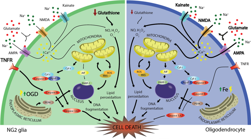
Figure 2. Molecular mechanisms of NG2 glia and oligodendrocyte cell death.
Both NG2 glia and oligodendrocytes are susceptible to oxidative and excitotoxic cell death. Low levels of glutathione render each cell vulnerable to reactive oxygen and nitrogen species that lead to mitochondrial-induced stress. Due to their high metabolic rates, oligodendrocytes have increased susceptibility to iron accumulation within their cell bodies and myelin membranes. Cell death pathways of greater severity are emphasized in bold. NMDA: N-methyl-Daspartate receptor, AMPA: α-amino-3-hydroxy-5-methyl-4-isoxazolepropionic acid receptor, TNFR: tumor necrosis factor receptor; NO: nitric oxide; Cyt c: cytochrome c; Apaf-1: apoptotic protease-activating factor 1; AIF: apoptosis-inducing factor, PARP-1: poly [ADP-ribose] polymerase 1, PERK: protein kinase R (PKR)-like endoplasmic reticulum kinase, ROS: reactive oxygen species; RNS: reactive nitrogen species; OGD: oxygen glucose deprivation.

