Abstract
Purpose
Provide a detailed assessment of peripheral refractive error and peripheral eye length in myopic children.
Methods
Subjects were 294 children aged 7 to 11 years with −0.75 to −5.00 diopter (D) of myopia by cycloplegic autorefraction. Peripheral refraction and eye length were measured at ±20° and ±30° horizontally and vertically, with peripheral refraction also measured at ±40° horizontally.
Results
Relative peripheral refraction became more hyperopic in the horizontal meridian and more myopic in the vertical meridian with increasing field angle. Peripheral eye length became shorter in both meridians with increasing field angle, more so horizontally than vertically with correlations between refraction and eye length ranging from −0.40 to −0.57 (all P < 0.001). Greater foveal myopia was related to more peripheral hyperopia (or less peripheral myopia), shorter peripheral eye lengths, and a consistent average asymmetry between meridians.
Conclusions
Peripheral refractive errors in children do not appear to exert strong local control of peripheral eye length given that their correlation is consistently negative and the degree of meridional asymmetry is similar across the range of refractive errors. The BLINK study will provide longitudinal data to determine whether peripheral myopia and additional peripheral myopic defocus from multifocal contact lenses affect the progression of myopia in children.
Translational Relevance
Local retinal control of ocular growth has been demonstrated numerous times in animal experimental myopia models but has not been explored in detail in human myopia development. These BLINK baseline results suggest that children's native peripheral optical signals may not be a strong stimulus for local growth responses.
Keywords: myopia, refractive error, peripheral refraction, contact lenses
Introduction
The prevalence of myopia has increased in recent decades in the Unites States and throughout the developed world, particularly in Asia.1–3 For example, 25% of adults in the United States were categorized as myopic in the National Health and Nutrition Examination Survey during 1971 and 1972, but that number rose to 33.1% when data were collected from 1999 to 2004.4 The prevalence of myopia was estimated to be 26.4% in Asian adults aged 30 to 39 years, but was 47.3% for those aged 20 to 29 years, according to a 2015 meta-analysis.2 The risk of pathological complications such as glaucoma, cataract, retinal detachment, and myopic maculopathy, as well as the economic impact and burden of healthcare associated with myopia, have strongly motivated researchers to explore treatments to reduce the risk of onset and the rate of myopic refractive error progression.5–7
Many previous treatments to control myopia progression were based on reducing hyperopic foveal defocus because exposure of the central retina to optical defocus is known to alter the refractive error development of young eyes in many animal species.8–12 Based on the findings of several clinical trials, bifocal spectacles or progressive addition lenses have not found widespread clinical use for myopia control, however, because myopic progression was usually only minimally reduced in treated children compared to controls, on average by less than 0.20 diopters (D).13–20 More recent management strategies for myopia progression involve treating the peripheral retina using optical defocus. This approach has received significant attention since work in primates showed that foveal refractive error could be manipulated through peripheral optical defocus without the involvement of an intact fovea.21 The connection between peripheral optics and central refractive error has a long history.22,23 It is well established that myopic eyes are associated with a hyperopic relative peripheral refractive error24–26 and a less oblate overall shape than emmetropic or hyperopic eyes.27,28 What is less clear is whether these differences represent the cause of myopic refractive error or whether they are the effect of differences between central and peripheral ocular growth. Arguing against a causative role, relative peripheral hyperopia appears to occur nearly simultaneously with the development of myopia.25,29 In addition, relative peripheral hyperopic defocus has not been shown to be a strong signal for myopic progression.29–31 The higher amounts of relative peripheral hyperopia that are associated with more foveal myopia seem more likely to be the effect of increases in axial length that outpace increases in equatorial diameter.24,32
Regardless of the role played by peripheral defocus in the etiology of refractive error, there is growing evidence that myopic defocus that extends into the retinal periphery while the fovea experiences acceptably focused images, typically created by optically based treatments such as multifocal soft contact lenses or overnight orthokeratology, may be an effective inhibitor of axial elongation in humans.33–38 The evidence is incomplete, however, primarily due to limited duration of follow-up, typically 2 years or less, and assessment of a limited set of covariates. The overall goal of our research is to test the hypothesis that relative peripheral myopic defocus from center-distance multifocal soft contact lenses will slow the progression of myopia in children over a 3-year period. The total defocus signal experienced by the peripheral retina during treatment requires an understanding of the underlying peripheral refractive error in combination with the optics of multifocal soft contact lenses. In addition, assessment of this optical signal and corresponding eye lengths in various quadrants will help to determine the spatial integration properties of that combined signal, whether peripheral refractive error influences eye growth in a quadrant-specific local fashion, or more globally across quadrants.39,40 The purpose of the current investigation is to describe the peripheral refractive error, the corresponding peripheral eye lengths, and the relationship between the two in a group of myopic children between the ages of 7 and 11 years prior to initiating myopia control treatment in a National Eye Institute/National Institutes of Health-supported randomized clinical trial of multifocal soft contact lenses, the Bifocal Lenses in Nearsighted Kids (BLINK) study.
Methods
Subjects
A total of 294 myopic children ages 7 to 11 years (inclusive) were enrolled between September 2014 and June 2016 in a double-masked, 3-year, randomized clinical trial at two sites (University of Houston College of Optometry and The Ohio State University College of Optometry). The purpose of the BLINK Study is to determine if children wearing multifocal soft contact lenses have a slower rate of myopia progression than those wearing single vision soft contact lenses. Eligible subjects had +0.10 logMAR or better best-corrected high-contrast distance visual acuity in each eye, −0.75 to −5.00 D of myopic refractive error in the most hyperopic meridian, 1.00 D or less astigmatism in each eye, and 2.00 D or less of anisometropia by cycloplegic autorefraction. All subjects were free of eye disease or binocular vision problems (e.g., strabismus, amblyopia, corneal disease, etc.) that could affect vision or contact lens wear and were free of systemic diseases that may affect vision, vision development, or contact lens wear (e.g., diabetes, Down syndrome, etc.). Subjects were not allowed to have had more than 1 month of any myopia control treatment or wear of gas permeable, soft bifocal, or orthokeratology contact lenses (no child had any prior experience with myopia control treatment). Subjects were excluded if they chronically used medications that might affect immunity, such as oral or ophthalmic corticosteroids. The research adhered to the tenets of the Declaration of Helsinki, was approved by the institutional review boards at the University of Houston and The Ohio State University, and was monitored by an independent data and safety monitoring committee. Assent from children and parental permission was obtained from each subject and subject's parent/guardian, respectively. Full baseline characteristics and methods have been previously reported;41 the details and methods relevant to this portion of the study are described below.
Cycloplegic Central and Peripheral Refractive Error
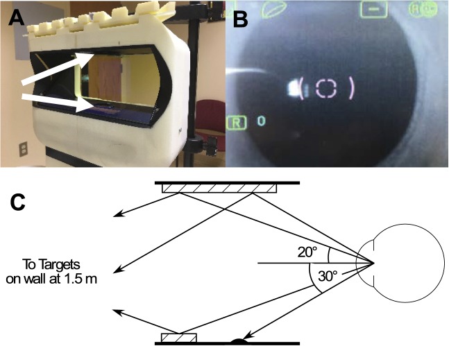
All measurements were taken at least 25 minutes after instillation of one drop of 0.5% proparacaine in each eye followed by two drops of 1.0% tropicamide instilled 5 minutes apart. Central and peripheral refractive error were measured on the right eye without spectacle or contact lens correction and with the left eye patched using the open-view Grand Seiko WAM-5500 binocular autorefractor/keratometer (AIT Industries, Bensenville, IL). Central refractive error was measured in primary gaze at 0°, then peripheral refractive error was measured at 20°, 30°, and 40° nasal and temporal gaze, and at 20° and 30° superior and inferior gaze. For horizontal readings the subject viewed spots on the wall 1.5 m from the subject created by a laser pointer held by a rigid tray with wells angled at 20°, 30°, and 40° nasally and temporally relative to the entrance pupil of the eye. The autorefractor instrument head was modified with a cutout at its edges in order to have an unobstructed peripheral view. The forehead rest was also removed and the subject's chin was placed on a custom chinrest that allowed the subject to turn the head (as opposed to the eyes) to maintain primary gaze for all peripheral measurements (Fig. 1). This method was chosen so that all peripheral study measurements, including those made during contact lens wear not reported here, would be done in a consistent manner but not be affected by lens decentration.42 Turning eyes compared to turning heads may help to avoid bias, although two reports show little difference between the two methods.22,43 All vertical targets were placed on the wall 1.5 m away except the 30° inferior gaze target, which was located within the housing of the autorefractor. For vertical readings, the forehead rest was used, and the subject turned the eyes to view each target in front-silvered mirrors within the autorefractor instrument head. At least five readings in which neither the sphere nor cylinder differed from the median by more than 1.00 D were recorded in each direction of gaze.
Figure 1.
(A) The experimental setup including the custom headrest to allow for rotation of a child's head in order to maintain fixation in primary gaze and the cutouts on the side to allow for targets at 40° eccentricity to be seen. White arrows point to the front-silvered mirrors for measurement of vertical peripheral refraction. (B) The appearance of the pupil with fixation at 40° eccentricity. The autorefractor is centered within the elliptical pupil without obstruction by the iris. (C) Schematic diagram of mirror placement within the autorefractor housing and angles to the illuminated targets placed on the wall at 1.5 m for 20° superior and inferior gaze and 30° superior gaze. Targets were beyond the rim of the autorefractor housing and not visible to the subject without the use of the mirrors (striped rectangles). For 30° inferior gaze, subjects fixated a target placed within the hood of the autorefractor in order to avoid a mirror obscuring its camera aperture. The autorefractor would be translated and re-focused during peripheral refraction to maintain alignment with the center of the entrance pupil.
Axial and Peripheral Eye Length
The Lenstar LS 900 (Haag-Streit USA, Mason, OH) optical biometer was used to measure central and peripheral eye length of the right eye. Measurements were made under cycloplegia. Central axial length was measured while covering the subject's contralateral eye with a patch while the subject fixated the internal red fixation light. Axial measurements were repeated until five readings without a poor-quality warning indicator or red highlight indicating implausible measurement were obtained. Peripheral measurements were obtained by having the subject turn the right eye (left eye patched) to look at small targets placed on the face of the instrument at 20° and 30° nasal and temporal gaze and 20° and 30° superior and inferior gaze. The peripheral measurements were also repeated until five reliable readings without red highlights indicating implausible measurements were obtained. Peripheral eye length measurements with the Lenstar LS 900 made away from the line of sight using these same methods have similar repeatability to measurements made along the line of sight.44
Peripheral refraction was calculated in absolute terms as the average spherical equivalent refractive error and in relative terms as the difference between the average peripheral minus central spherical equivalent. Relative peripheral refraction was also calculated for the most hyperopic meridian (sagittal) and the most myopic meridian (tangential). Eye length was calculated in absolute terms and in relative terms as the difference between the average peripheral minus central eye length. Left gaze (temporal field, nasal retina) was coded as a negative angle and right gaze (nasal field, temporal retina) as positive. For the vertical direction, upgaze (inferior field, superior retina) was coded as a negative angle and downgaze (superior field, inferior retina) was coded as a positive angle, consistent with the sign conventions used by Verkicharla et al.32 The relationship between peripheral refraction and peripheral eye length was explored using bivariate Pearson correlations. The group average refractive error and eye length as a function of gaze angle were fit by quadratic equations. To facilitate comparisons with adult data from Verkicharla et al.,32 these fits were also done on individual-level data with the coefficient for the quadratic term then plotted as a function of central spherical equivalent.
Results
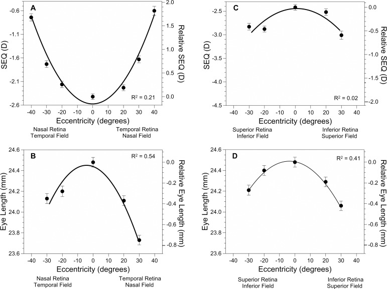
The baseline peripheral refraction and peripheral eye length data are given in Table 1. Figures 2A and 2B display these data for horizontal peripheral refraction and peripheral eye length. Refractive errors were myopic on average at all measured eccentricities across the visual field. Both lateral fields were less myopic than the mean ± SD central refractive error of −2.42 ± 1.04 D. The amount of relative peripheral hyperopia reached values of about +1.75 D at 40° with little nasal-temporal asymmetry. Peripheral eye length became shorter with greater eccentricity in the horizontal meridian relative to the central axial length of 24.48 ± 0.81 mm. There was about 0.4 mm of nasal-temporal asymmetry with the nasal retinal eye length shorter by roughly −0.4 mm and the temporal retinal eye length shorter by −0.8 mm at 30° compared to the central axial length. Figures 2C and 2D display data for vertical peripheral refraction and peripheral eye length. Peripheral refraction at 30° in the vertical meridian was more myopic compared to central refractive error by about −0.4 D for the superior retina and showed minimal asymmetry compared to the inferior retina (Table 1). Peripheral eye length became shorter with eccentricity to a lesser extent in the vertical meridian than in the horizontal meridian, as indicated by the shallower curve for vertical peripheral eye length in Figure 2D compared to horizontal eye length in Figure 2B. Peripheral eye length was only shorter by roughly −0.3 mm superiorly and −0.4 mm inferiorly at 30° compared to the central axial length.
Table 1.
Peripheral Refraction and Peripheral Eye Length Data (mean ± SD) as a Function of Eccentricity in the Horizontal and Vertical Meridians
| Eccentricity (°) |
n |
Peripheral Refraction (D) |
Most Hyperopic Meridian (D) |
Most Myopic Meridian (D) |
| Horizontal | ||||
| −40 (nasal retina) | 294 | −0.74 ± 1.32 | 0.00 ± 1.27 | −1.50 ± 1.47 |
| −30 | 294 | −1.73 ± 1.24 | −1.26 ± 1.25 | −2.20 ± 1.27 |
| −20 | 294 | −2.16 ± 1.17 | −1.77 ± 1.19 | −2.56 ± 1.20 |
| 0 | 294 | −2.42 ± 1.04 | −2.17 ± 1.03 | −2.68 ± 1.06 |
| 20 | 294 | −2.23 ± 1.08 | −1.70 ± 1.11 | −2.76 ± 1.10 |
| 30 | 294 | −1.63 ± 1.26 | −0.69 ± 1.23 | −2.59 ± 1.38 |
| 40 (temporal retina) | 294 | −0.60 ± 1.62 | 0.87 ± 1.43 | −2.08 ± 1.96 |
| Vertical | ||||
| −30 (superior retina) | 294 | −2.83 ± 1.22 | −1.73 ± 1.10 | −3.94 ± 1.44 |
| −20 | 294 | −2.88 ± 1.09 | −2.25 ± 1.01 | −3.52 ± 1.22 |
| 0 | 294 | −2.42 ± 1.04 | −2.17 ± 1.03 | −2.68 ± 1.06 |
| 20 | 294 | −2.52 ± 1.15 | −1.98 ± 1.09 | −3.06 ± 1.27 |
| 30 (inferior retina) | 292 | −3.01 ± 1.50 | −2.12 ± 1.41 | −3.91 ± 1.67 |
Columns labelled “relative” are measurements relative to the central value.
Figure 2.
Plots of both absolute and relative peripheral refraction (A) and absolute and relative peripheral eye length (B) in the horizontal meridian and absolute and relative peripheral refraction (C) and absolute and relative peripheral eye length (D) in the vertical meridian. Error bars represent the standard error of the mean. The best-fit parabola is shown for each. The equations for horizontal peripheral refraction and eye length, respectively, are y = 0.0012 x2 + 0. 0009 x − 2.57 and y = −0.00059 x2 − 0.0056x − 24.44. The equations for vertical peripheral refraction and eye length, respectively, are y = −0.00054 x2 + 0.0008 x − 2.44 and y = −0.00040 x2 − 0.0026x − 24.49. Each term is significantly different from 0 (P < 0.001) except for the linear term for peripheral refraction. The model R2 was computed by squaring the correlation between predicted and observed values.
Table 1.
Extended
| Eccentricity (°) |
Relative Peripheral Refraction (D) |
n |
Peripheral Eye Length (mm) |
Relative Peripheral Eye Length (mm) |
| Horizontal | ||||
| −40 (nasal retina) | 1.68 ± 1.07 | |||
| −30 | 0.69 ± 0.72 | 294 | 24.13 ± 0.84 | −0.36 ± 0.24 |
| −20 | 0.26 ± 0.55 | 294 | 24.20 ± 0.88 | −0.28 ± 0.26 |
| 0 | 0.00 | 294 | 24.48 ± 0.81 | 0.00 |
| 20 | 0.19 ± 0.55 | 294 | 24.11 ± 0.81 | −0.37 ± 0.17 |
| 30 | 0.78 ± 0.91 | 293 | 23.73 ± 0.79 | −0.76 ± 0.24 |
| 40 (temporal retina) | 1.82 ± 1.44 | |||
| Vertical | ||||
| −30 (superior retina) | −0.41 ± 0.83 | 294 | 24.21 ± 0.84 | −0.27 ± 0.25 |
| −20 | −0.47 ± 0.56 | 294 | 24.40 ± 0.82 | −0.09 ± 0.17 |
| 0 | 0.00 | 294 | 24.48 ± 0.81 | 0.00 |
| 20 | −0.10 ± 0.60 | 294 | 24.29 ± 0.81 | −0.19 ± 0.14 |
| 30 (inferior retina) | −0.59 ± 1.20 | 292 | 24.06 ± 0.78 | −0.43 ± 0.22 |
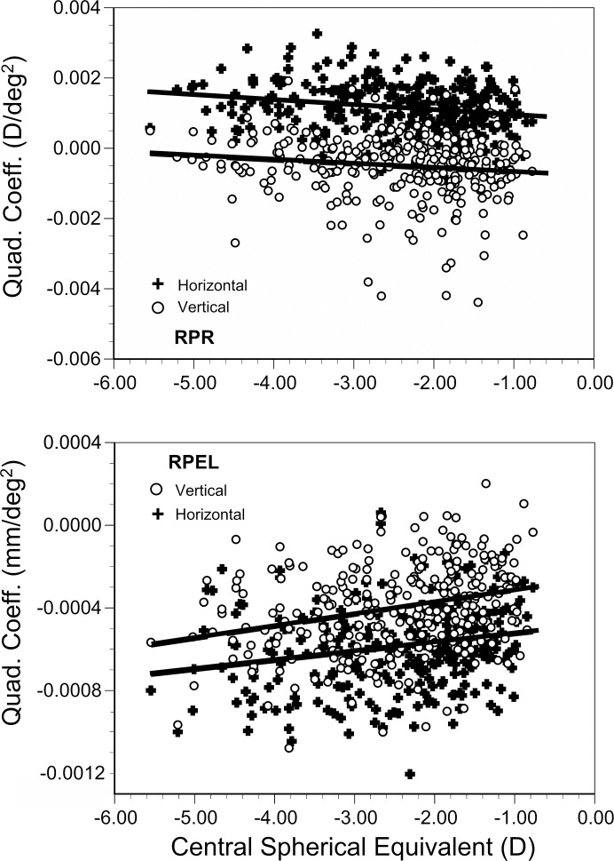
The coefficients for the parabolic fits of individual subject data for peripheral refraction and peripheral eye length are shown as a function of central spherical equivalent in Figures 3A and 3B. A positive sign for the coefficient indicates an upward-turned parabola (as opposed to negative being turned downward) and values farther from zero indicate a faster rate of change with eccentricity (as opposed to zero being a flat profile). Consistent with the upward-turned curve for peripheral refraction in Figure 2A and the downward-turned curve in Figure 2C, the average (±SD) coefficient for horizontal peripheral refraction was +0.0012 ± 0.0007, significantly different from the average (± SD) coefficient of −0.00053 ± 0.0010 for vertical peripheral refraction (P < 0.0001). The larger value of the coefficient for the horizontal meridian is consistent with the larger amount of relative peripheral hyperopia horizontally compared to relative peripheral myopia vertically (Figs. 2A, 2C). Negative coefficients were rare for horizontal peripheral refraction (4%) while some of the vertical coefficients were positive (26%). This finding indicates almost all myopic eyes had relative peripheral hyperopia in the horizontal meridian and the majority had relative peripheral myopia in the vertical meridian. The sign of the slope of the regression between the coefficients for the parabolic fits for peripheral refractive error and central refractive error was negative and significantly different from zero for both the horizontal and the vertical meridians (−0.00015 and −0.00013, P < 0.0001 and = 0.012, respectively) with no significant difference between the two (P = 0.78; test for interaction between meridian and refractive error). For the mostly positive coefficients horizontally, the negative slope indicates a greater amount of relative peripheral hyperopia when central refractive error was more myopic. For the mostly negative coefficients vertically, the negative slope indicates less relative peripheral myopia when central refractive error was more myopic.
Figure 3.
Plots of quadratic coefficients fit to subject-level data as a function of central spherical equivalent refractive error for peripheral refractive error (RPR) and peripheral eye length (RPEL). The horizontal meridian is represented by the plus (+) symbols and the vertical by the open circles (o). The equations for the best-fit regression lines for peripheral refraction in the horizontal and vertical meridians, respectively, are y = −0.00015x + 0.00080 (R2 = 0.05) and y = −0.00013x − 0.00086 (R2 = 0.02). The equations for the best-fit regression lines for peripheral eye length in the horizontal and vertical meridians, respectively, are y = 0.000044x − 0.00048 (R2 = 0.05) and y = 0.000056x − 0.00026 (R2 = 0.07).
Consistent with the downward-turned curve for peripheral eye length in Figures 2B and 2D, the average coefficients for the parabolic fits to horizontal and vertical peripheral eye length were both negative and significantly different from each other, −0.00059 ± 0.0002 and −0.00040 ± 0.0002, respectively (P < 0.0001). The more negative coefficient for the horizontal meridian is consistent with the shorter horizontal compared to vertical peripheral eye lengths (Figs. 2B, 2D). Almost no coefficients were positive for horizontal (1%) or vertical (2%) peripheral eye length meaning almost all peripheral eye length measurements were shorter than the central axial length. The sign of the slope of the regression between the coefficients for the parabolic fits to peripheral eye length and central refractive error was positive and significantly different from zero for both the horizontal and the vertical meridians (0.000044 and 0.000056, P = 0.0002 and <0.00001, respectively) with no significant difference between the two (P = 0.27; test for interaction between meridian and refractive error). The positive slope in each meridian indicates shorter peripheral eye lengths when central refractive error was more myopic. The opposite signs for the slopes in Figures 3A and 3B are to be expected; more peripheral hyperopia and less peripheral myopia are consistent with shorter peripheral eye lengths.
Peripheral refraction and peripheral eye length were moderately correlated at each eccentricity, both horizontally and vertically (Table 2). Correlations ranged from −0.41 to −0.57 (all P < 0.001) and were negative in sign, as expected, indicating a more hyperopic or less myopic refractive error centrally and peripherally with shorter eye length. The magnitude and sign of the correlations were similar for relative peripheral refraction and relative peripheral eye length, ranging from −0.33 to −0.60. Correlation coefficients were fairly consistent across eccentricities in both meridians, with the possible exception of 30° inferior retina (−0.33, Table 2). The consistency in this relationship across meridians can also be seen in the similar magnitude of the correlation between the coefficients for the parabolic fits for peripheral refraction and peripheral eye length (−0.44 and −0.48 for the horizontal and vertical meridians, respectively; both P < 0.0001). Correlations between peripheral eye length and refractive error in either the sagittal or the tangential meridians were also similar in magnitude to those for spherical equivalent, ranging from −0.38 to −0.56 (data not shown, all P < 0.001).
Table 2.
Correlations Between Peripheral Refractive Error and Peripheral Eye Length (absolute and relative, horizontal and vertical; all P < 0.001)
| Eccentricity (°) |
Horizontal Peripheral Refraction and Eye Length |
Horizontal Relative Peripheral Refraction and Relative Peripheral Eye Length |
Vertical Peripheral Refraction and Eye Length |
Vertical Relative Peripheral Refraction and Relative Peripheral Eye Length |
| −30 (nasal or superior retina) | −0.57 | −0.59 | −0.47 | −0.58 |
| −20 | −0.51 | −0.53 | −0.47 | −0.54 |
| 0 | −0.41 | −0.41 | ||
| 20 | −0.44 | −0.51 | −0.47 | −0.40 |
| 30 (temporal or inferior retina) | −0.44 | −0.58 | −0.41 | −0.33 |
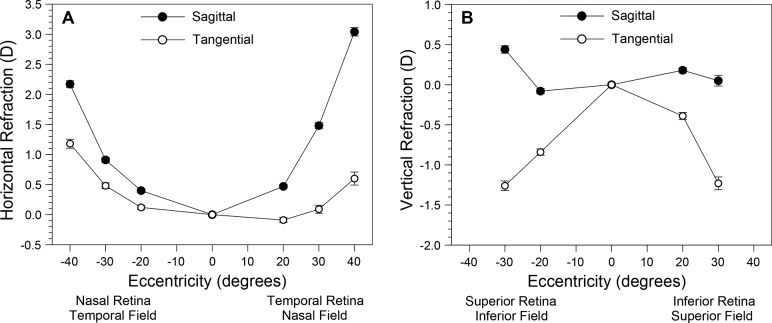
The sagittal and tangential meridians showed nasal-temporal asymmetry. The sagittal meridian became much more hyperopic than the minimal changes seen in the tangential meridian with increasing field angle for the temporal retina (Fig. 4). The nasal retina showed smaller increases in hyperopia in the sagittal meridian than the temporal retina, but greater increases than those in the tangential meridian. This nasal-temporal asymmetry in the sagittal and tangential meridians resulted in larger peripheral cylinder values for the temporal retina even though the nasal and temporal spherical equivalents were symmetric. The vertical meridian showed more symmetry between superior and inferior retina. The sagittal meridian changed little with field angle while the tangential meridian became more myopic.
Figure 4.
Relative peripheral refractive error in the sagittal and tangential meridians as a function of eccentricity and meridian: horizontal (A) and vertical (B). Error bars (some obscured) represent the standard error of the mean.
Discussion
Compared to central refractive error, myopic children in this study, on average, had a more hyperopic relative peripheral refractive error in the horizontal meridian and a more myopic relative peripheral refractive error in the vertical meridian. Compared to central axial length, peripheral eye length was shorter in both the horizontal and vertical meridians, but more so in the horizontal meridian, consistent with the relative hyperopia in that meridian. Both of these findings varied by the amount of refractive error. A more myopic central refractive error was associated with more positive coefficients for the parabolic fits for peripheral refraction and more negative coefficients for the parabolic fits for peripheral eye length, both horizontally and vertically. In other words, more foveal myopia was associated with more relative peripheral hyperopia horizontally and less relative peripheral myopia vertically. More foveal myopia was associated with shorter peripheral eye lengths in both the horizontal and vertical meridians. The simplest explanation for these effects is that the retinal profile is steeper when the amount of myopia is greater. The relationship between central refractive error and either the parabolic fit coefficients for peripheral refraction (Fig. 3A) or peripheral eye length (Fig. 3B) were not significantly different between the horizontal and the vertical meridians. These slopes were similar despite the underlying meridional asymmetry in peripheral refraction and eye length. Although these data are cross sectional and do not represent longitudinal development, the similarity of the slopes within Figure 3A and within Figure 3B suggests that the amount of retinal steepening with increased myopia occurs symmetrically for the two meridians. A 2-year longitudinal study did suggest that there was symmetric peripheral growth, finding that relative peripheral eye length did not change substantially in any quadrant in the growing eyes of children aged 7 to 11 years.45
These results expand on previous reports of peripheral retinal optical and structural properties in children in several ways. Data were collected from only one peripheral point, the nasal visual field/temporal retina at 30°, in the Orinda Longitudinal Study of Myopia and the Collaborative Longitudinal Evaluation of Ethnicity and Refractive Error.25,46 The Study of Theories about Myopia Progression reported on peripheral refraction in 85 myopic children aged 6 to 11 years from two horizontal and two vertical peripheral points with results that are generally consistent with the current findings at each location.47 The current study sampled from several more points: six peripheral points horizontally and 4 peripheral points vertically. The one peripheral point in common showed reasonable agreement, 0.50 D25 and 0.62 D47 of relative peripheral hyperopia compared to 0.79 D in the current study. The Peripheral Refraction in Preschool study measured refractive error at 4 peripheral points in 187 Asian children aged 3.4 to 15.8 years of age, but only in the horizontal meridian and without measures of peripheral eye length. Their results at 30° show more nasal-temporal asymmetry (1.0 D nasal field and 0.47 D temporal field) in peripheral hyperopia than the current study (0.79 D and 0.69 D, respectively) despite similar average central myopic refractive errors.29 Peripheral refractive error was also assessed in a very large number of Chinese children aged 7 and 14 years at four peripheral points, but again, only in the horizontal meridian and without eye length.31 The older children in that study had similar results to the current study: about 1.0 D of relative peripheral hyperopia at 30° without much nasal-temporal asymmetry. Interestingly, nasal-temporal asymmetry appeared after 1 year of follow-up with more relative peripheral hyperopia developing in the temporal than in the nasal visual field.31 Peripheral eye length data measured at 20° using a custom partial coherence biometer have been reported in a study of 140 children aged between 7 and 11 years, but peripheral refraction was not measured.45 Similar to the results in the current study, particularly in the fitted curves in Figures 2B and 2D, that study's baseline findings also showed meridional differences in peripheral eye length with the inferior retina steeper (a shorter peripheral eye length) than the superior, and the temporal retina steeper (shorter) than the nasal.45 In summary, the current study results are similar to those previously reported for myopic children but represent a unique dataset of peripheral refractive error and peripheral eye lengths taken on the same subjects at multiple points across the visual field in two meridians.
Detailed studies of peripheral refraction24 and peripheral eye length have been done in adults, with both measurements done on the same subjects in several studies.32,48–50 One such study where the average foveal myopia was −5.76 D showed larger amounts of relative peripheral hyperopia, about 2 D at 30°, compared to the current findings in children. Interestingly, about 1 D of relative peripheral hyperopia was seen in the vertical meridian48 in contrast to the relative peripheral myopia seen vertically in the current study and other studies of adults.24,32 As suggested by Figure 3, this may be due to the large amount of foveal myopia in that adult sample. A more typical amount of relative peripheral hyperopia at 40° in the horizontal meridian of adults would be about 1 to 1.5 D, similar to the current study. The amount of relative peripheral myopia in the vertical meridian of adults was about 0.75 to 1.00 D, slightly more than seen in the current study.24,32 Similar to the current results for children, studies of myopic adults find little asymmetry between the hemiretinas in peripheral refraction in either meridian.24,32,48
Peripheral eye length data do not seem to be as consistent as peripheral refraction data between adults and children. Peripheral eye lengths at 30° in adults are about 1.0 and 0.8 mm shorter than central lengths horizontally and vertically, respectively, clearly more than seen in the current results for children.24,32 Perhaps the more powerful crystalline lenses of children produce similar amounts of peripheral refractive error from smaller amounts of peripheral eye length differences. Studies of myopic adults tend to find little asymmetry between the hemiretinas in peripheral eye length in either meridian24,32 or only that the temporal retina was steeper than the nasal.49 In contrast, the inferior and temporal retina were both found to be steeper in children previously and in the current study.45 Differences in crystalline lens tilt or decentration, not measured in this study, may produce symmetric peripheral refraction profiles from asymmetric eye length contours. The strength of association between peripheral refractive error and peripheral eye length seems higher in adults than in the children. In contrast with the correlation coefficients ranging between −0.33 and −0.60 in the current study, the R2 values were between 0.5 and 0.7 in one study of adults49 and correlations between the parabolic coefficients for peripheral refraction and peripheral eye length were between −0.67 and −0.74 in another.32
The purpose of the BLINK Study is to investigate whether reducing relative peripheral hyperopia, or inducing relative peripheral myopia, using center-distance multifocal soft contact lenses alters the rate of myopic progression in children. Several clinical studies suggest that this approach will be effective in slowing the rate of progression.33–38 The presence of relative peripheral myopia in the vertical meridian in children and the suggestion of symmetric growth between the two meridians in these cross-sectional data is something of a challenge to the theory that underlies this approach to myopia control. If peripheral myopia is an effective “stop” signal, why doesn't the existing relative peripheral myopia vertically exert some effective myopia control or restrict the expansion of the globe in the vertical meridian relative to the horizontal meridian? One possibility is that the amount of relative peripheral myopia is too small in magnitude to be effective. Magnetic resonance imaging of the eye is consistent with the current study results, suggesting that the vertical meridian is the flatter, more expanded of the two.27,51 Inhibited ocular growth vertically would have produced the opposite result. The current baseline results suggest that children's native peripheral optical signals are not strong stimuli for local growth responses. Even small under-corrections of foveal refractive error by 0.50 to 0.75 D do not produce meaningful myopia control.52,53 Existing relative peripheral myopia vertically may not be sufficient to inhibit ocular expansion but perhaps additional myopic defocus would have this effect when applied over a larger amount of the peripheral visual field. Progressive addition lenses increase relative peripheral myopia when the eye is in primary gaze and have been shown to result in a small reduction in the rate of foveal myopic progression.15,16,18 There may be a dioptric threshold or critical amount of visual field required for the inhibitory effect. Once past this threshold, the inhibition may show dose-response behavior, such as in response to the two add strengths (+1.50 and +2.50 D) being evaluated in BLINK.41 Examining longitudinal results from the BLINK Study clinical trial with respect to the ocular expansion of the four measured quadrants in relation to the optical signal from the combination of the eye's own relative peripheral refractive error and the optics of the multifocal soft contact lenses may provide some insights into these issues.
Acknowledgments
Supported by the National Institutes of Health, National Eye Institute (U10-EY023204, U10-EY023206, U10-EY023208, U10-EY023210, P30-EY007551) and National Center for Advancing Translational Sciences (UL1TR002733).
Contact lens solutions for the study were provided by Bausch & Lomb.
ClinicalTrials.gov Registration: NCT02255474
Disclosure: D.O. Mutti, Bausch & Lomb (F), Johnson & Johnson Vision Care (F), Welch Allyn (advisory board) (C, R); L.T. Sinnott, Bausch & Lomb (F), Johnson & Johnson Vision Care (F); K.S. Reuter, Bausch & Lomb (F); M.K. Walker, Bausch & Lomb (F), Tangible Science (F), Alcon (R), Paragon Vision Sciences (R), Bausch & Lomb Specialty Vision Products (R), ABB Optical Group (R), Contamac (R); D.A. Berntsen, Bausch & Lomb (F), Visioneering Technologies (advisory board) (C, R); L.A. Jones-Jordan, Bausch & Lomb (F), Alcon (F), Contamac (F), Oculus (F); J.J. Walline, Bausch & Lomb (F), SightGlass (C, R)
BLINK Study Group
Executive Committee
Jeffrey J. Walline (Study Chair), David A. Berntsen (UH Clinic Principal Investigator), Donald O. Mutti, (OSU Clinic Principal Investigator), Lisa A. Jones-Jordan (Data Coordinating Center Director), Donald F. Everett (NEI Program Official)
Chair's Center
Kimberly J. Shaw (Study Coordinator), Juan Huang (Investigator), Bradley E. Dougherty (Survey Consultant)
Data Coordinating Center
Loraine T. Sinnott (Biostatistician), Pamela Wessel (Project Coordinator, 2014–2017), Jihyun Lee (Research Programmer, 2015–2017), Luke A. Bobay (Data Coordinator, 2017–2018), Tina Opoku (Clinical Research Coordinator, 2018-present)
University of Houston Clinic Site
Laura Cardenas (Clinic Coordinator), Krystal L. Schulle (Unmasked Examiner), Dashaini V. Retnasothie (Unmasked Examiner, 2014–2015), Amber Gaume Giannoni (Masked Examiner), Anita Tićak Gostović (Masked Examiner), Maria K. Walker (Masked Examiner), Moriah A. Chandler (Unmasked Examiner, 2016-present), Mylan Nguyen (Data Entry, 2016–2017), Lea Hair (Data Entry, 2017-present)
Ohio State University Clinic Site
Jill A. Myers (Clinic Coordinator), Alex D. Nixon (Unmasked Examiner), Katherine M. Bickle (Unmasked Examiner), Gilbert E. Pierce (Unmasked Examiner), Kathleen S. Reuter (Masked Examiner), Dustin J. Gardner (Masked Examiner, 2014–2016), Andrew D. Pucker (Masked Examiner, 2015–2016), Matthew Kowalski (Masked Examiner, 2016–2017), Ann Morrison (Masked Examiner, 2017-present), Danielle J. Orr (Unmasked Examiner, 2018–present)
Data Safety and Monitoring Committee
Janet T. Holbrook (Chair), Jane Gwiazda (Member), Timothy B. Edrington (Member), John Mark Jackson (Member), Charlotte E. Joslin (Member)
References
- 1.Vitale S, Sperduto RD, Ferris FL., III Increased prevalence of myopia in the United States between 1971–1972 and 1999–2004. Arch Ophthalmol. 2009;127:1632–1639. doi: 10.1001/archophthalmol.2009.303. [DOI] [PubMed] [Google Scholar]
- 2.Pan CW, Dirani M, Cheng CY, Wong TY, Saw SM. The age-specific prevalence of myopia in asia: a meta-analysis. Optom Vis Sci. 2015;92:258–266. doi: 10.1097/OPX.0000000000000516. [DOI] [PubMed] [Google Scholar]
- 3.Holden BA, Fricke TR, Wilson DA, et al. Global prevalence of myopia and high myopia and temporal trends from 2000 through 2050. Ophthalmology. 2016;123:1036–1042. doi: 10.1016/j.ophtha.2016.01.006. [DOI] [PubMed] [Google Scholar]
- 4.Vitale S, Ellwein L, Cotch MF, Ferris FL, III, Sperduto R. Prevalence of refractive error in the United States, 1999–2004. Arch Ophthalmol. 2008;126:1111–1119. doi: 10.1001/archopht.126.8.1111. [DOI] [PMC free article] [PubMed] [Google Scholar]
- 5.Saw SM, Gazzard G, Shih-Yen EC, Chua WH. Myopia and associated pathological complications. Ophthalmic Physiol Opt. 2005;25:381–391. doi: 10.1111/j.1475-1313.2005.00298.x. [DOI] [PubMed] [Google Scholar]
- 6.Vitale S, Cotch MF, Sperduto R, Ellwein L. Costs of refractive correction of distance vision impairment in the United States, 1999–2002. Ophthalmology. 2006;113:2163–2170. doi: 10.1016/j.ophtha.2006.06.033. [DOI] [PubMed] [Google Scholar]
- 7.Flitcroft DI. The complex interactions of retinal, optical and environmental factors in myopia aetiology. Prog Retin Eye Res. 2012;31:622–660. doi: 10.1016/j.preteyeres.2012.06.004. [DOI] [PubMed] [Google Scholar]
- 8.Irving EL, Sivak JG, Callender MG. Refractive plasticity of the developing chick eye. Ophthalmic Physiol Opt. 1992;12:448–456. [PubMed] [Google Scholar]
- 9.Smith EL, III, Hung LF. The role of optical defocus in regulating refractive development in infant monkeys. Vision Res. 1999;39:1415–1435. doi: 10.1016/s0042-6989(98)00229-6. [DOI] [PubMed] [Google Scholar]
- 10.Shen W, Sivak JG. Eyes of a lower vertebrate are susceptible to the visual environment. Invest Ophthalmol Vis Sci. 2007;48:4829–4837. doi: 10.1167/iovs.06-1273. [DOI] [PubMed] [Google Scholar]
- 11.Barathi VA, Boopathi VG, Yap EP, Beuerman RW. Two models of experimental myopia in the mouse. Vision Res. 2008;48:904–916. doi: 10.1016/j.visres.2008.01.004. [DOI] [PubMed] [Google Scholar]
- 12.Howlett MH, McFadden SA. Spectacle lens compensation in the pigmented guinea pig. Vision Res. 2009;49:219–227. doi: 10.1016/j.visres.2008.10.008. [DOI] [PubMed] [Google Scholar]
- 13.Fulk GW, Cyert LA, Parker DE. A randomized trial of the effect of single-vision vs. bifocal lenses on myopia progression in children with esophoria. Optom Vis Sci. 2000;77:395–401. doi: 10.1097/00006324-200008000-00006. [DOI] [PubMed] [Google Scholar]
- 14.Edwards MH, Li RW, Lam CS, Lew JK, Yu BS. The Hong Kong progressive lens myopia control study: study design and main findings. Invest Ophthalmol Vis Sci. 2002;43:2852–2858. [PubMed] [Google Scholar]
- 15.Gwiazda J, Hyman L, Hussein M, et al. A randomized clinical trial of progressive addition lenses versus single vision lenses on the progression of myopia in children. Invest Ophthalmol Vis Sci. 2003;44:1492–1500. doi: 10.1167/iovs.02-0816. [DOI] [PubMed] [Google Scholar]
- 16.Correction of Myopia Evaluation Trial 2 Study Group for the Pediatric Eye Disease Investigator Group. Progressive addition lenses versus single vision lenses for slowing progression of myopia in children with high accommodative lag and near esophoria. Invest Ophthalmol Vis Sci. 2011;52:2749–2757. doi: 10.1167/iovs.10-6631. [DOI] [PMC free article] [PubMed] [Google Scholar]
- 17.Walline JJ, Lindsley K, Vedula SS, et al. Interventions to slow progression of myopia in children. Cochrane Database Syst Rev. 2011. CD004916. [DOI] [PMC free article] [PubMed]
- 18.Berntsen DA, Sinnott LT, Mutti DO, Zadnik K. A randomized trial using progressive addition lenses to evaluate theories of myopia progression in children with a high lag of accommodation. Invest Ophthalmol Vis Sci. 2012;53:640–649. doi: 10.1167/iovs.11-7769. [DOI] [PMC free article] [PubMed] [Google Scholar]
- 19.Cheng D, Woo GC, Drobe B, Schmid KL. Effect of bifocal and prismatic bifocal spectacles on myopia progression in children: three-year results of a randomized clinical trial. JAMA Ophthalmol. 2014;132:258–264. doi: 10.1001/jamaophthalmol.2013.7623. [DOI] [PubMed] [Google Scholar]
- 20.Huang J, Wen D, Wang Q, et al. Efficacy comparison of 16 interventions for myopia control in children: a network meta-analysis. Ophthalmology. 2016;123:697–708. doi: 10.1016/j.ophtha.2015.11.010. [DOI] [PubMed] [Google Scholar]
- 21.Smith EL, Hung LF, Huang J. Relative peripheral hyperopic defocus alters central refractive development in infant monkeys. Vision Res. 2009;49:2386–2392. doi: 10.1016/j.visres.2009.07.011. [DOI] [PMC free article] [PubMed] [Google Scholar]
- 22.Ferree CE, Rand G, Hardy C. Refraction for the peripheral field of vision. Arch Ophthalmol. 1931;5:717–731. [Google Scholar]
- 23.Rempt F, Hoogerheide J, Hoogenboom W. Peripheral retinoscopy and the skiagram. Ophthalmologica. 1971;162:1–10. doi: 10.1159/000306229. [DOI] [PubMed] [Google Scholar]
- 24.Atchison DA, Pritchard N, Schmid KL. Peripheral refraction along the horizontal and vertical visual fields in myopia. Vision Res. 2006;46:1450–1458. doi: 10.1016/j.visres.2005.10.023. [DOI] [PubMed] [Google Scholar]
- 25.Mutti DO, Hayes JR, Mitchell GL, et al. Refractive error, axial length, and relative peripheral refractive error before and after the onset of myopia. Invest Ophthalmol Vis Sci. 2007;48:2510–2519. doi: 10.1167/iovs.06-0562. [DOI] [PMC free article] [PubMed] [Google Scholar]
- 26.Sng CC, Lin XY, Gazzard G, et al. Peripheral refraction and refractive error in singapore chinese children. Invest Ophthalmol Vis Sci. 2011;52:1181–1190. doi: 10.1167/iovs.10-5601. [DOI] [PubMed] [Google Scholar]
- 27.Atchison DA, Pritchard N, Schmid KL, et al. Shape of the retinal surface in emmetropia and myopia. Invest Ophthalmol Vis Sci. 2005;46:2698–2707. doi: 10.1167/iovs.04-1506. [DOI] [PubMed] [Google Scholar]
- 28.Singh KD, Logan NS, Gilmartin B. Three-dimensional modeling of the human eye based on magnetic resonance imaging. Invest Ophthalmol Vis Sci. 2006;47:2272–2279. doi: 10.1167/iovs.05-0856. [DOI] [PubMed] [Google Scholar]
- 29.Sng CC, Lin XY, Gazzard G, et al. Change in peripheral refraction over time in Singapore Chinese children. Invest Ophthalmol Vis Sci. 2011;52:7880–7887. doi: 10.1167/iovs.11-7290. [DOI] [PubMed] [Google Scholar]
- 30.Mutti DO, Sinnott LT, Mitchell GL, et al. Relative peripheral refractive error and the risk of onset and progression of myopia in children. Invest Ophthalmol Vis Sci. 2011;52:199–205. doi: 10.1167/iovs.09-4826. [DOI] [PMC free article] [PubMed] [Google Scholar]
- 31.Atchison DA, Li SM, Li H, et al. Relative peripheral hyperopia does not predict development and progression of myopia in children. Invest Ophthalmol Vis Sci. 2015;56:6162–6170. doi: 10.1167/iovs.15-17200. [DOI] [PubMed] [Google Scholar]
- 32.Verkicharla PK, Suheimat M, Schmid KL, Atchison DA. Peripheral refraction, peripheral eye length, and retinal shape in myopia. Optom Vis Sci. 2016;93:1072–1078. doi: 10.1097/OPX.0000000000000905. [DOI] [PubMed] [Google Scholar]
- 33.Aller TA, Liu M, Wildsoet CF. Myopia control with bifocal contact lenses: a randomized clinical trial. Optom Vis Sci. 2016;93:344–352. doi: 10.1097/OPX.0000000000000808. [DOI] [PubMed] [Google Scholar]
- 34.Anstice NS, Phillips JR. Effect of dual-focus soft contact lens wear on axial myopia progression in children. Ophthalmology. 2011;118:1152–1161. doi: 10.1016/j.ophtha.2010.10.035. [DOI] [PubMed] [Google Scholar]
- 35.Sankaridurg P, Holden B, Smith E, III, et al. Decrease in rate of myopia progression with a contact lens designed to reduce relative peripheral hyperopia: one-year results. Invest Ophthalmol Vis Sci. 2011;52:9362–9367. doi: 10.1167/iovs.11-7260. [DOI] [PubMed] [Google Scholar]
- 36.Walline JJ, Jones LA, Sinnott LT. Corneal reshaping and myopia progression. Br J Ophthalmol. 2009;93:1181–1185. doi: 10.1136/bjo.2008.151365. [DOI] [PubMed] [Google Scholar]
- 37.Cho P, Cheung SW. Retardation of myopia in Orthokeratology (ROMIO) study: a 2-year randomized clinical trial. Invest Ophthalmol Vis Sci. 2012;53:7077–7085. doi: 10.1167/iovs.12-10565. [DOI] [PubMed] [Google Scholar]
- 38.Santodomingo-Rubido J, Villa-Collar C, Gilmartin B, Gutierrez-Ortega R. Myopia control with orthokeratology contact lenses in Spain: refractive and biometric changes. Invest Ophthalmol Vis Sci. 2012;53:5060–5065. doi: 10.1167/iovs.11-8005. [DOI] [PubMed] [Google Scholar]
- 39.Wallman J, Adams JI. Developmental aspects of experimental myopia in chicks: susceptibility, recovery and relation to emmetropization. Vision Res. 1987;27:1139–1163. doi: 10.1016/0042-6989(87)90027-7. [DOI] [PubMed] [Google Scholar]
- 40.Smith EL, Huang J, Hung LF, et al. Hemiretinal form deprivation: evidence for local control of eye growth and refractive development in infant monkeys. Invest Ophthalmol Vis Sci. 2009;50:5057–5069. doi: 10.1167/iovs.08-3232. [DOI] [PMC free article] [PubMed] [Google Scholar]
- 41.Walline JJ, Gaume Giannoni A, Sinnott LT, et al. A randomized trial of soft multifocal contact lenses for myopia control: baseline data and methods. Optom Vis Sci. 2017;94:856–866. doi: 10.1097/OPX.0000000000001106. [DOI] [PMC free article] [PubMed] [Google Scholar]
- 42.Ticak A, Walline JJ. Peripheral optics with bifocal soft and corneal reshaping contact lenses. Optom Vis Sci. 2013;90:3–8. doi: 10.1097/OPX.0b013e3182781868. [DOI] [PMC free article] [PubMed] [Google Scholar]
- 43.Radhakrishnan H, Charman WN. Peripheral refraction measurement: does it matter if one turns the eye or the head? Ophthalmic Physiol Opt. 2008;28:73–82. doi: 10.1111/j.1475-1313.2007.00521.x. [DOI] [PubMed] [Google Scholar]
- 44.Schulle KL, Berntsen DA. Repeatability of on- and off-axis eye length measurements using the lenstar. Optom Vis Sci. 2013;90:16–22. doi: 10.1097/OPX.0b013e3182780bfd. [DOI] [PMC free article] [PubMed] [Google Scholar]
- 45.Schmid GF. Association between retinal steepness and central myopic shift in children. Optom Vis Sci. 2011;88:684–690. doi: 10.1097/OPX.0b013e3182152646. [DOI] [PubMed] [Google Scholar]
- 46.Mutti DO, Sholtz RI, Friedman NE, Zadnik K. Peripheral refraction and ocular shape in children. Invest Ophthalmol Vis Sci. 2000;41:1022–1030. [PubMed] [Google Scholar]
- 47.Berntsen DA, Barr CD, Mutti DO, Zadnik K. Peripheral defocus and myopia progression in myopic children randomly assigned to wear single vision and progressive addition lenses. Invest Ophthalmol Vis Sci. 2013;54:5761–5770. doi: 10.1167/iovs.13-11904. [DOI] [PMC free article] [PubMed] [Google Scholar]
- 48.Ehsaei A, Mallen EA, Chisholm CM, Pacey IE. Cross-sectional sample of peripheral refraction in four meridians in myopes and emmetropes. Invest Ophthalmol Vis Sci. 2011;52:7574–7585. doi: 10.1167/iovs.11-7635. [DOI] [PubMed] [Google Scholar]
- 49.Ehsaei A, Chisholm CM, Pacey IE, Mallen EA. Off-axis partial coherence interferometry in myopes and emmetropes. Ophthalmic Physiol Opt. 2013;33:26–34. doi: 10.1111/opo.12006. [DOI] [PubMed] [Google Scholar]
- 50.Faria-Ribeiro M, Queiros A, Lopes-Ferreira D, Jorge J, Gonzalez-Meijome JM. Peripheral refraction and retinal contour in stable and progressive myopia. Optom Vis Sci. 2013;90:9–15. doi: 10.1097/OPX.0b013e318278153c. [DOI] [PubMed] [Google Scholar]
- 51.Atchison DA, Jones CE, Schmid KL, et al. Eye shape in emmetropia and myopia. Invest Ophthalmol Vis Sci. 2004;45:3380–3386. doi: 10.1167/iovs.04-0292. [DOI] [PubMed] [Google Scholar]
- 52.Chung K, Mohidin N, O'Leary DJ. Undercorrection of myopia enhances rather than inhibits myopia progression. Vision Res. 2002;42:2555–2559. doi: 10.1016/s0042-6989(02)00258-4. [DOI] [PubMed] [Google Scholar]
- 53.Adler D, Millodot M. The possible effect of undercorrection on myopic progression in children. Clin Exp Optom. 2006;89:315–321. doi: 10.1111/j.1444-0938.2006.00055.x. [DOI] [PubMed] [Google Scholar]


