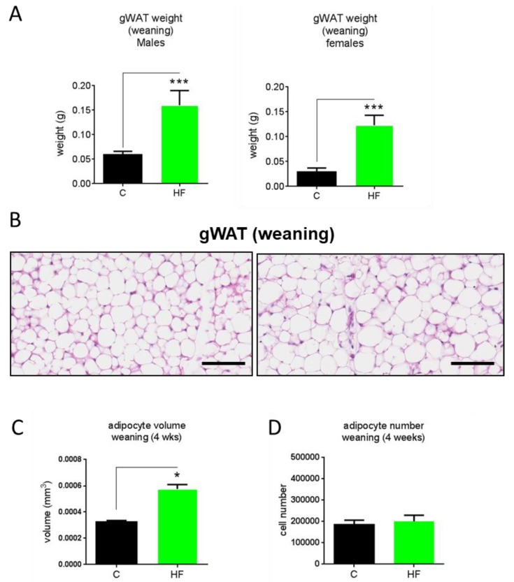
Figure 3.
Gonadal white adipose tissue weights at weaning (4 weeks old) in male and female offspring (A), representative gWAT histological images (H&E stain), scale bars 50 µM (B), calculated adipocyte volume (C) and number (D) of dams fed a chow (C) or high-fat (HF) diet for 6 weeks prior to pregnancy and during pregnancy and lactation. N = 5. * p < 0.05, *** p < 0.001.

