Abstract
A better understanding of the permittivity property of skeletal muscle is essential for the development of new diagnostic tools and approaches for neuromuscular evaluation. However, there remain important knowledge gaps in our understanding of this property in healthy and diseased skeletal muscle, which hinder its translation into clinical application. Here, we report the permittivity of gastrocnemius muscle in healthy wild type mice and murine models of spinal muscular atrophy, muscular dystrophy, diabetes, amyotrophic lateral sclerosis and in a model of myofiber hypertrophy. Data were measured ex vivo from 10 kHz to 1 MHz using the four-electrode impedance technique. Additional quantitative histology information were obtained. Ultimately, the normative data reported will offer the scientific community the opportunity to develop more accurate models for the validation and prediction of experimental observations in both pre-clinical and clinical neuromuscular disease research.
Subject terms: Neuromuscular disease, Computational biophysics, Mouse
| Design Type(s) | physiological data analysis objective • strain comparison design • ex vivo design |
| Measurement Type(s) | permittivity property |
| Technology Type(s) | impedance analyzer |
| Factor Type(s) | temporal_instant • frequency • Mouse Model • experimental condition |
| Sample Characteristic(s) | Mus musculus • skeletal muscle tissue |
Machine-accessible metadata file describing the reported data (ISA-Tab format)
Background & Summary
Electromagnetism constitutes a basic physical principle widely used in the field of biomedical engineering, designed to monitor and treat a broad spectrum of conditions including Parkinson’s disease1 and brain tumors2,3. Understanding how different biological tissues and fluids interact with electromagnetic fields is essential for improving the accuracy of existing analytical techniques as well as developing new diagnostic tools and therapies.
In electromagnetism, permittivity is one fundamental material parameter affecting the propagation of electromagnetic fields. When exposed to an electromagnetic field, the dipole moment of the material’s molecules opposes the external electric field and so the net electric field is reduced within the material. In other words, the permittivity is a measure of the ability to store an electric charge in the polarization of the material.
Basic and applied scientific endeavors have reported the permittivity property for well over 100 years in a collective effort to understand the propagation of electromagnetic fields in the human body4–13. The birth of biomedical engineering and the field of electrophysiology arose from these pioneers’ early multidisciplinary studies applying new theory, tools and methods. For example, voltage clamp and telegrapher’s equations made it possible to understand how ionic currents give rise to the action potential14. Another prominent contribution that emerged from studying tissues’ permittivity includes the development of the bidomain model to study cardiac muscle, which laid the foundation for the development of implantable cardiac pacemakers.
Previous studies have reported permittivity values of specific tissue as a function of the applied frequency. However, a recent meta-analysis revealed that major gaps in the knowledge of the frequency-dependence of the conductivity and relative permittivity in many tissues still exist15, especially for those tissues such as skeletal muscle, in which these properties are directionally dependent16–20. For example, prior authors acknowledged the technical limitations of their reported paravertebral muscle permittivity values nominally measured along and across the muscle fiber directions21.
In addition, some previous studies of the permittivity property of biological specimens did not specify the state of the tissue examined, even though it is known that the permittivity values change postmortem22 and with temperature23, nor did they specify the extent of disease, if any, present, and some did not include healthy control tissue for comparison. Other noteworthy factors that have not been exhaustively evaluated include the variation of tissues’ permittivity with age, gender, and disease progression. This missing information highlights critical gaps in our understanding of the factors that affect the permittivity property of biological tissues required to aid in identification of clinically abnormal results in pathological tissue.
The present study is motivated by the need to obtain permittivity values from both healthy and diseased skeletal muscle to aid in the diagnosis and monitoring of patients with neuromuscular disorders (NMDs). Analysis of muscle histology performed over the course of disease progression has shown that the composition and structure of skeletal muscle tissue changes as a function of the specific NMD. For example, in amyotrophic lateral sclerosis (ALS) muscle fibers tend to atrophy over time24, whereas in Duchenne muscular dystrophy, the lack of dystrophin protein25, causes a loss of muscle fibers and their progressive substitution by fat and fibrous tissue26.
Changes in the permittivity property in diseased muscle establishes the underlying scientific premise of electrical impedance myography (EIM)27,28, a relatively new electrodiagnostic technique that is gradually finding its niche for the assessment of NMD progression and the success of therapeutic intervention29. EIM is a non-destructive technique that is based on the measurement of the electrical impedance of individual muscles or groups of muscles. The permittivity property of the muscle can be extracted from the EIM values by accounting for the experimental setup. To date, however, EIM studies that have reported permittivity values of diseased muscle are scarce and often the data are incomplete, evaluating permittivity values at only a single frequency30. For EIM to achieve its full potential, it is paramount to first establish normative permittivity values in the frequency range where the histological alterations in disease would be expected to have an effect.
Here, using EIM technique, we report the conductivity and relative permittivity values of mouse muscle in healthy and four disease models (i.e., spinal muscular atrophy, muscular dystrophy, diabetes, amyotrophic lateral sclerosis) and in a drug-induced model of myofiber hypertrophy, which represents an all-encompassing effort by our group over the course of the last three years. The objective of this report is to equip the reader with a comprehensive database of the permittivity properties of healthy and diseased skeletal muscle from 10 kHz to 1 MHz, in both longitudinal and transverse directions. We also include additional quantitative histology data and periodic measurements obtained during disease progression. Providing the permittivity of healthy and diseased muscle using these various mouse models will open new venues for the development and improvement of the clinical diagnosis and monitoring of patients with NMD.
Methods
Terminology and definitions
The permittivity ε determines the dielectric behavior of materials when exposed to an applied electric field and it is defined as (dimensionless)
| 1 |
where εr is the relative permittivity (dimensionless), σ is the total conductivity of the material (S m−1), ω is the (angular) frequency of the field measured (rad s−1), ε0 = 8.85 · 10−12 is the permittivity of the vacuum (F m−1), and i is the imaginary unit (dimensionless).
In skeletal muscle, due to its highly organized cellular and fascicular structure, the permittivity is different along and perpendicular to the direction determined by the myofibers orientation31. Here, we calculated the longitudinal and transverse conductivity and relative permittivity from longitudinal and transverse resistance R and reactance X muscle data measured using a dielectric cell with the four-electrode technique,
| 2 |
where K (m−1) is a geometrical factor determined from measurements in saline solution.
Ethical approval and informed consent
All animal procedures were carried out in strict accordance with the recommendations in the Guide for the Care and Use of Laboratory Animals of the NIH and approved by the Institutional Animal Care and Use Committee at Beth Israel Deaconess Medical Center (Protocol #087–2016).
Animal experimentation
Animal experimentation was the same for all animals. All animal procedures were carried out in strict accordance with the recommendations in the Guide for the Care and Use of Laboratory Animals of the NIH and approved by the Institutional Animal Care and Use Committee at Beth Israel Deaconess Medical Center (Protocol #087–2016). Mice were given ad libitum access to food (Formulab Diet 5008, LabDiet, MO, USA) and water. Animals were allowed to acclimate at least 72 h prior to testing. Prior to measurements, animals were humanely euthanized with CO2 and the gastrocnemius muscle harvested from both left and right legs. Muscle impedance measurements were made immediately after with a heating pad under the dielectric cell to maintain a constant temperature 37 °C. Unless otherwise stated, the animals were obtained from Jackson Laboratories (JAX, Bar Harbor, ME, USA).
Spinal muscular atrophy (SMA) mice
Five spinal muscular atrophy (SMA) Model (“Smn 2B/2B-Neo”) (B6.129-Smn1tm1.1Cdid/tm1Cdid) mice were generated by intercrossing SMN 2B mice with SMN 2B-Neo mice. The official name of the Smn 2B allele is B6.129-Smn1tm1.1Cdid. 2B mice were generated from the progenitor line Smn 2B-Neo (B6.129-Smn1tm1Cdid) through removal of the flox-neo cassette32. Germline mice were subsequently crossed to C57BL/6J mice (JAX stock #000664) for at least 3 generations prior to use in these studies. The median survival of 2B/2B-Neo mice is ~13 months for males and 24 months for females. Details of this model will be reported elsewhere. Mice were studied at 40 weeks of age. Five wild type littermates served as controls for EIM analysis.
Muscular dystrophy (MDX) mice
The D2.B10 (DBA/2-congenic) Dmdmdx mouse (also referred in the literature as DBA/2J-mdx or D2-mdx mice) was chosen as a model of Duchenne muscular dystrophy model as it recapitulates several of the human characteristics of DMD myopathology including lower hind limb muscle weight, fewer myofibers, increased fibrosis and fat accumulation, and muscle weakness relative to strains with this mutant allele on other genetic backgrounds33–35. These genetically altered mice develop the disease without additional intervention and live at least one year. Fifteen male D2.B10-Dmdmdx/J mice hemizygous for Dmdmdx (6–9 weeks of age, JAX strain #013141) and studied at various ages from 6 to 43 weeks. Fifteen male wild type mice (DBA/2J, JAX strain #000671) served as controls (5 mice per time point).
Obese/Diabetic mice
The DB/DB mouse (BKS.Cg-Dock7m +/+ Leprdb/J) was chosen as a model of diabetes type II and obesity36–39. Mice homozygous for the diabetes spontaneous mutation (Leprdb) become obese at approximately three to four weeks of age. Elevations of plasma insulin begin at 10 to 14 days and elevations of blood sugar at four to eight weeks. Homozygous mutant mice are polyphagic, polydipsic, and polyuric. These mice are well known for their obesity and for developing substantial intramuscular fat deposition by approximately 8 weeks of age. Ten male mice (5 weeks, JAX strain #000642) were studied at 6 and 20 weeks (5 mice per time point). Ten WT type C57BLKS/J (JAX strain #000662) served as controls (5 mice per time point).
Amyotrophic lateral sclerosis (ALS) mice
Breeding pairs of ALS B6SJL-Tg(SOD1*G93A)1Gur/J mice (JAX strain #002726) were obtained and bred to obtain 37 animals (approximately half female and half male). To study varying fiber size40, animals were studied at various ages ranging from 8–18 weeks (approximately 6–7 animals per fortnight, at 8, 10, 12, 14, 16, and 18 weeks).
Mice with myofiber hypertrophy
Twenty male wild type mice (C57BL/6J, 8 weeks of age, JAX strain #000664) were obtained. Starting at 9 weeks of age, mice were divided randomly into two groups of 10 mice per group. Mice were treated twice weekly with subcutaneous injections of either phosphate-buffered saline (PBS) or the myostatin ligand trap ActRIIB-mFc (Acceleron Pharma, Cambridge, MA, USA) at a dose of 3.3 mg/kg. ActRIIB-mFc (also termed RAP-031)41 is a protein comprised of a form of the extracellular domain of ActRIIB fused to a mouse Fc that acts as a ligand trap to inhibit myostatin signaling. Animals were weighed weekly with an analytical balance (AS64, Adventurer SL, Ohaus Corporation, Pine Brook, NJ, USA) to ensure correct dosing throughout the course of the study. All procedures were performed after 5 weeks of treatment with ActRIIB-mFc. Dose-dependent impedance and functional data were published elsewhere42.
Ex vivo analysis of excised gastrocnemius muscle
Following euthanasia via CO2 inhalation, the gastrocnemius muscle was excised at its proximal extent just below the knee and distally by cutting the gastrocnemius tendon at its insertions, after removing the bicep femoris muscle. Each gastrocnemius muscle was trimmed with a scalpel to 5 mm (width) × 5 mm (length) centered at the belly of the muscle in order to fit into the dielectric measuring cell.
Dielectric cell
We used the dielectric cell described elsewhere43. Briefly, the dielectric cell was made of two flat plate stainless steel electrodes for applying electrical current side to side of the slab of muscle. The voltage electrodes were two monopolar EMG needles in contact the top surface of the muscle (Carefusion #902-DMG50-TP). The geometrical dimensions of the dielectric cell were 5 mm (width) × 5 mm (length), the muscle height measured with a caliper. The distance between the voltage electrodes was 4 mm. The slab of muscle was inserted into the cell first with the muscle fibers oriented parallel (longitudinal) and then perpendicular (transverse) to the current electrodes. The geometric factor K was determined from measurements of saline solution with the corresponding height of each tissue sample.
Four-electrode impedance measurements
Muscle resistance and reactance were collected from 9 to 862 kHz using a commercial impedance analyzer (EIM1103, Myolex, Inc., Boston, MA, USA).
Statistical analysis
Student’s t-tests, two-tailed, significance p = 0.05 was used to statistically examine the myofiber cross sectional area difference between healthy and diseased muscle tissue as quantified through histological assessment of the muscle.
Histological analysis
After ex vivo EIM measurements were completed, the left gastrocnemius was fixed in 10% formalin, embedded in paraffin and sectioned into 10 μm slices. Sections were stained with collagen VI antibodies (ab6588; Abcam, Cambridge, MA, USA) to identify the cell membrane and with 4′,6-diamidino- 2-phenylindole to stain nuclei. Individual sections were viewed at 20X and photographed with an epifluorescence microscope (Axio Imager M1, Zeiss, Oberkochen, Germany). Myofiber area was identified and analyzed with FIJI (ImageJ, NIH) using the muscle morphometry plug-in (Anthony Sinadinos using Eclipse IDE) to calculate the cross sectional area of the myofibers. On average, ~250 fibers were counted per muscle. Careful attention was paid to measure all cells that appeared to be myofibers, with the goal of measuring representative fields that accurately reflected the overall status of the muscle.
Data Records
The dataset described is freely available in PhysioNet44. The format of the data files is the same for healthy and diseased muscle conditions. The anisotropic permittivity property measured in longitudinal and transverse directions is organized in columns while the frequency dependence is organized in rows. For each time point measured, which also is organized in columns, the permittivity average value and the standard error of the mean are reported.
Technical Validation
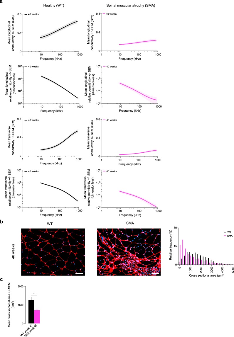
Our murine data on SMA (Fig. 1), MDX (Fig. 2), DB/DB (Fig. 3), ALS (Fig. 4), and a model of myofiber hypertrophy (Fig. 5) fill the gap of previous studies published in the literature. A single-frequency summary of conductivity values is provided in Table 1 (multi-frequency data are available in PhysioNet). Regarding the relative permittivity (not shown), we found the longitudinal values were greater than in transverse direction in the low frequencies, an observation that had been previously attributed to the polarization of the t-tubule muscle system20,31,45.
Fig. 1.
Permittivity in a mice model of spinal muscular atrophy. (a) Longitudinal and transverse conductivity and relative permittivity of healthy wild type (WT) and spinal muscular atrophy (SMA) mice. Mean ± standard error of the mean (SEM). (b) Representative histology images from the gastrocnemius muscle. Scale bar: 50 μm. Quantification of the myofiber cross sectional area (CSA) in WT and SMA mice. (c) Mean CSA. *p < 0.05.
Fig. 2.
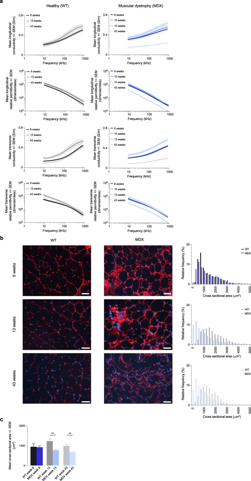
Permittivity in a mice model of muscular dystrophy. (a) Longitudinal and transverse conductivity and relative permittivity of healthy wild type (WT) and muscular dystrophy (MDX) mice. Mean ± standard error of the mean (SEM). (b) Representative histology images from the gastrocnemius muscle. Scale bar: 50 μm. Quantification of the myofiber cross sectional area (CSA) in WT and MDX mice. (c) Mean CSA. **p < 0.01.
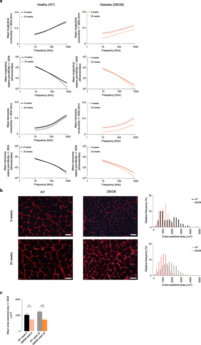
Fig. 3.
Permittivity in a mice model of diabetes. (a) Longitudinal and transverse conductivity and relative permittivity of healthy wild type (WT) and diabetes (DB/DB) mice. Mean ± standard error of the mean (SEM). (b) Representative histology images from the gastrocnemius muscle. Scale bar: 50 μm. Quantification of the myofiber cross sectional area (CSA) in WT and DB/DB mice. (c) Mean CSA. **p < 0.01; ***p < 0.001.
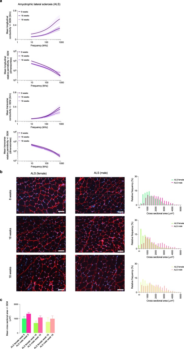
Fig. 4.
Permittivity in a mice model of amyotrophic lateral sclerosis. (a) Longitudinal and transverse conductivity and relative permittivity of male and female amyotrophic lateral sclerosis (ALS) mice. Mean ± standard error of the mean (SEM). (b) Representative histology images from the gastrocnemius muscle. Scale bar: 50 μm. Quantification of the myofiber cross sectional area (CSA) in male and female ALS mice. (c) Mean CSA.
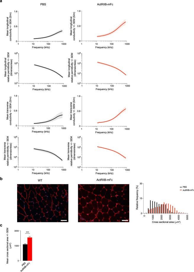
Fig. 5.
Permittivity in a mice model of myofiber hypertrophy. (a) Longitudinal and transverse conductivity and relative permittivity of healthy wild type (WT) mice treated with phosphate-buffered saline (PBS) and myostatin ligand trap ActRIIB-mFc. Mean ± standard error of the mean (SEM). (b) Representative histology images from the gastrocnemius muscle. Scale bar: 50 μm. Quantification of the myofiber cross sectional area (CSA) in WT treated with PBS and ActRIIB-mFc. (c) Mean CSA. ***p < 0.001.
Table 1.
Summary of skeletal muscle conductivity values reported at 50 kHz.
| Model (strain) |
Time point | Muscle | Direction | Conductivity (S m−1) |
|
|---|---|---|---|---|---|
| Li et al.30 | WT (C57Bl/6N) | 10 weeks | Mouse gastrocnemius | L | 0.5 |
| T | 0.15 | ||||
| ALS (SOD1*G93A) | 18 weeks | L | 0.5 | ||
| T | 0.3 | ||||
| MDX (10ScSn-DMDmdx/J) | 10 weeks | L | 0.7 | ||
| T | 0.4 | ||||
| Epstein et al.31 | WT (n.a.) | n.a. | Canine adductor magnus et brevis | L | 0.7 |
| T | 0.1 | ||||
| Sanchez et al.43 | WT (Wistar) | 14 weeks | Rat gastrocnemius | L | 0.6 |
| T | 0.25 | ||||
| Rush et al.46 | WT (Mongrel) | 14–30 kg | Canine longissimus dorsi, spinalis dorsi et cervicis | L | 0.35–0.81 |
| T | 0.04–0.1 | ||||
| Ahad et al.47 | WT (Wistar) | 140–160 g | Rat immature gastrocnemius | L | 0.44 ± 0.04 |
| T | 0.12 ± 0.01 | ||||
| 420–480 g | Rat mature gastrocnemius | L | 0.38 ± 0.02 | ||
| T | 0.15 ± 0.01 | ||||
| ALS (SOD1*G93A) | 8 weeks | Mouse gastrocnemius | L | 0.23 ± 0.05 | |
| T | 0.11 ± 0.03 | ||||
| 18 weeks | L | 0.11 ± 0.02 | |||
| T | 0.10 ± 0.01 | ||||
| D2-mdx (DBA/2-congenic) | 6 weeks | Mouse gastrocnemius | L | 0.36 ± 0.03 | |
| T | 0.24 ± 0.03 | ||||
| 10 weeks | L | 0.37 ± 0.04 | |||
| T | 0.25 ± 0.01 | ||||
| 13 weeks | L | 0.42 ± 0.03 | |||
| T | 0.33 ± 0.02 | ||||
| 43 weeks | L | 0.17 ± 0.01 | |||
| T | 0.11 ± 0.01 | ||||
| This work | WT for D2-mdx (DBA/2J) | 6 weeks | Mouse gastrocnemius | L | 0.28 ± 0.02 |
| T | 0.17 ± 0.01 | ||||
| 13 weeks | L | 0.31 ± 0.04 | |||
| T | 0.26 ± 0.03 | ||||
| 43 weeks | L | 0.27 ± 0.02 | |||
| T | 0.13 ± 0.01 | ||||
| SMA (Smn 2B/2B-Neo) | 40 weeks | Mouse gastrocnemius | L | 0.16 ± 0.02 | |
| T | 0.06 ± 0.01 | ||||
| DB/DB (BKS.Cg-Dock7m +/+ Leprdb/J) | 6 weeks | Mouse gastrocnemius | L | 0.31 ± 0.04 | |
| T | 0.20 ± 0.04 | ||||
| 20 weeks | L | 0.21 ± 0.01 | |||
| T | 0.09 ± 0.02 | ||||
| WT for DB/DB (C57BLKS/J) | 6 weeks | Mouse gastrocnemius | L | 0.34 ± 0.02 | |
| T | 0.20 ± 0.03 | ||||
| 20 weeks | L | 0.33 ± 0.01 | |||
| T | 0.14 ± 0.02 | ||||
| Mice (C57BL/6J) treated with ActRIIB-m Fc | 14 weeks | Mouse gastrocnemius | L | 0.34 ± 0.06 | |
| T | 0.26 ± 0.02 |
Abbreviations: L, longitudinal; T, transverse; n.a., not available.
In the absence of in vivo human muscle permittivity values, mouse muscle permittivity data reported here allow the user to approximate, through the use of analytic models, EIM values for the conditions studied. It also gives the user the ability to predict the temporal evolution with disease progression using the permittivity from different time points. For example, EIM phase angle θ can be computed in the longitudinal and transverse direction as follows
where f is the frequency in Hz. The reader can also use the permittivity reported to perform more accurate numerical simulation studies using finite element models for example.
ISA-Tab metadata file
Acknowledgements
This research was supported by grant NIH/NINDS 1R01NS091159 to S.B.R. and NIH/NINDS R01NS060926 to C.J.D. and with additional support from the Muscular Dystrophy Association (MDA418685) and CureSMA (DID1719) to C.J.D.
Author Contributions
Conceived and designed the experiments: J.A.N., C.J.D., S.B.R., B.S. Performed the experiments: J.A.N. Analyzed the data: J.A.N., B.S. Wrote the paper: B.S. All authors revised and approved the manuscript.
Code Availability
The algorithm used in this work to convert impedance data to permittivity is described in Eq. 2.
Competing Interests
Dr. Rutkove serves as scientific advisor and consultant to Myolex, Inc., a company that commercializes impedance technology. Dr. Rutkove is also a member of the company’s Board of Directors. Dr. Sanchez serves as a consultant to Myolex, Inc., and Impedimed, Inc., a company that develop impedance technology for research and clinical use. Dr. Sanchez and Dr. Rutkove are Co-Founders of Haystack Diagnostics, Inc., a company that commercializes needle impedance technology. Haystack Diagnostics, Inc., has the option to license patented needle impedance technology of which Dr. Sanchez and Dr. Rutkove are named inventors.
Footnotes
Publisher’s note: Springer Nature remains neutral with regard to jurisdictional claims in published maps and institutional affiliations.
ISA-Tab metadata
is available for this paper at 10.1038/s41597-019-0045-2.
References
- 1.Sabatini U, et al. Cortical motor reorganization in akinetic patients with Parkinson’s disease. A functional MRI study. Brain. 2000;123:394–403. doi: 10.1093/brain/123.2.394. [DOI] [PubMed] [Google Scholar]
- 2.Kirson ED, et al. Alternating electric fields arrest cell proliferation in animal tumor models and human brain tumors. Proc. Natl. Acad. Sci. 2007;104:10152–10157. doi: 10.1073/pnas.0702916104. [DOI] [PMC free article] [PubMed] [Google Scholar]
- 3.Rehman AA, Elmore KB, Mattei TA. The effects of alternating electric fields in glioblastoma: current evidence on therapeutic mechanisms and clinical outcomes. Neurosurg. Focus. 2015;38:E14. doi: 10.3171/2015.1.FOCUS14742. [DOI] [PubMed] [Google Scholar]
- 4.Maxwell, J. C. Electricity and magnetism (Oxford, 1892).
- 5.Fricke H. A Mathematical Treatment of the Electric Conductivity and Capacity of Disperse Systems ii. The Capacity of a Suspension of Conducting Spheroids Surrounded by a Non-Conducting Membrane for a Current of Low Frequency. Phys. Rev. 1924;24:678–681. doi: 10.1103/PhysRev.24.575. [DOI] [Google Scholar]
- 6.Fricke H, Morse S. The electric resistance and capacity of blood for frequencies between 800 and 4(1/2) million cycles. J. Gen. Physiol. 1925;9:153–67. doi: 10.1085/jgp.9.2.153. [DOI] [PMC free article] [PubMed] [Google Scholar]
- 7.Kaufman W, Johnston F. The electrical conductivity of the tissues near the heart and its bearing on the distribution of the cardiac action currents. Am. Heart J. 1943;26:42–54. doi: 10.1016/S0002-8703(43)90050-X. [DOI] [Google Scholar]
- 8.Schwan HP, Kay CF. The conductivity of living tissues. Ann. N. Y. Acad. Sci. 1957;65:1007–13. doi: 10.1111/j.1749-6632.1957.tb36701.x. [DOI] [PubMed] [Google Scholar]
- 9.Geddes LA, Costa CP, Wise G. The impedance of stainless-steel electrodes. Med. Biol. Eng. 1971;9:511–521. doi: 10.1007/BF02474708. [DOI] [PubMed] [Google Scholar]
- 10.Schwan HP. Electrical and acoustic properties of biological materials and biomedical applications. IEEE Trans. Biomed. Eng. 1984;31:872–8. doi: 10.1109/TBME.1984.325251. [DOI] [PubMed] [Google Scholar]
- 11.Surowiec A, Stuchly S, Keaney M, Swarup A. Dielectric Polarization of Animal Lung at radio Frequencies. IEEE Trans. Biomed. Eng. 1987;34:62–67. doi: 10.1109/TBME.1987.326016. [DOI] [PubMed] [Google Scholar]
- 12.Pethig R. Dielectric properties of body tissues. Clin. Phys. Physiol. Meas. 1987;8(Suppl A):5–12. doi: 10.1088/0143-0815/8/4A/002. [DOI] [PubMed] [Google Scholar]
- 13.Gabriel S, Lau RW, Gabriel C. The dielectric properties of biological tissues: II. Measurements in the frequency range 10 Hz to 20 GHz. Phys. Med. Biol. 1996;41:2251–2269. doi: 10.1088/0031-9155/41/11/002. [DOI] [PubMed] [Google Scholar]
- 14.Hodgkin AL, Huxley AF, Katz B. Measurement of current-voltage relations in the membrane of the giant axon of Loligo. J. Physiol. 1952;116:424–448. doi: 10.1113/jphysiol.1952.sp004716. [DOI] [PMC free article] [PubMed] [Google Scholar]
- 15.Gabriel C, Peyman A, Grant EH. Electrical conductivity of tissue at frequencies below 1 MHz. Phys. Med. Biol. 2009;54:4863–78. doi: 10.1088/0031-9155/54/16/002. [DOI] [PubMed] [Google Scholar]
- 16.Rush S. Methods of measuring the resistivities of anisotropic conducting media in situ. J. Res. Natl. Bur. Stand. Sect. C Eng. Instrum. 1962;66C:217. doi: 10.6028/jres.066C.018. [DOI] [Google Scholar]
- 17.Fatt P. An analysis of the transverse electrical impedance of striated muscle. Proc. R. Soc. London. Ser. B, Biol. Sci. 1964;159:606–51. doi: 10.1098/rspb.1964.0023. [DOI] [PubMed] [Google Scholar]
- 18.Clerc L. Directional Differences of Impulse Spread in Trabecular Muscle from Mammalian Heart. J. Physiol. 1976;255:335–346. doi: 10.1113/jphysiol.1976.sp011283. [DOI] [PMC free article] [PubMed] [Google Scholar]
- 19.Plonsey R, Barr RC. A critique of impedance measurements in cardiac tissue. Ann. Biomed. Eng. 1986;14:307–322. doi: 10.1007/BF02367405. [DOI] [PubMed] [Google Scholar]
- 20.Eisenberg, R. S. Impedance measurement of the electrical structure of skeletal muscle. In Handb. Physiol. Skelet. Muscle, chap. 11, 301–323 (John Wiley & Sons, Inc., 2010).
- 21.Gabriel, C. Compilation of the dielectric properties of body tissues at RF and microwave frequencies. Tech. Rep 1–273 (1996).
- 22.Surowiec A, Stuchly SS, Swarup A. Postmortem changes of the dielectric properties of bovine brain tissues at low radiofrequencies. Bioelectromagnetics. 1986;7:31–43. doi: 10.1002/bem.2250070105. [DOI] [PubMed] [Google Scholar]
- 23.Rossmanna C, Haemmerich D. Review of temperature dependence of thermal properties, dielectric properties, and perfusion of biological tissues at hyperthermic and ablation temperatures. Crit. Rev. Biomed. Eng. 2014;42:467–92. doi: 10.1615/CritRevBiomedEng.2015012486. [DOI] [PMC free article] [PubMed] [Google Scholar]
- 24.Tsitkanou, S., Gatta, P. A. & Russell, A. P. Skeletal muscle satellite cells, mitochondria, and MicroRNAs: Their involvement in the pathogenesis of ALS. Front Physiol.7, 403 (2016). [DOI] [PMC free article] [PubMed]
- 25.Hoffman EP, Brown RH, Kunkel LM. Dystrophin: The protein product of the duchenne muscular dystrophy locus. Cell. 1987;51:919–928. doi: 10.1016/0092-8674(87)90579-4. [DOI] [PubMed] [Google Scholar]
- 26.Deconinck N, Dan B. Pathophysiology of Duchenne Muscular Dystrophy: Current Hypotheses. Pediatr Neurol. 2007;36(1):1–7. doi: 10.1016/j.pediatrneurol.2006.09.016. [DOI] [PubMed] [Google Scholar]
- 27.Sanchez B, Rutkove SB. Electrical impedance myography and its applications in neuromuscular disorders. Neurotherapeutics. 2017;14:107–118. doi: 10.1007/s13311-016-0491-x. [DOI] [PMC free article] [PubMed] [Google Scholar]
- 28.Sanchez B, Rutkove SB. Present Uses, Future Applications, and Technical Underpinnings of Electrical Impedance Myography. Curr. Neurol. Neurosci. Rep. 2017;17:86. doi: 10.1007/s11910-017-0793-3. [DOI] [PubMed] [Google Scholar]
- 29.Rutkove SB, et al. Electrical impedance myography for assessment of Duchenne muscular dystrophy. Ann. Neurol. 2017;81:622–632. doi: 10.1002/ana.24874. [DOI] [PMC free article] [PubMed] [Google Scholar]
- 30.Li J, Jafarpoor M, Bouxsein M, Rutkove SB. Distinguishing neuromuscular disorders based on the passive electrical material properties of muscle. Muscle Nerve. 2015;51:49–55. doi: 10.1002/mus.24270. [DOI] [PMC free article] [PubMed] [Google Scholar]
- 31.Epstein BR, Foster KR. Anisotropy in the dielectric properties of skeletal muscle. Med. Biol. Eng. Comput. 1983;21:51–55. doi: 10.1007/BF02446406. [DOI] [PubMed] [Google Scholar]
- 32.Hammond SM, et al. Mouse survival motor neuron alleles that mimic SMN2 splicing and are inducible rescue embryonic lethality early in development but not late. PLoS One. 2010;5:e15887. doi: 10.1371/journal.pone.0015887. [DOI] [PMC free article] [PubMed] [Google Scholar]
- 33.Fukada S-I, et al. Genetic Background Affects Properties of Satellite Cells and mdx Phenotypes. Am. J. Pathol. 2010;176:2414–2424. doi: 10.2353/ajpath.2010.090887. [DOI] [PMC free article] [PubMed] [Google Scholar]
- 34.Coley WD, et al. Effect of genetic background on the dystrophic phenotype in mdx mice. Hum. Mol. Genet. 2016;25:130–145. doi: 10.1093/hmg/ddv460. [DOI] [PMC free article] [PubMed] [Google Scholar]
- 35.Rodrigues M, Echigoya Y, Fukada S-I, Yokota T. Current Translational Research and Murine Models For Duchenne Muscular Dystrophy. J. Neuromuscul. Dis. 2016;3:29–48. doi: 10.3233/JND-150113. [DOI] [PMC free article] [PubMed] [Google Scholar]
- 36.Hummel KP, Dickie MM, Coleman DL. Diabetes, a new mutation in the mouse. Science. 1966;153:1127–8. doi: 10.1126/science.153.3740.1127. [DOI] [PubMed] [Google Scholar]
- 37.Wang X, Hu Z, Hu J, Du J, Mitch WE. Insulin Resistance Accelerates Muscle Protein Degradation: Activation of the Ubiquitin-Proteasome Pathway by Defects in Muscle Cell Signaling. Endocrinology. 2006;147:4160–4168. doi: 10.1210/en.2006-0251. [DOI] [PubMed] [Google Scholar]
- 38.Ostler JE, et al. Effects of insulin resistance on skeletal muscle growth and exercise capacity in type 2 diabetic mouse models. Am. J. Physiol. Metab. 2014;306:E592–E605. doi: 10.1152/ajpendo.00277.2013. [DOI] [PMC free article] [PubMed] [Google Scholar]
- 39.Qiu S, et al. Increasing Muscle Mass Improves Vascular Function in Obese (db/db) Mice. J. Am. Heart Assoc. 2014;3:e000854. doi: 10.1161/JAHA.114.000854. [DOI] [PMC free article] [PubMed] [Google Scholar]
- 40.Kapur K, Nagy JA, Taylor RS, Sanchez B, Rutkove SB. Estimating Myofiber Size With Electrical Impedance Myography: a Study In Amyotrophic Lateral Sclerosis MICE. Muscle Nerve. 2018;58:713–717. doi: 10.1002/mus.26187. [DOI] [PMC free article] [PubMed] [Google Scholar]
- 41.Sanchez B, et al. Evaluation of Electrical Impedance as a Biomarker of Myostatin Inhibition in Wild Type and Muscular Dystrophy Mice. PLoS One. 2015;10:e0140521. doi: 10.1371/journal.pone.0140521. [DOI] [PMC free article] [PubMed] [Google Scholar]
- 42.Nagy JA, Kapur K, Taylor RS, Sanchez B, Rutkove SB. Electrical impedance myography as a biomarker of myostatin inhibition with ActRIIB-mFc: a study in wild-type mice. Futur. Sci. OA. 2018;4:FSO308. doi: 10.4155/fsoa-2018-0002. [DOI] [PMC free article] [PubMed] [Google Scholar]
- 43.Sanchez B, Li J, Bragos R, Rutkove SB. Differentiation of the intracellular structure of slow- versus fast-twitch muscle fibers through evaluation of the dielectric properties of tissue. Phys. Med. Biol. 2014;59:1–12. doi: 10.1088/0031-9155/59/10/2369. [DOI] [PMC free article] [PubMed] [Google Scholar]
- 44.Sanchez, B. Permittivity of Healthy and Diseased Skeletal Muscle. Physio Net, 10.13026/C23H3B (2019).
- 45.Cole, K. S. Membranes, Ions, and Impulses: A Chapter of Classical Biophysics (University of California Press, 1968).
- 46.Rush S, Abildskov JA, McFee R. Resistivity of body tissues at low frequencies. Circ. Res. 1963;12:40–50. doi: 10.1161/01.RES.12.1.40. [DOI] [PubMed] [Google Scholar]
- 47.Ahad MA, Fogerson PM, Rosen GD, Narayanaswami P, Rutkove SB. Electrical characteristics of rat skeletal muscle in immaturity, adulthood and after sciatic nerve injury, and their relation to muscle fiber size. Physiol. Meas. 2009;30:1415–27. doi: 10.1088/0967-3334/30/12/009. [DOI] [PMC free article] [PubMed] [Google Scholar]
Associated Data
This section collects any data citations, data availability statements, or supplementary materials included in this article.
Supplementary Materials
Data Availability Statement
The algorithm used in this work to convert impedance data to permittivity is described in Eq. 2.


