

Abstract
Patient: Female, 9
Final Diagnosis: Multifocal osteomyelitic tuberculosis at rare locations with metastatic tuberculosis abscess
Symptoms: Lumps at the left elbow joint • lateral side the left hand • lateral side of the left feet
Medication: —
Clinical Procedure: —
Specialty: Pediatrics and Neonatology
Objective:
Unusual clinical course
Background:
Multifocal tuberculosis (TB) with more than 1 tuberculous osteoarticular lesion is rare. Furthermore, metastatic tuberculous abscess (MTA) is also a very rare manifestation of cutaneous TB in children. A non-specific, often subtle, early clinical presentation in conjunction with a low prevalence rate constitute obstacles for diagnosis.
Case Report:
A 9-years old female patient was referred to Hasan Sadikin Pediatric Respirology Outpatient clinic from the Orthopedic Department with lumps at the left elbow joint, lateral side of the left hand, and lateral side of the left foot. Fine needle aspiration biopsy of the lumps suggested a chronic inflammation due to TB. The patient was then started with a course of anti-TB drugs consisting of rifampicin, isoniazid, pyrazinamide, and ethambutol. During the treatment course, she experienced anti-TB drug-induced hepatotoxicity (ADIH). We then switched the regimen to streptomycin and ethambutol for 2 weeks, then reintroduced treatment with the modified British Thoracic Society guidelines regimen. The nodules appeared shrunken after 3 months of treatment with anti-TB drugs.
Conclusions:
Increased awareness of unusual manifestations of TB will likely allow for proper diagnosis and management of this common infection. Accordingly, timely diagnosis and management will prevent further debilitating sequelae.
MeSH Keywords: Abscess, Osteomyelitis, Tuberculosis, Pediatrics
Background
Extraspinal musculoskeletal tuberculosis (TB) is uncommon, affecting about 2–5% of all TB cases [1]. It results from lympho-hematogenous dissemination of Mycobacterium tuberculosis from the lungs [2,3]. The diagnosis can be challenging because of the absence of classical symptoms of pulmonary TB such as fever, cough, night sweats, and weight loss [1,2]. Cutaneous TB occurs in 1% of all TB cases. Metastatic tuber-culous abscess (MTA) is a rare form of skin TB. It is characterized by nodule and abscess formation throughout the body [4]. Straightforward and timely use of appropriate antimicrobial therapy has virtually eliminated mortality and long-term sequel for this condition. However, the incidence of chronicity, deformity, and disability is substantial when diagnosis and management are delayed or incorrect.
Case Report
A 9-year-old female patient was referred to our Pediatric Pulmonology Outpatient Unit from the Orthopedic Department with lumps on the lateral side of her left hand, the lateral side of her left foot, and her left elbow (Figures 1–3). The lump on her hand first appear about 2 months before her admission. It started small, the size of a marble, and grew bigger and tender each day. At 3 weeks prior to admission, lumps on her left foot and left elbow started to appear. The complaint was accompanied with a low-grade fever. Weight loss were also noted by the parents about a month prior to admission. Her weight was 28 kg at 2 months prior to admission. There were no symptoms of cough, night sweat, or dyspnea. This was the first time the patient had any of these symptoms. Previously, she had been diagnosed as having pulmonary TB at 1-year of age and treated with anti-TB drugs for 6 months. The treatment was done at a local primary health care center, and consisted of isoniazid, rifampicin, and pyrazinamide in separate loose powder. We had no information regarding the dosage, but she had no anti-TB drug-induced hepatotoxicity (ADIH) during that treatment. There was no history of a similar complaint in the family, however, her father was previously diagnosed with extrapulmonary TB (lymphadenitis TB).
Figure 1.

Lump on the lateral side of the left hand.
Figure 2.

Lump on the lateral side of the left foot.
Figure 3.

Lump on the left elbow.
Upon admission, the patient’s vital signs were normal. She had normal body mass index (BMI) with short stature (height < −2 SD on the World Health Organization [WHO] growth standards [5]), her weight was 23 kg. The cervical lymph nodes were palpable, 0.5–1 cm in diameter, multiple, soft, bilateral, and non-tender. The lumps were 10×8×8 cm, 2×2×1 cm, and 3×3×2 cm in size, on the elbow, hand, and foot, respectively. The lumps on the foot and hand looked swollen and red with tenderness on palpation, while the lump on the elbow was not painful nor red. Blood work at the time of admission was within normal limits. Liver function test, namely aspartate aminotransferase (AST) 28 U/L and alanine aminotransferase (ALT) 18 U/L were within normal limits (normal reference value at our institution for AST: 35 U/L; ALT: 45 U/L). From the clinical findings, we made differential diagnosis of extrapulmonary TB (joint, bones, and skin) and bacterial infection, most likely staphylococcal or other mixed bacteria. Tuberculin skin test was 12 mm in induration. GenXpert result from induced sputum yielded no detected M. tuberculosis. Chest x-ray revealed pulmonary TB with lymphadenopathy on the right supra-hilar (Figure 4). X-ray of the hand and elbow showed an osteomyelitis of the proximal left ulnar (Figure 5) and 5th metacarpal of the left hand, with a spina ventosa depiction on the 5th meta-carpal (Figure 6). Fine needle aspiration biopsy (FNAB) of the lumps suggested a chronic inflammation caused by TB. Human immunodeficiency virus (HIV) test was negative.
Figure 4.

Chest x-ray showed signs of tuberculosis: lymphadenopathy on the right supra-hilar (arrow).
Figure 5.
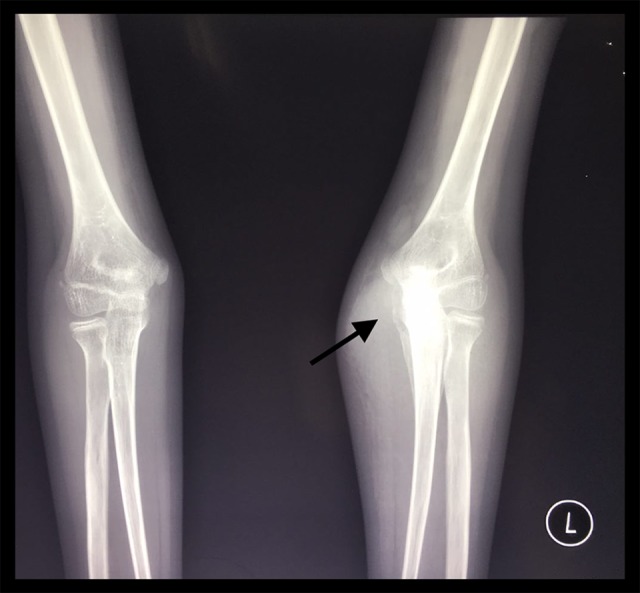
X-ray of the left elbow showed osteomyelitis of the proximal left ulnar (arrow).
Figure 6.

X-ray of the left hand showed osteomyelitis of the 5th metacarpal along with spina ventosa depiction.
During hospitalization, the lump on the elbow was evacuated and yielded about 20–30 mL of pus; it was cultured, and the results were negative for specific bacteria. Skin biopsy was also done from the surface of the lump. Histopathological examination, PCR MTB, and GenXpert from skin biopsy results were positive for M. tuberculosis rifampicin sensitive. With all the clinical findings, histopathology, PCR, and GenXpert results, the patient was then diagnosed with multifocal osteomyelitis TB with metastatic tuberculous abscess. The patient then started with a course of anti-TB drugs which consisted of isoniazid (10 mg/kg/day), rifampicin (15 mg/kg/day), pyrazinamide (35 mg/kg/day), and ethambutol (20 mg/kg/day). During the treatment course, the patient experienced ADIH. The liver function test results showed an increase, with AST 120 U/L and ALT 164 U/L. We then switched the regimen to streptomycin (15 mg/kg/day) and ethambutol (20 mg/kg/day) for 2 weeks and reintroduced the rifampicin and isoniazid according to the modified British Thoracic Society (BTS) guidelines regimen. During reintroduction, the liver function test results remain normal, therefore isoniazid and rifampicin was continued. The nodules appeared shrunken after 3 months of treatment with anti-TB drugs. The elbow showed no sign of inflammation with good range of motion. The nodules on the hand and foot were no longer palpable, but there was some scarring from where the nodules were (Figures 7, 8). Her weight gradually increased, her weight was 26 kg after 3 months of therapy. After she finally completed the treatment of 12 months, her weight increased to 28 kg.
Figure 7.

Picture of the left hand after 3 months of anti-tuberculosis therapy.
Figure 8.

Picture of the left foot after 3 months of anti-tuberculosis therapy.
Discussions
TB can be classified into pulmonary TB and extrapulmonary TB, with bone and joint TB accounting for up to 35% of extra-pulmonary TB [6]. Extraspinal musculoskeletal TB and cutaneous TB is among the least common manifestations of TB, with a frequency of about 1–2% for each [7,8]. Children age less than 15 years present higher risk of M. tuberculosis dissemination and extrapulmonary disease. The global impact of pediatric extrapulmonary TB remains unknown, due to limitations in pediatric TB case detection and poor notification rates [9].
Even though mechanisms for extrapulmonary dissemination remain largely unknown, the host-pathogen interaction appears to play an important role in establishing the site of disease presentation and dissemination; some risk factors identified in previous studies were: HIV infection, malnutrition, female gender, low socio-economic status, and underlying immune condition [9,10]. Our case came from a low-income family. Even though the BMI was normal for her age, her height to age was below < −3 SD on the WHO growth standard reflecting poor nutritional supports.
Multifocal tuberculosis osteomyelitis, defined as the involvement of 2 or more noncontiguous skeletal regions, is a rare condition estimated to occur in 10–15% of musculoskeletal tuberculosis cases [11]. The pathogenesis includes hematogenous spread of the tubercle bacillus from pulmonary foci and in some cases by the lymphatic route [2,7]. Although a pulmonary focus is often presumed, active pulmonary TB is seen in less than 50% of the patients [7]. After the spread, the initial focus starts in the metaphysis in childhood or at the end of the bone in adults. In bones with superficial cortical surfaces (such as the meta-carpals, metatarsals, phalanges, tibia, and ulna) the lesions may produce reactive subperiosteal new bone formation surrounding lytic areas [2]. The synovium, in response to the bacterial invasion, produce an inflammatory reaction with granulation and pannus formation, and cicatrisation followed by the erosion of the cartilage and bone. Plain radiography of the joint typically show non-specific changes in early stages, including joint effusion soft tissue swelling, juxta-articular osteopenia, and subchondral erosions [1,2]. In our patient case, based on evidence from the histopathology examination, PCR test results and GenXpert results, we concluded that the bacilli invade the bones and joints, resulting in osteomyelitis of the hand and foot, with arthritis of the elbow.
The diagnosis of osteomyelitis and osteoarticular TB can be difficult. The most consistent symptoms include a gradual onset of joint pain associated with swelling and a decreased range of motion. Systemic symptoms are usually absent. Radiological findings in osteoarticular TB are non-specific and require aspiration or synovial biopsy for definitive diagnosis [1,6]. Microscopy and cultures of synovial fluid yield positive results in up to 80% of patients with osteoarticular TB [6].
Cutaneous TB is an infrequent first sign of disseminated TB. Single or multiple metastatic tuberculous abscess may develop on extremities and trunk by hematogenous spread from a primary focus of infection during a period of decreased immunity, particularly in malnourished and immunosuppressed children [4].
The differential diagnosis includes staphylococcal abscess, other mixed bacterial infections, sporotrichosis, nocardiosis, chromomycosis, leishmaniasis, atypical mycobacterial infections, deep fungi infections, syphilitic gumma, leprosy, and all forms of panniculitis. Confirmation of the clinical diagnosis is obtained by histopathology, and bacterial or fungal culture [8,12].
Therapy with anti-TB drugs and early mobilization is the goal of management. Most of the patients respond with conservative antitubercular therapy with reasonable joint movement [1,6]. In our institution, anti-TB drug induced hepatoxicity is quite frequent, accounting for 3.5% of pediatric TB patient. Accordingly, a pediatric ADIH patient was treated using modified BTS guidelines [13].
Conclusions
We have to consider TB as the etiology in any lesion especially in a high burden country like ours. Increased awareness of unusual manifestation of TB will allow for proper diagnosis and management of this common infection. Accordingly, timely diagnosis and management will prevent further debilitating sequelae.
Footnotes
Statement
The written consent form for conducting procedures, treatment, and publication were signed by the parents.
Conflicts of interest
None.
References:
- 1.Kushwaha SS, Shantanu K, Kumar D, et al. Elbow tuberculosis mimicking chronic osteomyelitis – a case report. Sch Acad J Biosci. 2017;5:200–2. [Google Scholar]
- 2.Tuli S. General principles of osteoarticular tuberculosis. Clin Orthop Relat Res. 2002;(398):11–19. doi: 10.1097/00003086-200205000-00003. [DOI] [PubMed] [Google Scholar]
- 3.Teo HEL, Peh WCG. Skeletal tuberculosis in children. Pediatr Radiol. 2004;34:853–60. doi: 10.1007/s00247-004-1223-7. [DOI] [PubMed] [Google Scholar]
- 4.Sezgin B, Atilganoglu U, Yigit O, et al. Concomitant cutaneous metastatic tuberculous abscesses and multifocal skeletal tuberculosis. Indian J Dermatol. 2008;53:149–53. doi: 10.4103/0019-5154.43208. [DOI] [PMC free article] [PubMed] [Google Scholar]
- 5.World Health Organization . Global Tuberculosis Report 2017. Geneva: 2017. [Google Scholar]
- 6.Sagoo RS, Lakdawala A, Subbu R. Tuberculosis of the elbow joint. JRSM Short Rep. 2011;2:17. doi: 10.1258/shorts.2011.010130. [DOI] [PMC free article] [PubMed] [Google Scholar]
- 7.De Backer AI, Mortelé KJ, Vanhoenacker FM, et al. Imaging of extraspinal musculoskeletal tuberculosis. Eur J Radiol. 2006;57:119–30. doi: 10.1016/j.ejrad.2005.07.005. [DOI] [PubMed] [Google Scholar]
- 8.Bravo FG, Gotuzzo E. Cutaneous tuberculosis. Clin Dermatol. 2007;25:173–80. doi: 10.1016/j.clindermatol.2006.05.005. [DOI] [PubMed] [Google Scholar]
- 9.Santiago-garcía B, Blázquez-gamero D, Baquero-artigao F, et al. Pediatric extrapulmonary tuberculosis: Clinical spectrum, risk factors and diagnostic challenges in a low prevalence region. Pediatr Infect Dis J. 2016;35:1175–81. doi: 10.1097/INF.0000000000001270. [DOI] [PubMed] [Google Scholar]
- 10.Qian X, Nguyen DT, Lyu J, et al. Risk factors for extrapulmonary dissemination of tuberculosis and associated mortality during treatment for extra-pulmonary tuberculosis. Emerg Microbes Infect. 2018;7:102. doi: 10.1038/s41426-018-0106-1. [DOI] [PMC free article] [PubMed] [Google Scholar]
- 11.Lebowitz D, Wolter L, Zenklusen C, et al. TB determined: tuberculous osteomyelitis. Am J Med. 2014;127:198–201. doi: 10.1016/j.amjmed.2013.12.001. [DOI] [PubMed] [Google Scholar]
- 12.Vanmarsenille J, De Berg B, Houssiau FA, et al. Unusually prolonged course of tuberculous dactylitis with osteitis. Joint Bone Spine. 2003;70:535–37. doi: 10.1016/s1297-319x(03)00068-x. [DOI] [PubMed] [Google Scholar]
- 13.Nataprawira HM, Hannah RA, Kartika HH. Hospitalized pediatric antituberculosis drug induced hepatotoxicity: Experience of an Indonesian referral hospital. Asian Pacific J Trop Dis. 2017;7:276–79. [Google Scholar]