Abstract
Background:
Various biochemical and histological methods are available for human age determination which are invasive and may require extraction of teeth. The present study aims to assess the accuracy of age estimation from tooth-coronal index (TCI) of known age and sex individuals and to present a noninvasive method for age estimation.
Materials and Methods:
This retrospective study comprised 88 patients, which included 54 males and 34 females. An orthopantomogram of these individuals were taken, and premolars and molars in the same were evaluated. The height of the crown (coronal height [CH]) and the height of the coronal pulp cavity (coronal pulp cavity height [CPCH]) was digitally measured on the computer screen. The TCI given by Ikeda et al. in 1985 (TCI = [CPCH × 100]/CH.) was computed on each tooth and regressed on real age of the sample. The mean, median, range, and standard deviation of the computed index were calculated. The correlation between the actual age and the estimated age was calculated using t-test. P < 0.05 was considered significant.
Results:
Results revealed that there is a significant correlation between the TCI with age. Increase in TCI observed with age; however, it showed no significant sex difference.
Conclusion:
TCI is a precise, noninvasive and easily used reliable biomarker for age estimation and is applicable to both living and dead individuals.
Key Words: Age, estimation, forensic science, noninvasive
INTRODUCTION
Age estimation is an important part of one's identification process in the discipline of forensic science. Declaration of age is not only important for legal, ethical issues, and death reports but also an essential for living persons to clarify criminal and civil liability and social issues.[1] Human teeth proved to be the most reliable biological marker in forensic science as they withstand death and sustain themselves for many thousands of years without changes.[2,3]
Various morphological, biochemical, and histological methods are available for human age determination with the help of teeth.[4,5,6] These methods are invasive and require extraction of the teeth. With the advancement of radiology in forensic science, radiographic images can be utilized for age estimation in vivo and in vitro, with the advantage of being noninvasive.[7,8] Progressive modification of the coronal pulp cavity throughout the life occurs due to continuous deposition of the secondary dentin by odontoblasts. The secondary dentin formation is diverse for different teeth, for example, in molars the greatest dentin formation occurs on the floor of the pulp chamber and lesser amounts are deposited on the occlusal and lateral walls.[9]
The outermost covering of the tooth crown, enamel and the outermost covering of the surface of the root, cementum followed by dentin underneath, are hard tissues resistant to decomposition. The innermost soft core of pulp is protected by these structures.[9,10] Radiographic examination of the teeth gives affirmative information regarding the size of pulp chamber which can be correlated with the chronological age.[7] Kvaal et al. used deposition of secondary dentin by measuring pulp radiolucency and correlated it to age.[11] On the basis of measurements on various radiographic and morphological parameters discussed in many studies, formulae on such basis were developed.[7,12] As the values may be different for individuals from different ethnical groups, the reproducibility of these parameters is uncertain.[13,14]
We decided to carry out a study to correlate the size of the pulp chamber in premolar and molar teeth with the chronological age of the patients. On the other hand, the tooth-coronal index (TCI) (TCI = Length of the coronal pulp cavity/Length of the crown × 100) was measured by Ikeda et al. and then was computed for each tooth and regressed on real age.[9] The present study aims to assess the accuracy of age estimation from TCI of mandibular second premolar and first molar of right quadrant using panoramic radiographs of known age and sex individuals and to develop regression equations that can be used in the Indian population.
Aims and objectives
The aim of this study is to evaluate the TCI on panoramic radiographs free of technical errors and correlate it with the chronologic age. The objective of this study is to present a method for assessing and evaluating the change in the pulp dimension at the level of cementoenamel junction and its correlation to different age groups and to assess the accuracy of digital panoramic radiographs as a simple, noninvasive tool for age estimation in adult.
MATERIALS AND METHODS
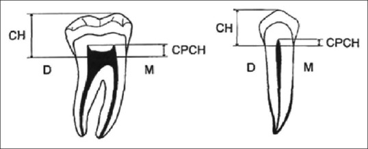
This retrospective study sample consisted of 88 patients, comprising 54 males, 34 females. Subjects aged between 20 and 40 years and belonged to the same geographical population, Navi Mumbai, Maharashtra, India. This was a retrospective study conducted after obtaining approval from the Institutional Ethics Committee. The principal investigator was blinded about the identity of the cases regarding age and sex. A panoramic radiograph of known sex and age were obtained and second premolar and first molar of the right mandibular quadrant were evaluated. This study included 176 intact teeth (88 premolars and 88 molars) from 88 Indian individuals (54 males and 34 females). Measurements on all orthopantomograms with a fully visible pulp cavity were taken. However, orthopantomograms with teeth that were fractured, restored, and teeth with gross evidence of hypercementosis or grossly decayed were excluded. The height of the crown (coronal height [CH]) and the height of the coronal pulp cavity (coronal pulp cavity height [CPCH]) was digitally measured Dicom software (Digital Imaging and communication in medicine) on the computer screen. With this measurement, the coronal tooth cavity index was calculated for each tooth with the help of following formula, as suggested by Ikeda et al.[9] TCI = (CPCH × 100)/CH [Figure 1].
Figure 1.

Schematic representation of measurements taken of panoramic radiographs.
To ensure the accuracy of the technique used for measuring TCI the detailed reference points used were cervical line that connect two landmarks to be measured, the mesial and distal cemento-enamel junction points, and divides the tooth into crown and root. Crown height is the maximum perpendicular distance from the cervical line to the tip of the highest cusp of teeth. While coronal pulp cavity height is the distance from the cervical line to the coronal tip of the pulp chamber. All these measurements were analyzed by two observers and the mean recorded to minimize intra- and inter-observer errors. The TCI was calculated using mean values and subjected to regression analysis.
Statistical analysis
The data collected was statistically analyzed using the SPSS (Statistical package for Social Sciences) After the TCI was computed for each tooth, regression analysis on the real age of the sample was done. The mean, median, range and standard deviation of the computed index was calculated. The correlation between the actual age and the estimated age was calculated using the t-test.
RESULTS
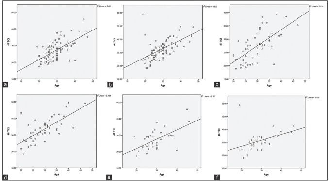
The analysis of the studied number subjects is summarized in Table 1. Mean of the comparison of the age of both males and females using unpaired t-test with t = 0.353 and P = 0.725 is summarized in Table 1. The mean value of CH, CPCH, and TCI in premolars and molar for males and females were significant statistically as shown in Tables 2 and 3. The comparison of CH, CPCH, and TCI in different age groups in premolars and molars for males and females were highly significant statistically as shown in Tables 4 and 5. The correlation of TCI in the different age group of both sexes was highly significant for premolars and molars as shown in Tables 6 and 7 and Graph 1 and was concluded that TCI can be a good predictor of age in both premolars and molar.
Table 1.
Comparison of age (mean±standard deviation) of males and females using unpaired t-test
| Gender | Number of samples | Mean±SD |
|---|---|---|
| Males | 54 | 29.59±5.8 |
| Females | 34 | 29.12±6.6 |
| t | - | 0.353 |
| P | - | 0.725 |
SD: Standard deviation
Table 2.
Comparison of coronal pulp cavity height, coronal height and tooth-coronal index (mean±standard deviation) in male and female subjects using unpaired test (45-2nd premolar)
| Gender | Number of samples | Mean±SD |
||
|---|---|---|---|---|
| CPCH | CH | TCI | ||
| Males | 54 | 1.95±0.4 | 7.27±0.7 | 27.18±5.4** |
| Females | 34 | 1.91±0.4 | 7.06±0.8 | 27.11±6.2 |
| T | - | 0.592** | 1.295 | 0.062 |
| P | - | 0.555* | 0.199* | 0.951* |
*P>0.05 - Non significant, **P<0.05 - Significant. SD: Standard deviation; CPCH: Coronal pulp cavity height (mm); CH: Coronal height; TCI: Tooth-coronal index
Table 3.
Comparison of coronal pulp cavity height, coronal height, and tooth-coronal index (mean±standard deviation) in male and female subjects using unpaired test (46-1st molar)
| Gender | Number of samples | Mean±SD |
||
|---|---|---|---|---|
| CPCH | CH | TCI | ||
| Males | 54 | 2.58±0.5 | 7.82±1.0 | 33.29±6.8 |
| Females | 34 | 2.32±0.5 | 7.53±0.9 | 31.14±7.6 |
| t | - | 2.275 | 1.230 | 1.383 |
| P | - | 0.025** | 0.222* | 0.170* |
*P>0.05 - Non significant, **P<0.05 - Significant. SD: Standard deviation; CPCH: Coronal pulp cavity height (mm); CH: Coronal height; TCI: Tooth-coronal index
Table 4.
Comparison of coronal pulp cavity height, coronal height, and tooth-coronal index (mean±standard deviation) in different age groups using unpaired test (45-2nd premolar)
| Age groups | Number of samples | Mean±SD |
||
|---|---|---|---|---|
| CPCH | CH | TCI | ||
| ≤30 years | 56 | 1.81±0.4 | 7.32±0.7 | 24.72±4.3 |
| >30 years | 32 | 2.15±0.3 | 6.95±0.8 | 31.39±5.3 |
| t | - | 4.366 | 2.218 | 6.435 |
| P | - | <0.001** | 0.029* | <0.001** |
*P<0.05 - Significant, **P<0.001 - Highly significant. SD: Standard deviation; CPCH: Coronal pulp cavity height (mm); CH: Coronal height; TCI: Tooth-coronal index
Table 5.
Comparison of coronal pulp cavity height, coronal height, and tooth-coronal index (mean±standard deviation) in different age groups using unpaired test (46-1st molar)
| Age groups | Number of samples | Mean±SD |
||
|---|---|---|---|---|
| CPCH | CH | TCI | ||
| ≤30 years | 56 | 2.33 (0.5) | 7.88 (0.8) | 25.65 (6.5) |
| >30 years | 32 | 2.74 (0.5) | 7.41 (1.3) | 37.38 (5.3) |
| t | - | 3.779 | 2.119 | 5.714 |
| P | - | <0.001** | 0.037* | <0.001** |
*P<0.05 - Significant, **P<0.001 - Highly significant. SD: Standard deviation; CPCH: Coronal pulp cavity height (mm); CH: Coronal height; TCI: Tooth-coronal index
Table 6.
Correlation coefficient and P value in the total sample
| Teeth (TCI) | Combined group |
|
|---|---|---|
| r (correlation coefficient) | P | |
| 45-2nd premolar | 0.634 | <0.001** |
| 46-1st molar | 0.568 | <0.001** |
**P<0.001 - Highly significant. TCI: Tooth-coronal index
Table 7.
Correlation coefficient and P value in males and females
| Teeth (TCI) | Males |
|
|---|---|---|
| r (correlation coefficient) | P | |
| 45-2nd premolar | 0.657 | <0.001** |
| 46-1st molar | 0.703 | <0.001** |
| Teeth (TCI) | Females |
|
| r (correlation coefficient) | P | |
| 45-2nd premolar | 0.606 | <0.001** |
| 46-1st molar | 0.397 | 0.020* |
*P<0.05 - Significant, **P<0.001 - Highly significant. TCI: Tooth-coronal index
Graph 1.

(a) Scattered plot showing regression line correlation between tooth coronal index and age in all the subjects (combined group) (45 – 2nd premolar). (b) Scattered plot showing regression line correlation between tooth coronal index and age in all the subjects (combined group) (46 – 1st molar). (c) Scattered plot showing regression line Correlation between tooth coronal index and age in all the subjects (males) (45 – 2nd premolar). (d) Scattered plot showing regression line Correlation between tooth coronal index and age in all the subjects (Males) (46 – 1st molar). (e) Scattered plot showing regression line Correlation between tooth coronal index and age in all the subjects (Males) (46 – 1st molar). (f) Scattered plot showing regression line Correlation between tooth coronal index and age in all the subjects (Females) (46 – 1st molar).
Followed by the calculations, a simple linear regression was carried out with significant correlation by regressing the proportional coronal pulp cavity length (TCI) on the actual age for each group of teeth, for the combined sample.
DISCUSSION
In adults, age estimation would be challenging as the development of dentition completes by this age and there is no clue which could be reliable to assess the age.[15] The two criteria that can be utilized for age determination in adults are assessment of the volume of pulp cavity and of third molar development.[15] The length of the coronal pulp cavity shows a significant correlation with individual chronological age. The reduction in the size of the pulp cavity resulting from a deposition of secondary dentine with aging can be assessed by radiographs.[16] Assessment of the pulp/tooth index is an indirect quantification of secondary dentin deposition.[17] This study measures the morphometric values of pulp on digital panoramic radiographs for the mandibular second premolar and first molar of the right quadrant, thereby calculating the TCI for each tooth. The aim of this study is to evaluate the TCI on panoramic radiographs and correlate it with the chronologic age.
The panoramic radiograph has the advantage of displaying all the mandibular and maxillary teeth on a single film.[18] The computer-assisted image analysis avoids the bias inherent in observer subjectivity, which supplies a relatively exact method of measurements, improves reliability and consequently the statistical data analysis.[11] The present study is based on the nondestructive method suggested by Ikeda et al. found a high correlation that the extent of pulp cavity is visible in premolars and molars in panoramic radiographs as shown by Drusini and El Morsi.[14,15]
The present study reveals that age correlation and TCI in different age groups are sizeable in both premolars and molars. The study results are in agreement with the study results done by Drusini et al.,[19] Zadinska et al.,[20] Shrestha.,[6] Khattab et al.,[21] and Karkhanis et al.[22] that there is no sex difference in TCI. As stated by these studies, sex of individual appears to have no significant influence on age estimation, so that sex-specific formulae are not necessary for age estimation in specimens of unknown sex. This result is contradictory to that of Agematsu et al.[23] Igbigbi and Nyirenda[24] who mentioned that due to influence of estrogen on the formation of secondary dentin, gender has a significant influence on age estimation using TCI and hence there is need for sex- specific formulae in the sampled population.[11]
However, the results obtained prove that there is a positive correlation between TCI in the present work and age and the correlation is more in females than males, i.e., the index increases with increasing age. This is in favor of the study by Shrestha[6] in India which mentioned that TCI increases with increasing age. However, negative correlation between TCI and age was given by Drusini et al.[19] Zadinska et al.[20] Igbigbi and Nyirenda[24] and Karkhanis et al.[22] This could be explained by the relatively young age of the present work samples as the age ranged from 20 to 40 years with mean age of 25.51 ± 0.84 years; so the decrease in pulp cavity due to dentin deposition is not evident.
Moreover, the study shows that the mean TCI of premolars is larger than the molars and it is higher in males than in females, respectively. These findings are supportive to that of Igbigbi and Nyirenda[24] who mentioned that the TCI for molars were lower than those of premolars. This has been explained by the fact that the mandibular molars have morphological diversity than premolars. Hence, in different age groups, the difference in the decrease in pulp chamber value was not clear in molars than in premolars.
The present study devices a simple, practical, noninvasive, cost-effective method for the morphometric analysis of the coronal pulp chamber reduction using TCI on the digital panoramic radiographs. The results of the study should be viewed with caution as the study sample was small. Hence, there is a definite need for similar studies with large Indian population.
CONCLUSION
TCI is an excellent forensic tool for age estimation. The potential of TCI using digital panoramic radiography is useful as a biomarker of aging with increasing availability of digital radiographic systems in the dental institutes and offices. Thus from the present work, it could be concluded that TCI is a precise, noninvasive and not time-consuming; and does not require highly specialized equipment and is applicable to both living and dead individuals. Furthermore, the result concluded that gender has no effect on TCI.
Financial support and sponsorship
Nil.
Conflicts of interest
The authors of this manuscript declare that they have no conflicts of interest, real or perceived, financial or nonfinancial in this article.
REFERENCES
- 1.Willems G. A review of the most commonly used dental age estimation techniques. J Forensic Odontostomatol. 2001;19:9–17. [PubMed] [Google Scholar]
- 2.Ranganathan K, Rooban T, Lakshmminarayan V. Forensic odontology: A review. J Forensic Odontol. 2008;1:4–12. [Google Scholar]
- 3.Achary AB, Sivapathasundharan B. Forensic odontology. In: Rajendran R, Sivapathasundharan B, editors. Shafer's Textbook of Oral Pathology. India: Elsevier Private Ltd; 2009. pp. 871–92. [Google Scholar]
- 4.Ohtani S, Yamamoto K. Age estimation using the racemization of amino acid in human dentin. J Forensic Sci. 1991;36:792–800. [PubMed] [Google Scholar]
- 5.Takasaki T, Tsuji A, Ikeda N, Ohishi M. Age estimation in dental pulp DNA based on human telomere shortening. Int J Legal Med. 2003;117:232–4. doi: 10.1007/s00414-003-0376-5. [DOI] [PubMed] [Google Scholar]
- 6.Shrestha M. Comparative evaluation of two established age estimation techniques (Two histological and radiological) by image analysis software using single tooth. J Forensic Res. 2014;5:1–6. [Google Scholar]
- 7.Panchbhai AS. Dental radiographic indicators, a key to age estimation. Dentomaxillofac Radiol. 2011;40:199–212. doi: 10.1259/dmfr/19478385. [DOI] [PMC free article] [PubMed] [Google Scholar]
- 8.Limdiwala PG, Shah JS. Age estimation by using dental radiographs. J Forensic Dent Sci. 2013;5:118–22. doi: 10.4103/0975-1475.119778. [DOI] [PMC free article] [PubMed] [Google Scholar]
- 9.Ikeda N, Umetsu K, Kashimura S, Suzuki T, Oumi M. Estimation of age from teeth with their soft X-ray findings. Nihon Hoigaku Zasshi. 1985;39:244–50. [PubMed] [Google Scholar]
- 10.Sharma R, Srivastava A. Radiographic evaluation of dental age of adults using Kvaal's method. J Forensic Dent Sci. 2010;2:22–6. doi: 10.4103/0974-2948.71053. [DOI] [PMC free article] [PubMed] [Google Scholar]
- 11.Kvaal SI, Kolltveit KM, Thomsen IO, Solheim T. Age estimation of adults from dental radiographs. Forensic Sci Int. 1995;74:175–85. doi: 10.1016/0379-0738(95)01760-g. [DOI] [PubMed] [Google Scholar]
- 12.Cameriere R, Brogi G, Ferrante L, Mirtella D, Vultaggio C, Cingolani M, et al. Reliability in age determination by pulp/tooth ratio in upper canines in skeletal remains. J Forensic Sci. 2006;51:861–4. doi: 10.1111/j.1556-4029.2006.00159.x. [DOI] [PubMed] [Google Scholar]
- 13.Saxena S. Age estimation of Indian adults from orthopantomographs. Braz Oral Res. 2011;25:225–9. doi: 10.1590/s1806-83242011005000009. [DOI] [PubMed] [Google Scholar]
- 14.El Morsi DA, Rezk HM, Aziza A, El-Sherbiny M. Tooth coronal pulp index as a tool for age estimation in Egyptian population. J Forensic Sci Criminol. 2015;3:201. [Google Scholar]
- 15.Drusini AG, Toso O, Ranzato C. The coronal pulp cavity index: A biomarker for age determination in human adults. Am J Phys Anthropol. 1997;103:353–63. doi: 10.1002/(SICI)1096-8644(199707)103:3<353::AID-AJPA5>3.0.CO;2-R. [DOI] [PubMed] [Google Scholar]
- 16.Veera SD, Kannabiran J, Suratkal N, Chidananada DB, Gujjar KR, Goli S. Coronal pulp biomarker: A lesser known age estimation modality. J Indian Acad Oral Med Radiol. 2014;26:398. [Google Scholar]
- 17.Grover S, Marya CM, Avinash J, Pruthi N. Estimation of dental age and its comparison with chronological age: Accuracy of two radiographic methods. Med Sci Law. 2012;52:32–5. doi: 10.1258/msl.2011.011021. [DOI] [PubMed] [Google Scholar]
- 18.Huda TF, Bowman JE. Age determination from dental microstructure in juveniles. Am J Phys Anthropol. 1995;97:135–50. doi: 10.1002/ajpa.1330970206. [DOI] [PubMed] [Google Scholar]
- 19.Manigandan SC, Sivagami AV. Age estimation with dental radiographs. Res J Pharm Biol Chem Sci. 2014;5:1370–6. [Google Scholar]
- 20.Zadinska E, Drusini AG, Carrara N. The comparison between two age estimation methods based on human teeth. Anthropol Rev. 2000;63:95–101. [Google Scholar]
- 21.Khattab NA, Marzouk HM, AbdelWahab TM. Application of tooth coronal index for age estimation among adult Egyptians. Schoolary Res. 2013:1–15. [Google Scholar]
- 22.Karkhanis S, Mack P, Franklin D. Age estimation standards for a Western Australian population using the coronal pulp cavity index. Forensic Sci Int. 2013;231:412.e1–6. doi: 10.1016/j.forsciint.2013.04.004. [DOI] [PubMed] [Google Scholar]
- 23.Agematsu H, Someda H, Hashimoto M, Matsunaga S, Abe S, Kim HJ, et al. Three-dimensional observation of decrease in pulp cavity volume using micro-CT: Age-related change. Bull Tokyo Dent Coll. 2010;51:1–6. doi: 10.2209/tdcpublication.51.1. [DOI] [PubMed] [Google Scholar]
- 24.Igbigbi PS, Nyirenda SK. Age estimation of Malawian adults from dental radiographs. West Afr J Med. 2005;24:329–33. doi: 10.4314/wajm.v24i4.28227. [DOI] [PubMed] [Google Scholar]


