

Abstract
Umbilical hernias are estimated to occur in up to 20% of patients with long-standing cirrhosis and ascites. Complications such as strangulation and infarction of the bowel or rupture of the bowel loops and/or hernia sac are uncommon but potentially life-threatening consequences. Often these events happen in association with abdominal trauma or after procedures such as paracentesis. Spontaneous rupture is more likely in cases where strangulation and subsequent necrosis of the bowel has weakened and thinned the incarcerated intestinal wall.
We report a case of a 63-year-old male with a history of alcohol abuse complicated by cirrhosis and ascites who was found dead in his home approximately 30 minutes after last being seen alive. He was found at rest in a seated position without evidence of recent trauma. At autopsy, an ulcerated umbilical hernia was present containing a loop of perforated, necrotic small bowel. There was evidence of peritonitis including one liter of yellow-red fluid accumulation in the peritoneal cavity, multifocal areas of fibrinopurulent debris adherent to the small bowel, and a dull gray discoloration to the peritoneal lining. The case was signed out as death due to peritonitis and spontaneous perforation of small intestine due to strangulated and infarcted small intestinal umbilical hernia and the manner, natural. Although spontaneous perforation of the bowel within a hernia sac is uncommon, it may contribute to sudden death in patients lacking a history of trauma or recent medical procedures and physicians certifying these deaths should be aware of the possibility.
Keywords: Forensic pathology, Small bowel perforation, Umbilical hernia
Introduction
Cirrhosis of the liver is a major cause of morbidity and mortality in the United States with an excess of 36 000 deaths per year, 18 000 of which are alcohol-related (1). Complications due to cirrhosis are numerous and can vary from benign to fatal. Of interest in this case is the presence of an incarcerated umbilical hernia in the setting of ascites. This combination of findings predisposes a patient to rupture of the hernia sac or bowel which carries a high mortality rate (2).
The umbilicus on the anterior abdominal wall is an anatomic remnant from the attachment of the umbilical cord during development, which persists into adulthood as a cutaneous scar. The network of ligaments underlying the umbilicus forms a fibrous ring centered beneath the scar. There is little to no subcutaneous tissue or muscle at this spot, which creates a weak spot in the abdominal wall. Other anatomical weak points include the femoral canal and sites of prior surgical scars (known as incisional hernias). Umbilical hernias are common in infants before the fibrous ring has time to fully develop and while the abdominal muscles are weak. Most umbilical hernias at this age involute spontaneously within a few years (3). In adults, umbilical hernias are estimated to occur in up to 3% of the general population, but can be seen in as many as 20% of patients with recurrent or longstanding ascites (4).
Umbilical hernias are more common in patients with ascites due to the accumulation of fluid in the abdominal cavity, leading to a marked increase in intra-abdominal pressure. The increased intra-abdominal pressure causes the fibrous ring below the umbilicus to dilate and allows herniation of the peritoneum (5). Over time, if the umbilical ring becomes large enough, a loop of small bowel can also extrude into the hernia sac. Subsequent reductions in intra-abdominal pressure and partial closure of the umbilical ring can cause incarceration of the loop of bowel within the hernia sac. This has been reported to happen secondary to therapeutic paracentesis, portal venous shunting, or medical diuresis with large volume ascites fluid removal and decompression of the abdomen (6-9). The entrapped bowel loop now has a compromised vascular supply, leading to ischemia and edema, with necrosis and potential rupture if the hernia is not reduced (10).
The entrapped loop of bowel is also in a compromised anatomic position, rendering it vulnerable to trauma. Direct blows to hernia sacs have resulted in rupture of inguinal hernias and incarcerated loops of bowel (11). Indirect trauma can cause a shearing-type injury to the intestine due to the presence of an anchor point where the bowel enters the hernia sac (12). Finally, even minor increases in abdominal pressure such as with coughing (13) or straining (14) have been reported to cause rupture of hernia sacs and transection of incarcerated bowel loops. Cutaneous disorders such as maceration, cellulitis, and other local complications lead to ulceration and predispose the underlying hernia sac and bowel loop to infection and possible rupture, especially in cirrhotic patients with poor nutrition and wound healing (3).
Case Description
A 63-year-old male with a history of alcohol abuse, cirrhosis, and smoking was found expired on his couch. He had last spoken to his mother approximately 30 minutes prior and had no specific complaints. An investigator responded to the scene and found him seated on the couch in the same position as when he was last seen. He was wearing a shirt and shorts that were soaked anteriorly with blood-tinged fluid (Image 1). His abdomen was noted to be distended and firm. Beneath the waistband of the shorts was a large, protruding, ulcerated mass at the umbilicus which was found to be the source of the fluid. A large area of red-black skin discoloration was seen surrounding the mass.
Image 1:

Image on scene showing protruding abdominal mass and blood-tinged fluid soaking the decedent's clothing.
At autopsy, the mass was revealed to be an umbilical hernia containing perforated and necrotic small bowel. A 22.9 × 10.2 cm area of red-black macerated skin surrounded the base of the ulcer (Image 2). On the surface of the hernia sac was a 3.8 × 3.8 cm transcutaneous ulcer that communicated with the peritoneal cavity and allowed the leakage of ascites fluid (Image 3). Peritonitis was evident by diffuse, yellow-grey fibrinopurulent exudates and a dull grey discoloration of the peritoneum (Image 4). The peritoneum surrounding the base of the hernia was black-gray and necrotic (Image 5). The loop of entrapped bowel had an approximately 5.1 cm area of transmural necrosis with perforation (Image 6). The liver was shrunken, firm, and showed bridging fibrosis histologically.
Image 2:
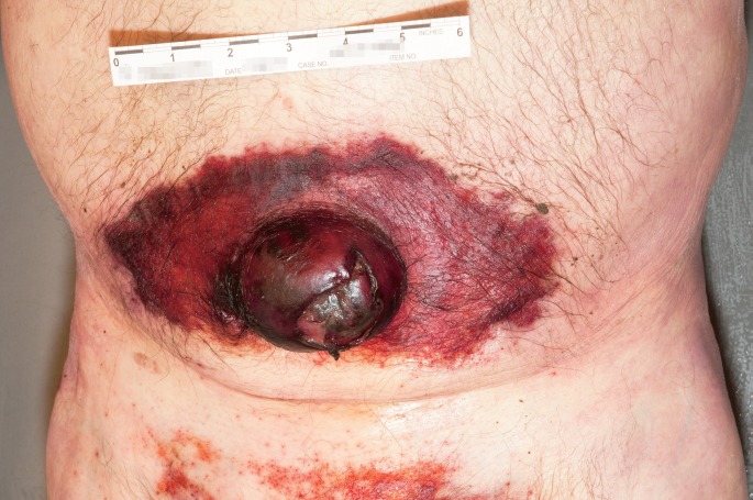
At autopsy, maceration surrounding the umbilical hernia was observed.
Image 3:
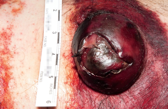
Close-up image of transcutaneous ulcer.
Image 4:

The abdominal cavity showing peritonitis as evidenced by diffuse fibrinopurulent exudates.
Image 5:

The peritoneal cavity at the base of the hernia showing a black-gray rim of necrosis.
Image 6:

The loop of bowel present in the umbilical hernia was necrotic with a transmural perforation.
The case was certified as peritonitis and spontaneous perforation of small intestine due to strangulated and infarcted small intestinal umbilical hernia with other conditions of chronic ethanol abuse with cirrhosis and ascites. The manner was natural.
Discussion
Our patient had a history of long-standing and recurrent ascites and had undergone a prior therapeutic paracentesis within the last year, but had not been seen by a doctor for some time. The skin overlying his hernia was ulcerated, which allowed the leakage of ascites fluid and caused maceration of a large area of his abdominal skin. This likely led to local inflammation and subsequent cellulitis of the skin immediately surrounding the base of the hernia. The strangulated loop of bowel within the hernia sac become ischemic, necrotic, and then perforated. The peritoneal cavity was then compromised to the outside environment through the cutaneous ulcer and to the intraluminal contents of the bowel. Seeding of the ascites fluid by bacteria from either source could have led to the development of peritonitis.
During the investigation, the decedent's family members did not provide a history of trauma to the abdomen or elsewhere. In addition, his last therapeutic paracentesis was approximately six months prior and likely unrelated to his current situation. Interestingly, despite the active diffuse peritonitis, the decedent had no specific complaints shortly before his death and he was found in the same seated and resting position in which he was last seen alive. Since no specific antecedent event was identified, we consider this a case of spontaneous perforation of the small bowel secondary to strangulation and infarction in the setting of an ulcerated umbilical hernia. Physicians who have the opportunity to certify these deaths should be aware of the potential perimortem triggering events such as trauma or therapeutic interventions, but should not hesitate to ascribe the cause of death to spontaneous rupture of the bowel if appropriate to the case.
Footnotes
Financial Disclosure
The authors have indicated that they do not have financial relationships to disclose that are relevant to this manuscript
ETHICAL APPROVAL
As per Journal Policies, ethical approval was not required for this manuscript
STATEMENT OF HUMAN AND ANIMAL RIGHTS
This article does not contain any studies conducted with animals or on living human subjects
STATEMENT OF INFORMED CONSENT
No identifiable personal data were presented in this manuscsript
DISCLOSURES & DECLARATION OF CONFLICTS OF INTEREST
This work was presented at the 2015 NAME Annual Meeting. The authors, reviewers, editors, and publication staff do not report any relevant conflicts of interest
References
- 1.CDC FastStats [Internet]. Atlanta: Centers for Disease Control and Prevention; c2015. Chronic liver disease and cirrhosis; 30 May 2013. [cited 2015 Aug 6]. Available from: http://www.cdc.gov/nchs/fastats/liver-disease.htm. [Google Scholar]
- 2.Kirkpatrick S., Schubert T. Umbilical hernia rupture in cirrhotics with ascites. Dig Dis Sci. 1988. Jun; 33(6): 762–5. PMID: 3286159. 10.1007/bf01540442. [DOI] [PubMed] [Google Scholar]
- 3.Shaw A. Pediatric surgery. 4th ed. Chicago: Year Book Medical Publishers; 1986. Chapter 15, Disorders of the umbilicus; p. 731–9. [Google Scholar]
- 4.Chapman C.B., Snell A.M., Rowntree L.G. Decompensated portal cirrhosis. JAMA. 1931. Jul 25; 97(4): 237–44. 10.1001/jama.1931.02730040019008. [DOI] [Google Scholar]
- 5.Belghiti J., Durand F. Abdominal wall hernias in the setting of cirrhosis. Semin Liver Dis. 1997; 17(3): 219–26. PMID: 9308126. 10.1055/s-2007-1007199. [DOI] [PubMed] [Google Scholar]
- 6.Baron H.C. Umbilical hernia secondary to cirrhosis of the liver. Complications of surgical correction. N Engl J Med. 1960. Oct 27; 263: 824–8. PMID: 13687191. 10.1056/nejm196010272631702. [DOI] [PubMed] [Google Scholar]
- 7.Eisenstadt S. Symptomatic umbilical hernias after peritoneovenous shunts. Arch Surg. 1979. Dec; 114(12): 1443 PMID: 534464. 10.1001/archsurg.1979.01370360097016. [DOI] [PubMed] [Google Scholar]
- 8.Lemmer J.H., Strodel W.E., Eckhauser F.E. Umbilical hernia incarceration: a complication of medical therapy of ascites. Am J Gastroenterol. 1983. May; 78(5): 295–6. PMID: 6846308. 10.3941/jrcr.v9i9.2614. [DOI] [PubMed] [Google Scholar]
- 9.Tan H.K., Chang P.E. Acute abdomen secondary to incarcerated umbilical hernia after treatment of massive cirrhotic ascites. Case Reports Hepatol. 2013; 2013: 948172 PMID: 25374722. PMCID: PMC4208442. 10.1155/2013/948172. [DOI] [PMC free article] [PubMed] [Google Scholar]
- 10.Lutwak N., Dill C. Incarcerated umbilical hernia leading to small bowel ischemia. BMJ Case Rep. 2011. Sep 19; 2011: bcr0620114370 PMID: 22679256. PMCID: PMC3185400. 10.1136/bcr.06.2011.4370. [DOI] [PMC free article] [PubMed] [Google Scholar]
- 11.Shahin Y., Sahota G., Hotouras A. et al. Small bowel perforation due to blunt trauma to an inguinal hernia: a case report and literature review. Hernia. 2012. Jun; 16(3): 349–50. PMID: 21125305. 10.1007/s10029-010-0755-z. [DOI] [PubMed] [Google Scholar]
- 12.Payson B.A., Mage S. Role of inguinal hernia in acute perforation of the small intestine secondary to blunt abdominal trauma. Ann Surg. 1962. Dec; 156: 944–50. PMID: 13942182.PMCID: PMC1466353. 10.1097/00000658-196212000-00012. [DOI] [PMC free article] [PubMed] [Google Scholar]
- 13.Hakiman H., Delibero J., Pham T. et al. Coughing-induced bowel transection in a patient with an incarcerated inguinal hernia: a case report. J Med Case Rep. 2013. Feb 15; 7(1): 47 PMID: 23414609. PMCID: PMC3582600. 10.1186/1752-1947-7-47. [DOI] [PMC free article] [PubMed] [Google Scholar]
- 14.Good D.W., Royds J.E., Smith M.J. et al. Umbilical hernia rupture with evisceration of omentum from massive ascites: a case report. J Med Case Rep. 2011. May 3; 5(1): 170 PMID: 21539740. PMCID: PMC3095553. 10.1186/1752-1947-5-170. [DOI] [PMC free article] [PubMed] [Google Scholar]