

Abstract
Recovering bodies from water is a common task for any medical examiner or coroner office. Unfortunately, there will be a significant postmortem interval before many of these remains are found. A thorough scene investigation must be undertaken to determine if the location of the death and that of the body recovery are the same. Decomposition in a wet environment differs from that in other settings, both in the changes that occur and the rate at which they occur. It is essential that the forensic pathologist or medicolegal death investigator recognize and appreciate the uniqueness of immersed and submerged remains. The typical decomposition changes proceed more slowly in the water, primarily due to cooler temperatures and the anaerobic environment. However, once a body is removed from the water, putrefaction will likely be accelerated. Postmortem changes are not only affected by water temperature, but also by current as well as obstacles and structures, both natural and man-made, that may interact with the remains. The anaerobic nature of decomposition for wet or submerged remains may result in adipocere formation, a unique and fascinating process that results from incomplete transformation of lipids by bacteria. Insect and animal species feeding on the remains are different for submerged bodies. Postmortem predation may cause external defects that mimic injuries and should be interpreted with care. Forensic pathologists and medicolegal death investigators must be aware of the postmortem changes that may occur with submerged and immersed bodies.
Keywords: Forensic pathology, Water, Submersion, Drowning, Adipocere, Postmortem changes
Introduction
The accurate interpretation of postmortem changes is an essential skill for any forensic pathologist or medicolegal death investigator. That being said, postmortem changes can assist or hinder a death investigation. Perhaps the most beneficial feature of postmortem change is in assisting with estimating the postmortem interval, though the imprecision of this estimation is well recognized and should be taken into account. Characteristic changes that occur to a body after death follow a somewhat predictable timetable and placed into the context of the death scene and witness accounts, as available, provide the investigator with a rough estimate of the time of death. On the other hand, postmortem changes alter the appearance of the body, making wounds and other evidence of the body's interaction with the environment more difficult to interpret or even recognize. Artifacts introduced by decomposition obscure external morphologic features of the individual and putrefaction alters key autopsy observations such as organ weights and tissue integrity. Advanced decomposition may completely preclude any detailed gross or histologic examination of major organs.
Establishing cause and manner of death for bodies recovered from a liquid environment, typically from water, is challenging enough without the additional complexity of interpreting postmortem changes. One must first establish that the recovery location is the primary death scene. For example, a death may occur on land the body subsequently placed in the water as a means of disposal. Alternatively, a strong current or tidal activity may move the body a considerable distance from where the decedent entered the water. Particular attention should be paid to anything weighting the body down, external wrappings like blankets, curtains or sheets, clothing on the body, and the presumed circumstances that would have placed that body in the water environment. Once that is accomplished, attention may be focused on the condition of the remains, evidence of interaction between the body and the surroundings, and proper interpretation of postmortem animal predation.
Discussion
Decomposition in Water
Decomposition progresses far differently in a liquid medium compared to what occurs in air. Similar to the usual decomposition process that occurs in a dry environment, postmortem changes in water are affected by temperature, animal predation, clothing, and microorganisms. Additional variables such as current and the physical changes brought about by saturation of the tissue will alter the appearance of a body located in water. The author and others have observed that postmortem decomposition proceeds rapidly after the body is removed from the liquid environment. For that reason it is recommended that the postmortem examination not be delayed for any significant length of time after a body is recovered from the water (1).
Early Postmortem Changes and Signs of Immersion
If a body of water is the primary death scene and the body has been immersed for only a short period of time, the position of the body will be affected by clothing and any personal effects on the body (Image 1). If the individual has drowned, typically the body will initially submerge and assume what has been called the “drowning position.” This is where the anterior aspect of the individual faces the bottom of the body of the water and the extremities and head hang downward toward the bottom while the individual's back is toward the surface (Image 2) (2). In shallow water, the hands, knees, dorsal aspect of the feet, and the forehead may drag along the bottom, creating postmortem cutaneous abrasions that may be difficult to differentiate from antemortem injuries (Image 3). These abrasions will be exaggerated in a strong current. As putrefaction progresses and gases are formed from bacterial activity, the body will typically surface unless entangled or the buoyancy is altered by clothing or personal effects. In a strong current or rough sea state the remains may strike rocks or brush with enough force to create the appearance of significant external trauma to the body.
Image 1:
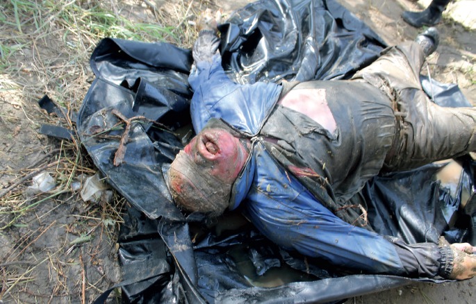
Clothing on a drowning victim will alter the buoyancy and the progression of decomposition. It may also be misleading as in this case where an item of clothing has the appearance of a blindfold.
Image 2:

A body in the water in the standard “drowning position” with the anterior aspect of the body facing the bottom of the river. As the body enters shallow water the distal extremities and forehead are frequently dragged along the bottom.
Image 3:
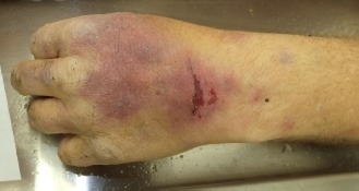
As the hands and feet drag along the bottom, abrasions occur on the extensor surfaces. Differentiating antemortem injuries from postmortem changes may be difficult.
Perhaps the most well-known external change that immersion in liquid has on the body is wrinkling of the skin, particularly involving the hands and feet (3). Traditionally this has been called “washerwoman's hands” or “washerwoman's changes,” though a better designation on the autopsy report would be cutaneous changes of immersion (Image 4). Cutis anserina or goose flesh is another cutaneous change of immersion and is caused by rigor of the erector pilli muscles within the skin. Both of these changes, wrinkling and cutis anserina, will occur as a postmortem change and do not require the individual to be alive upon entering the water (2). The usual postmortem changes of vascular marbling, dark discoloration of skin and soft tissue, bloating, and putrefaction occur in the water as they do on land though at a different rate, particularly in cold water (4). Sloughing of the skin, particularly involving the hands and feet, is common with prolonged immersion (Image 5).
Image 4:

Cutaneous changes of immersion with marked wrinkling of the skin and eventual sloughing of skin, also known as “washerwoman changes.”
Image 5:
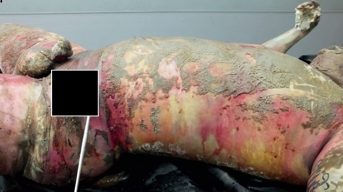
Typical postmortem changes combined with mud and debris as well as sloughing of the skin of the hands and feet are typical for bodies recovered from the water.
Drowning victims frequently have fluid collections in the pleural cavities at autopsy regardless of the postmortem interval. While some of the pleural fluid may represent true effusion occurring as part of the drowning process, fluid accumulation in the pleural spaces is also commonly present in bodies recovered from the water that have undergone decomposition irrespective of the cause of death. A similar phenomenon is seen with the presence of dirt and vegetation in the respiratory tract. Some aspiration of foreign material may occur during the drowning process, though water and debris may also enter the respiratory tree in the postmortem period, particularly in turbulent water. Compared to nondecomposed bodies recovered from water, bodies that have undergone decomposition and recovered from water have been found to have increased pleural fluid accumulation, increased animal predation, and more commonly have dirt and vegetation in the lower respiratory tract (5). Rigor mortis and livor mortis are typically present in bodies recovered from the water though the onset and waning of these classic postmortem changes may be altered by water temperature, current, changing of body position due to movement, and level of activity prior to death. Pink discoloration of the teeth and gums, an observation once thought to be a sign of drowning, is likely due to lividity in these tissues occurring while the body is in the aforementioned drowning position (6).
Temperature and Current
In most cases, the temperature of the water will be cooler than the ambient air temperature. Cooler temperatures generally slow the decomposition process. Exceptions include hot tubs and tropical bodies of water. Current has primarily a mechanical effect on bodies in water. The body itself may be dragged for a distance, creating artifacts that can be mistaken for injuries. The remains or clothing may also get caught on rocks, branches and other objects in the water, creating artifacts that require proper interpretation (Image 6). Not only will a strong current transport the remains for a moderate or even long distance, but other objects in the water can get caught up in the current and come into contact with the remains in a similar manner. In the ocean or fast running rivers and streams the body may strike rocks or brush creating postmortem abrasions and lacerations. Actual injuries may be difficult to appreciate due to leaching of blood from the wounds by the liquid environment. A strong current will enhance the leaching process and a careful assessment for any vital reaction is required to distinguish postmortem from antemortem trauma on the body. Water temperature and current will affect the rate of cooling for a body in a liquid environment. The core temperature of the body at the time of recovery is even less helpful in determining postmortem interval when the body is recovered from water.
Image 6:
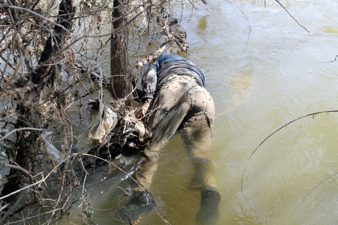
The current may cause the body to come into contact with rocks or drag a body into brush, creating postmortem changes that require proper interpretation.
Adipocere
Adipocere formation may occur in wet or immersed bodies. Adipocere is a yellow-brown, waxy material composed of long chain hydrocarbons such as oleic, palmitic, and stearic acids. It is produced by the conversion of neutral lipids to these compounds as part of the putrefaction process. Both enzymes in the body and within bacteria contribute to the conversion of lipids present in the body to the components of adipocere. Inadequate oxygen combined with a surplus of lipids results in insufficient microbial degradation. Adipocere has a characteristic appearance and is generally resistant to further decomposition (Images 7 and 8). The formation of adipocere usually occurs over a somewhat lengthy postmortem period, typically several months (7-9). However, relatively rapid formation of adipocere has been described (10).
Image 7.
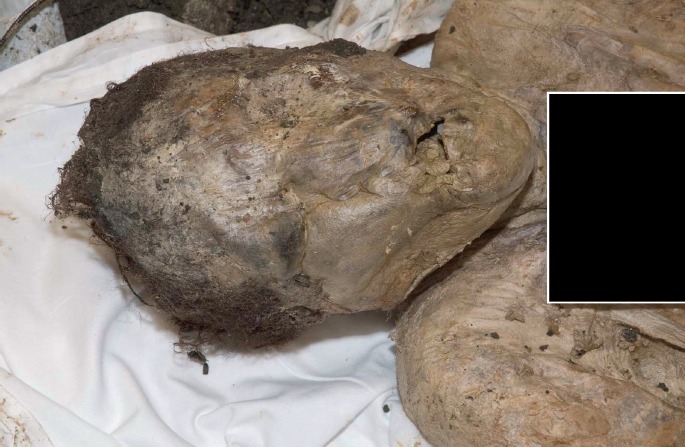
Adipocere formation in remains recovered from a moist environment.
(Image courtesy of Krista Timm MD, Denver Office of the Medical Examiner).
Image 8:
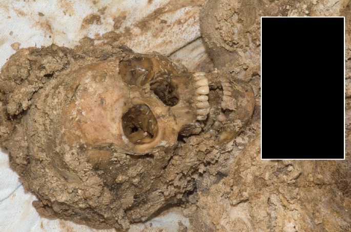
Adipocere formation and partial skeletonization in remains recovered from a moist environment.
(Image courtesy of Krista Timm MD, Denver Office of the Medical Examiner).
A similar putrefaction change may be observed on the surfaces of solid organs, particularly the liver, and on the surfaces of mucous membranes. White spots that have a somewhat miliary appearance have been observed in bodies that have been submerged for prolonged periods. This is presumed to be a breakdown of lipids in a process similar to adipocere formation or saponification (Image 9).
Image 9:

White spots on a mucosal surface from a drowning victim are part of the decomposition process.
Animal Predation
Animal predation, including insect activity, is very different in the water environment. In some cases, the body will be floating on the surface and the usual arthropod predators such as blowflies and carrion beetles will have access to exposed tissue. The immersed portion of the body will be subject to different predators. Aquatic insects may alter the appearance and condition of the remains. Large animals such as turtles, large fish, and large crustaceans will cause tissue damage that in some cases may mimic trauma to the body. Smaller fishes, crabs, shrimp, and invertebrates prey on soft tissue and if given the opportunity can completely deflesh exposed parts of the body. Fish, turtles, and other animals may aggressively feed on remains and in the ocean environment, large carnivores such as sharks will create postmortem artifacts. It is not unusual for small fish and crustaceans to gain access to the interior of the body through skin and soft tissue defects or even normal body orifices (11,12). Examples of rapid skeletonization of remains have been noted in tropical waters where carnivorous predators are abundant, such as the Amazon region of South America (Image 10). In the ocean, several species of sharks and other large carnivorous fish commonly feed on human remains. Large portions of human tissue, including entire extremities, have been recovered from the stomachs of sharks with some frequency. Sharks possess several rows of teeth and commonly, the teeth may be recovered from a bite wound. In most cases it will be concluded that the shark fed on the remains after the individual was deceased.
Image 10:

Rapid skeletonization of remains may occur in bodies of water in tropical areas due to water temperature and carnivorous fish species. This individual reportedly went missing only a few days prior to recovery.
(Image courtesy of Sergio Viegas, Divers Alert Network Brazil).
Conclusion
Determining the cause and manner of death for bodies recovered from water can be challenging. The challenge becomes even greater as the postmortem interval increases. The progression of decomposition changes in a liquid environment is altered by temperature, current, interaction between the remains and the physical environment, and animal predation. While postmortem putrefaction takes place as it does in a dry environment, differences in bacterial flora and an anaerobic atmosphere alter the usual chemical processes and with significant postmortem intervals may result in the conversion of fats to adipocere. Forensic pathologists and medicolegal death investigators must be familiar with the expected postmortem changes that occur in immersed and submerged bodies as well as postmortem artifacts such as animal predation that may be misinterpreted as antemortem injuries.
Footnotes
Financial Disclosure
The author has indicated that he does not have financial relationships to disclose that are relevant to this manuscript
ETHICAL APPROVAL
As per Journal Policies, ethical approval was not required for this manuscript
STATEMENT OF HUMAN AND ANIMAL RIGHTS
This article does not contain any studies conducted with animals or on living human subjects
STATEMENT OF INFORMED CONSENT
No identifiable personal data were presented in this manuscsript
DISCLOSURES & DECLARATION OF CONFLICTS OF INTEREST
The author, reviewers, editors, and publication staff do not report any relevant conflicts of interest
References
- 1.Dolinak D., Matshes E.W., Lew E.O. Forensic pathology, principles and practice. 1st ed. San Diego: Academic Press; c2005. Chapter 24, Postmortem changes; p. 527–54. [Google Scholar]
- 2.DiMaio D., DiMaio V.J.M. Forensic pathology. 2nd ed. Boca Raton: CRC Press; c2001, Chapter 15, Drowning; p. 399–407. [Google Scholar]
- 3.Reh H. [Early postmortem course of washerwoman's skin of the fingers]. Z Rechtsmed. 1984; 92(3): 183–8. German. PMID: 6741294. [DOI] [PubMed] [Google Scholar]
- 4.Karhunen P.J., Goebeler S., Winberg O., Tuominen M. Time of death of victims found in cold water environment. Forensic Sci Int. 2008. Apr 7; 176(2-3): e17–22. PMID: 17935919. 10.1016/j.forsciint.2007.06.014 [DOI] [PubMed] [Google Scholar]
- 5.Ambade V.N., Kukde H.G., Malani A. et al. Decomposed and nondecomposed bodies retrieved from water: a comparative approach. Med Sci Law. 2013. Jan; 53(1): 12–8. PMID: 23155119. 10.1258/msl.2012.012037. [DOI] [PubMed] [Google Scholar]
- 6.Borrman H., Du Chesne A., Brinkmann B. Medico-legal aspects of postmortem pink teeth. Int J Legal Med. 1994; 106(5): 225–31. PMID: 8068567. 10.1007/bf01225410. [DOI] [PubMed] [Google Scholar]
- 7.Schoenen D., Schoenen H. Adipocere formation—the result of insufficient microbial degradation. Forensic Sci Int. 2013. Mar 10; 226(1-3): 301.e1–6. 10.1016/j.forsciint.2013.01.023. [DOI] [PubMed] [Google Scholar]
- 8.Ubelaker D.H., Zarenko K.M. Adipocere: what is known after over two centuries of research. Forensic Sci Int. 2011. May 20; 208(1-3): 167–72. PMID: 21185137. 10.1016/j.forsciint.2010.11.024. [DOI] [PubMed] [Google Scholar]
- 9.O'Brien T.G., Kuehner A.C. Waxing grave about adipocere: soft tissue change in an aquatic context. J Forensic Sci. 2007. Mar; 52(2): 294–301. PMID: 17316224. 10.1111/j.1556-4029.2006.00362.x. [DOI] [PubMed] [Google Scholar]
- 10.Mohan Kumar T.S., Monteiro F.N., Bhagavath P., Bakkannavar S.M. Early adipocere formation: a case report and review of literature. J Forensic Leg Med. 2009. Nov; 16(8): 475–7. PMID: 19782320. 10.1016/j.jflm.2009.07.004. [DOI] [PubMed] [Google Scholar]
- 11.Duband S., Forest F., Clemenson A. et al. Postmortem injuries inflicted by crawfish: morphological and histological aspects. Forensic Sci Int. 2011. Mar 20; 206(1-3): e49–51. PMID: 20813472. 10.1016/j.forsciint.2010.08.006. [DOI] [PubMed] [Google Scholar]
- 12.Duband S., Forest F., Gaillard Y. et al. Macroscopic, histological and toxicological aspects of early Gammarus pulex scavenging. Forensic Sci Int. 2011. Jun 15; 209(1-3): e16–22. PMID: 21497468. 10.1016/j.forsciint.2011.03.025. [DOI] [PubMed] [Google Scholar]