

Abstract
Forensic identification of human remains has long been a core contribution of forensic anthropologists to death investigations. The array and scientific robusticity of the identification methods employed by the anthropologist has evolved in the last several decades, and as with other nonidentification methods, anthropologists have embraced the progression toward the use of validated and statistically defensible methods for identification. This article presents an overview of the role that the forensic anthropologist plays in the identification of human remains and the evolution of anthropological methods of identification.
Keywords: Forensic pathology, Forensic anthropology, Human identification
Introduction
Forensic identification of the deceased is of primary importance in the medicolegal investigation of death. This is true in the daily investigation of individual unexpected deaths and in investigations of mass fatality incidents involving large numbers of human remains and/or fragments of human remains. Since Krogman's 1939 article in the Federal Bureau of Investigation Newsletter entitled “A Guide to the Identification of Human Skeletal Material,” the anthropologist has maintained a significant and expanding role in identification (1). Much has changed since the days of anecdotal comparison of skeletal features for identification with advancements in the development of standard and validated methods for identification in the wake of the Daubert v. Merrell Dow ruling (2) and the more recent National Academy of Sciences (NAS) report (3). The NAS report called on the forensic sciences to validate existing methods and to develop new methods that can be validated via assurance of repeatability, reliability, and accuracy. Recognition by forensic anthropologists of the need for standardized, validated methods for identification predates the NAS report (4–6), as does much of the research aimed at validation of anthropological methods of identification. Anthropologists have continued to contribute to this discussion since the NAS report was published (7–11).
Discussion
Anthropological Methods for Identification
The contribution of the forensic anthropologist to the identification process varies by jurisdiction, in large part due to whether or not the anthropologist is a full-time employee of the medicolegal jurisdiction rather than a contractor. Those who are embedded in a medical examiner/coroner operation are typically involved in a greater variety of components of the identification process than those who consult, including both the application of anthropological methods and administrative contributions (12). This article focuses on the core contribution of the forensic anthropologist to identification: the direct comparison of antemortem radiographs attributable to suspected deceased persons to postmortem radiographic images collected from the deceasent by the medicolegal agency. The anthropologist also impacts the identification process via the extraction of biological indicators of age, sex, ancestry, and stature from unidentified human remains that allow for the development of a biological profile that limits the number of people to whom a decedent bears resemblance (13). Facial approximation and superimposition are methods employed by some anthropologists that are generally useful as tools for eliminating identification candidates or for narrowing candidate pools rather than for identification. These methods are thus not discussed in any detail in this article.
Forensic anthropologists leverage their detailed familiarity with the skeleton and its variability to assess the likelihood that the similarity between two images of the same skeletal feature constitutes identification. This comparison involves consideration of overall bone morphology and contour, trabecular bone patterns, sinus morphology, and the presence and morphology of orthopedic or surgical devices. In general, the greater the morphological complexity, the higher the likelihood that a particular bony feature will have discriminatory value. Thus, most of the bones of the body have the potential to contribute to identification, but certain elements of the skeleton (e.g., frontal sinuses, petrous temporal bone, dentition) are preferable because of their morphological complexity. Some elements that have complex morphology have compromised utility because of their location in the body and their appearance on antemortem imaging. The vertebral column, for example, has very complex morphology, particularly in aged individuals with degenerative changes, but is often viewed by the anthropologist in anterior-posterior chest radiographs and is thus viewed through the anterior ribs, sternum, and thoraco-abdominal organs. Another consideration is the prevalence of imaging in the antemortem record. For example, chest radiographs, though difficult to use for identification for the above stated reason, are commonly used for identification because of their high level of availability. In the author's experience, there also seem to be distinctions between the lay population and the deceased medicolegal population (e.g., higher incidence of interaction with the healthcare system and/or criminal justice system) in terms of antemortem record prevalence, which can benefit the anthropologist seeking antemortem records.
The first account of comparison of radiographs for the purpose of forensic identification of the deceased was described in the third decade of the last century (14). The authors used the morphology of the nasal accessory sinuses and the mastoid processes to confirm the identity of a heavily decomposed person. The subsequent literature, particularly between 1995 and 2008, is replete with published attempts to find means of extracting diagnostic information directly from skeletal remains via comparison of ante- and postmortem radiographs. Numerous publications describe individual identifications made using radiographic imaging of various parts of the postcranial skeleton including the leg and foot (15), chest (11, 16), clavicles (17, 18), pelvis (19, 20), vertebral column (9, 21), and the hand and wrist (6, 22), as well as features of the cranium, including the mastoid sinuses (23), nasal sinuses (14), cranial suture patterns (24), frontal sinuses (25–34), and orthopedic/surgical devices from various parts of the body (35, 36).
The frontal sinuses have received considerable attention as a highly variable and individualistic character of the human skull (25–31), and provide a synopsis of the progression of the identification literature from anecdotal accounts of comparisons to the development of methods for comparison of images complete with statistical measures of likelihood. An exhaustive review of the literature pertaining to identification methods based on every part of the skeleton is beyond the scope of this article. The following paragraph uses the frontal sinus as an example of the development of, problems with, and considerations associated with radiograph identification methods.
Articles discussing the radiographic evaluation of the frontal sinuses in personal identification have been published on several occasions (25, 27, 28, 37, 38). However, the majority of the early publications are case reports and do not present techniques for future application, nor do they satisfy the requirements of the Daubert guidelines or address the limitations identified in the NAS report. For example, Quatrehomme and Fronty suggests simple superimposition of ante- and postmortem conventional radiograph images as a viable technique for personal identification from the frontal sinus (39). The authors acknowledge the considerable difficulty associated with the correct orientation of the skull for postmortem imaging, but do not address the subjectivity associated with the method. Quatrehomme and Fronty's method is typical of these articles, which present a case, or series of cases, in which “identifications” were made based on matches perceived by the observer (25, 28,38). Kirk et al. published the results of a survey of 39 cases in which identifications were made in Ontario, Canada, based on nonmetric comparison of frontal sinus configuration (40). Only three of the 39 cases were considered unidentifiable using the frontal sinus because of poor film quality and the remaining 36 produced “conclusive postmortem to antemortem pattern matches” (40). The identifications were made based on uniqueness, as perceived by the viewer, between superimposed ante- and postmortem anteroposterior radiographs of the skull. They also refer to the “quantitative” association of 16 cases. This quantification involved measurement of the maximum vertical and horizontal dimensions of the sinuses. “Any discrepancy between antemortem and postmortem values greater than 5 mm was classified as a metric nonmatch” (40). Although less subjective than the identifications made solely on the basis of simple superimposition of ante- and postmortem imaging, this technique does not provide either a statistical measure of the strength of the associations or error rates associated with its use. More comprehensive systems for classifying and/or identifying crania according to variability of the frontal sinuses have been presented by Yoshino et al. (29), Yoshino et al. (41), Schuller (26), Reichs (31), and Reichs and Dorion (30). Yoshino et al. developed a potentially repeatable methodology for personal identification from conventional radiographs of the frontal sinuses (29). The method involved the development of a code number from the classification of six independent characteristics of the frontal sinuses. The code was a series of numbers that was statistically unlikely to be derived from more than a single individual by virtue of its length and the number of possible combinations represented by all of its possible variations. Yoshino used the skulls of 35 Japanese adults (21 males and 14 females) to develop the technique. They tested the technique on a single forensic case and concluded that the technique is of “considerable value in cases in which dental remains are not available” (29). Reichs and Dorion (30) and Reichs (31) modified this technique for use with axial computed tomography (CT) scans of the skull. Their argument is essentially that future use of CT imaging will far outweigh the use of fronto-occipital radiographs, and that identification techniques based on radiographs will someday become obsolete. Thus instead of conventional radiography, they developed a method similar to that of Yoshino et al. using axial CT imaging (29). They evaluated a slightly different series of characteristics than Yoshino et al. that were more effectively imaged on CT scans, including bilateral dimension, bilateral asymmetry, superiority of side, distribution of partial bony septations, number of partial bony septations, distribution of complete bony cells, and number of complete bony cells (29). They suggested that scoring these attributes on several consecutive CT slices added to the length of the code derived, and therefore, to its statistical uniqueness.
Development of Quantitative Methods for Identification
There has been a surge in publications regarding the validation of all aspects of forensic anthropological analysis in the last ten years, particularly since the publication of the NAS report. For many of the analyses conducted by forensic anthropologists, including sex and age estimation, this involves the statistical validation of existing anthropological methods like pubic symphysis or sternal rib end aging. On the other hand, identification of decedents via radiograph comparison has been historically subjective. The discriminatory value of subjective comparison of skeletal radiographs is valuable and these methods are likely to persist as an identification tool for use by the forensic anthropologist. This is true in part because the anthropologist cannot validate radiograph identification methods for every bone in the body, nor can we anticipate what bone(s) may be available for comparison in a given case. However, the importance of the development of validated identification methods is well recognized. “Because forensic anthropologists are trained first and foremost as physical anthropologists, they emphasize estimation problems at the expense of evidentiary problems” (42). Steadman and colleagues outline the application of Bayesian statistics, in particular likelihood ratios, in the validation of anthropological methods of identification. They explain the conceptual and practical differences between appropriate reference samples and the population at large. The authors identify several databases that can be used as references for population at large estimates, as well as novel data that can be used to calculate the frequency of specific identifiers within the population at large.
The primary problem with historical usage of radiograph comparison is the real or perceived lack of repeatability of the comparisons. Repeatability is dependent on the objectivity of the methodology employed, and objectivity is difficult to establish when using methods dependent on the investigator's individual assessment of subjective characteristics, such as a particular shape, level of symmetry, or side dominance. For example, the features of the frontal sinuses evaluated in the publications by both Yoshino et al. (29) and Reichs and Dorion (30) are arbitrary. There is a need for more easily reproducible character evaluations. Some authors have tried to increase the objectivity of radiographic methods by various means. As mentioned above, Kirk et al. attempted to add quantitative robusticity to the radiographic analysis of frontal sinuses by recording the maximum width and length of the sinuses in a sample of 39 crania, in addition to the more subjective variables used in other investigations (40). Reipert et al. completed an investigation that aimed to “improve the objectivity of X-ray comparison for the identification of unknown individuals” by using a computer program named FoXSIS to reduce subjectivity in the analysis of the frontal sinuses (43). Computed tomography data were collected for 30 skulls, and were converted to virtual X-ray representations of the skulls. According to the authors, the primary advantage of the program was that its use of digital measurements of the features of the skull allowed for a quantification of the “probability of identity” without reliance on the more subjective, and less repeatable measures of identity such as side dominance, shape, and symmetry levels.
There is also a problem with techniques that are based on the assumption that it is statistically improbable for two individuals to have the same or similar codes just because there are a large number of possible combinations. This is problematic (at least with the techniques outlined above) because no effort is made to weigh the various characters relative to one another. For example, it is much more likely for an individual to have a single or very few septations of the frontal sinus than for them to have six or seven, thereby increasing dramatically the statistical probability that two individuals may indeed have the same code for that particular trait. This is a problem with many of the characteristics considered by both Reichs (31) and Yoshino et al. (29). Christensen published the most objective and statistically robust analysis of the frontal sinuses to date (32). She used elliptical Fourier analysis (EFA), a geometric morphometric technique that imposes a set of coordinate data points to a closed curve, to quantify the superior border of the frontal sinuses. The coordinate data were then used to quantify the outlines by generating a set of coefficients that represent a numerical reproduction of the curve. She traced the superior border of the frontal sinuses of 808 individuals and closed the curve inferiorly by connecting the inferior-most extensions of the left and right sides of the curve with a straight horizontal line. The Euclidean distances between the EFA-generated outlines were compared as a means to test the accuracy with which they could be used to associate the correct images. The Euclidean distances between repeat images from the same individual were significantly smaller than the distances between individuals. She used typicality statistics to evaluate the statistical strength of the associations with good results. The probability of making an incorrect association between two outlines using her method is very small, and she concludes that “there is a quantifiable and significant difference between the shapes of individual frontal sinus outlines” (32).
Wiersema developed a method for comparison of ante- and postmortem axial CT images of the petrous portion of the temporal bone (44). The data used in the study were collected from repeat axial head CT images of 115 individuals, and the Euclidean distance comparisons were made between images of the same individual and images from different individuals. Two-dimensional coordinate data from 36 landmarks on each of the CT images were calculated and the distances between each of the coordinate points were captured to generate the data used in the statistical analyses. Measurement subsets were developed based on two separate models, the first of which used anatomical criteria identified by the study author and the second used principal components factor analysis to identify the subset with the most statistical significance (Images 1-3).
Image 1.
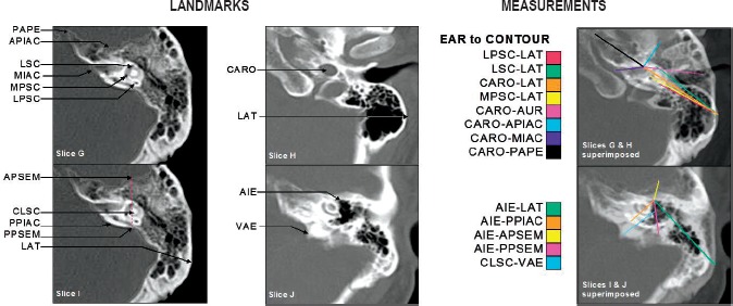
The first two columns illustrate the landmark locations in the inner and middle ear segments of the petrous part of the temporal bone used in this research. The third column illustrates the measurements between the landmarks in the inner and middle ear segments. The measurements are color-coded according to the legend located between columns 2 and 3.
Image 2.
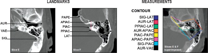
The first two images (labeled slices E&F) illustrate the locations of the landmarks around the contour of the petrous part of the temporal bone. The third image illustrates the measurements between the landmarks around the contour of the petrous part of the temporal bone. The measurements are color-coded according to the legend located between the second and third images.
Image 3.
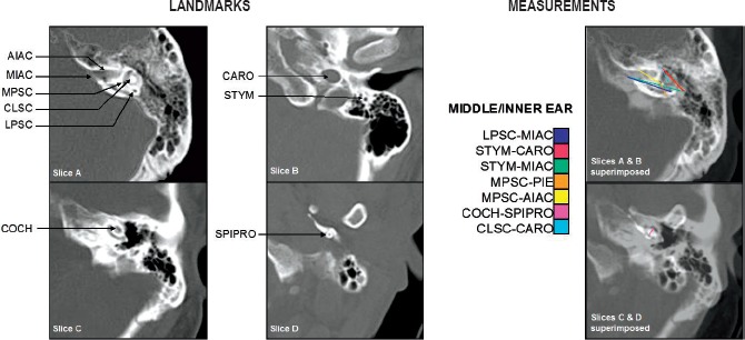
The first two columns illustrate the remaining landmark locations in both the inner and middle ear, and the contour segments of the petrous part of the temporal bone. The third column illustrates the measurements between the measurements that extend between the landmarks of the inner and middle ear, and the contour of the petrous part of the temporal bone. The measurements are color coded according to the legend located between columns 2 and 3.
The measurement sets of both models were then compared to one another using nearest neighbor analysis, to test their relative efficiency in matching replicate images to one another. The results of both models were highly accurate. Three incorrect nearest neighbor matches resulted from the biological model and five from the principal components factor analysis model. The errors appear to have been the result of variation in the axial plane between the first and second scans. The results of the nearest neighbor comparisons were then considered within the context of Bayes' Theorem by calculating likelihood ratios and posterior probabilities. The likelihood ratios and posterior probabilities were very high for both models, indicating that 1) there is significant individual variability in the measurements of the petrous portion used in this research and 2) this variation represents a high level of potential accuracy in the application of this method in the identification of forensic remains.
Derrick et al. describe an automated method for comparison of lateral images of the cervical spine that was mathematically validated for use by medical examiner's/coroner's offices (9). The software quantifies the likelihood that any two of the radiographs in an array of cervical spine images (Image 4) contain matching vertebral body morphology. Six validations were conducted to evaluate the repeatability, reliability, and sensitivity of the method. The authors report a 92-100% success rate in matching the test image to the correct image in the array. The success rate for cervical vertebrae is particularly high, less so for lumbar vertebral bodies (which tend to be less morphologically distinctive and often partially obstructed by intermediate objects).
Image 4.
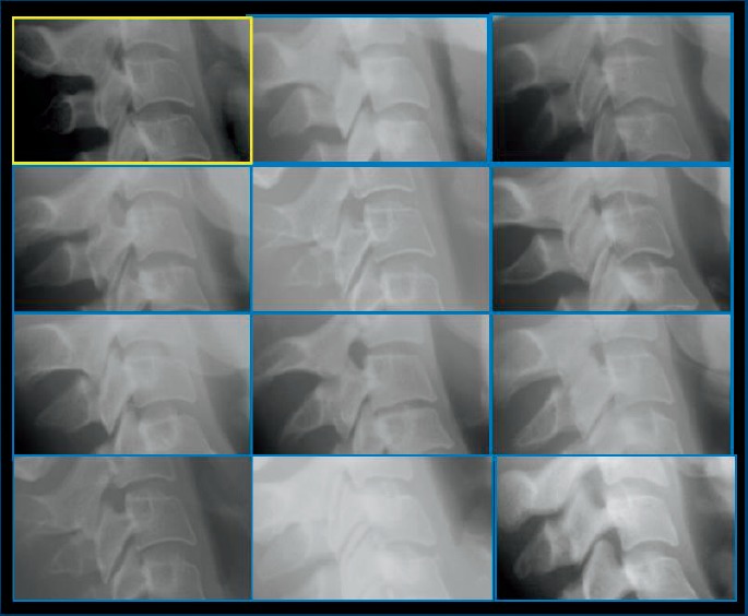
Sample test array.
The limitation of this particular method is the limited availability of antemortem lateral cervical spine radiographs. The utility of the method would be greatly enhanced if antemortem lateral CT scout images of the head and neck were deemed appropriate for comparison.
Forensic Anthropological Identification in the Mass Fatality Context
The role of the anthropologist in the mass fatality context is discussed at length elsewhere (45). However, that article does not address the methodological issues associated with the identification of fragmentary remains, or of common, unidentifiable tissue that is sometimes present following a mass fatality incident. Many of the radiographic techniques employed for daily cases are not useful for postdisaster fragmentary remains. Some of the more diagnostic portions of the skeleton, including the frontal sinus, are at considerable risk of destruction. Focus of current/future methods for use in the mass fatality context should involve segments of the skeleton that resist taphonomic destruction, including the petrous portion of the temporal bone. It may also be difficult to employ radiographic techniques due to availability of antemortem radiographs, as the victims of mass fatality incidents are often more similar to the lay population rather than the deceased medicolegal population.
Conclusion
The forensic sciences have undergone a considerable transformation in recent decades toward a more robust network of services that are based on validated methods and accredited laboratories. Anthropological methods have evolved concomitantly from the anecdotal to the statistically validated, accurate, and repeatable methods that are being developed today. The need for more subjective anthropological evaluation of radiographs will continue to have a place in forensic anthropology for identification, but validation of these methods is as important as it is for other anthropological methods (sex, age and ancestry estimation).
Footnotes
Disclosures
The author has indicated that he does not have financial relationships to disclose that are relevant to this manuscript
ETHICAL APPROVAL
As per Journal Policies, ethical approval was not required for this manuscript
STATEMENT OF HUMAN AND ANIMAL RIGHTS
This article does not contain any studies conducted with animals or on living human subjects
STATEMENT OF INFORMED CONSENT
No identifiable personal data were presented in this manuscsript
DISCLOSURES & DECLARATION OF CONFLICTS OF INTEREST
The authors, reviewers, editors, and publication staff do not report any relevant conflicts of interest
References
- 1).Krogman W.M. A guide to the identification of human skeletal material. FBI Law Enforc Bullet. 1939; 8(8): 3–31. [Google Scholar]
- 2).Daubert v. Merrell Dow Pharmaceuticals. U.S. 1993. p. 579.
- 3).National Research Council. Strengthening forensic science in the United States: a path forward. Washington: National Academies Press; 2009. 352 p. [Google Scholar]
- 4).Christensen A.M. The impact of Daubert: implications for testimony and research in forensic anthropology (and the use of frontal sinuses in personal identification). J Forensic Sci. 2004. May; 49(3): 427–30. PMID: 15171154. 10.1520/jfs2003185. [DOI] [PubMed] [Google Scholar]
- 5).Christensen A.M., Crowder C.M. Evidentiary standards for forensic anthropology. J Forensic Sci. 2009. Nov; 54(6): 1211–6. PMID: 19804520. 10.1111/j.1556-4029.2009.01176.x. [DOI] [PubMed] [Google Scholar]
- 6).Koot M.G., Sauer N.J., Fenton T.W. Radiographic human identification using bones of the hand: a validation study. J Forensic Sci. 2005. Mar; 50(2): 263–8. PMID: 15813535. 10.1520/jfs2004229. [DOI] [PubMed] [Google Scholar]
- 7).Christensen A.M., Crowder C.M., Ousley S.D., Houck M.M. Error and its meaning in forensic science. J Forensic Sci. 2014. Jan; 59(1): 123–6. PMID: 24111751. 10.1111/1556-4029.12275. [DOI] [PubMed] [Google Scholar]
- 8).Niespodziewanski E., Stephan C.N., Guyomarc'h P., Fenton T.W. Human identification via lateral patella radiographs: a validation study. J Forensic Sci. 2016. Jan; 61(1): 134–40. PMID: 26234529. 10.1111/1556-4029.12898. [DOI] [PubMed] [Google Scholar]
- 9).Derrick S.M., Raxter M.H., Hipp J.A. et al. Development of a computer-assisted forensic radiographic identification method using the lateral cervical and lumbar spine. J Forensic Sci. 2015. Jan; 60(1): 5–12. PMID: 24961154. 10.1111/1556-4029.12531. [DOI] [PubMed] [Google Scholar]
- 10).Ross A.H., Lanfear A.K., Maxwell A.B. Establishing standards for side-by-side radiographic comparisons. Am J Forensic Med Pathol. 2016. Jun; 37(2): 86–94. PMID: 26999427. 10.1097/paf.0000000000000223. [DOI] [PubMed] [Google Scholar]
- 11).Stephan C.N., Winburn A.P., Christensen A.F., Tyrrell A.J. Skeletal identification by radiographic comparison: blind tests of a morphoscopic method using antemortem chest radiographs. J Forensic Sci. 2011. Mar; 56(2): 320–32. PMID: 21306373. 10.1111/j.1556-4029.2010.01673.x. [DOI] [PubMed] [Google Scholar]
- 12).Crowder C.M., Wiersema J.M., Adams B.J. et al. The utility of forensic anthropology in the medical examiner's office. Acad Forensic Pathol. 2016. Sep; 6(3): 349–60. [DOI] [PMC free article] [PubMed] [Google Scholar]
- 13).Austin D., King R.E. The biological profile of unidentified human remains in a forensic context. Acad Forensic Pathol. 2016. Sep; 6(3): 370–90. [DOI] [PMC free article] [PubMed] [Google Scholar]
- 14).Culbert W.L., Law F.M. Identification by comparison with roentgenograms of nasal accessory sinuses and mastoid processes. JAMA. 1927; 88: 1634–6. 10.1001/jama.1927.02680470020009. [DOI] [Google Scholar]
- 15).Owsley D.W., Mann R.W. Positive identification based on radiographic examination of the leg and foot. A case report. J Am Podiatr Med Assoc. 1989. Oct; 79(10): 511–3. PMID: 2585283. 10.7547/87507315-79-10-511. [DOI] [PubMed] [Google Scholar]
- 16).Kuehn C.M., Taylor K.M., Mann F.A. et al. Validation of chest X-ray comparisons for unknown decedent identification. J Forensic Sci. 2002. Jul; 47(4): 725–9. PMID: 12136980. 10.1520/jfs15450j. [DOI] [PubMed] [Google Scholar]
- 17).Stephan C.N., Amidan B., Trease H. et al. Morphometric comparison of clavicle outlines from 3D bone scans and 2D chest radiographs: a shortlisting tool to assist radiographic identification of human skeletons. J Forensic Sci. 2014. Mar; 59(2): 306–13. PMID: 24313347. 10.1111/1556-4029.12324. [DOI] [PubMed] [Google Scholar]
- 18).Stephan C.N., Guyomarc'h P. Quantification of perspective-induced shape change of clavicles at radiography and 3D scanning to assist human identification. J Forensic Sci. 2014. Mar; 59(2): 447–53. PMID: 24313366. 10.1111/1556-4029.12325. [DOI] [PubMed] [Google Scholar]
- 19).Moser R.P. Jr., Wagner G.N. Nutrient groove of the ilium, a subtle but important forensic radiographic marker in the identification of victims of severe trauma. Skeletal Radiol. 1990; 19(1): 15–9. PMID: 2326650. 10.1007/bf00197922. [DOI] [PubMed] [Google Scholar]
- 20).Owsley D.W., Mann R.W. Positive personal identity of skeletonized remains using abdominal and pelvic radiographs. J Forensic Sci. 1992. Jan; 37(1): 332–6. PMID: 1545209. 10.1520/jfs13238j. [DOI] [PubMed] [Google Scholar]
- 21).Mundorff A.Z., Vidoli G., Melinek J. Anthropological and radiographic comparison of vertebrae for identification of decomposed human remains. J Forensic Sci. 2006. Sep; 51(5): 1002–4. PMID: 17018076. 10.1111/j.1556-4029.2006.00233.x. [DOI] [PubMed] [Google Scholar]
- 22).Greulich W.W. Skeletal features visible on the roentgenogram of the hand and wrist which can be used for establishing individual identification. Am J Roentgenol Radium Ther Nucl Med. 1960. Apr; 83: 756–64. PMID: 13829223. [PubMed] [Google Scholar]
- 23).Rhine S., Sperry K. Radiographic identification by mastoid sinus and arterial pattern. J Forensic Sci. 1991. Jan; 36(1): 272–9. PMID: 2007877. 10.1520/jfs13029j. [DOI] [PubMed] [Google Scholar]
- 24).Chandra-Sekharan P. Identification of skull from its suture pattern. Forensic Sci Int. 1985. Mar; 27(3): 205–14. PMID: 3988197. 10.1016/0379-0738(85)90156-2. [DOI] [PubMed] [Google Scholar]
- 25).Mayer J. Identification by sinus prints. Virginia Med Mon. 1935; 62: 517–519. [Google Scholar]
- 26).Schuller A. A note on the identification of skulls by x-ray pictures of the frontal sinuses. Med J Australia. 1943; 1: 554–6. [Google Scholar]
- 27).Asherson N. Identification by frontal sinus prints: a forensic medical pilot survey. London: H.K. Lewis; 1965. 80 p. [Google Scholar]
- 28).Ubelaker D.H. Human identification: case studies in forensic anthropology. Springfield (IL): C.C. Thomas; c1984. Chapter 29, Positive identification from the radiographic comparison of frontal sinus patterns; p. 399–411. [Google Scholar]
- 29).Yoshino M., Miyasaka S., Sato H., Seta S. Classification system of frontal sinus patterns by radiography. Its application to identification of unknown skeletal remains. Forensic Sci Int. 1987. Aug; 34(4): 289–99. PMID: 3623370. 10.1016/0379-0738(87)90041-7. [DOI] [PubMed] [Google Scholar]
- 30).Reichs K.J., Dorion R.B.J. The use of computed tomography (CT) scans in the comparison of frontal sinus configurations. Can Soc Forensic Sci J. 1992; 25(1): 1–16. 10.1080/00085030.1992.10756997. [DOI] [Google Scholar]
- 31).Reichs K.J. Quantified comparison of frontal sinus patterns by means of computed tomography. Forensic Sci Int. 1993. Oct; 61(2-3): 141–68. PMID: 8307523. 10.1016/0379-0738(93)90222-v. [DOI] [PubMed] [Google Scholar]
- 32).Christensen A. Assessing the variation in individual frontal sinus outlines. Am J Phys Anthropol. 2005. Jul; 127(3): 291–5. PMID: 15584070. 10.1002/ajpa.20116. [DOI] [PubMed] [Google Scholar]
- 33).David M.P., Saxena R. Use of frontal sinus and nasal septum patterns as an aid in personal identification: A digital radiographic pilot study. J Forensic Dent Sci. 2010. Jul; 2(2): 77–80. PMID: 21731344. PMCID: PMC3125957. 10.4103/0975-1475.81286. [DOI] [PMC free article] [PubMed] [Google Scholar]
- 34).Patil N., Karjodkar F.R., Sontakke S. et al. Uniqueness of radiographic patterns of the frontal sinus for personal identification. Imaging Sci Dent. 2012. Dec; 42(4): 213–7. PMID: 23301206. PMCID: PMC3534174. 10.5624/isd.2012.42.4.213. [DOI] [PMC free article] [PubMed] [Google Scholar]
- 35).Wilson R.J., Bethard J.D., DiGangi E.A. The use of orthopedic surgical devices for forensic identification. J Forensic Sci. 2011. Mar; 56(2): 460–9. PMID: 21342187. 10.1111/j.1556-4029.2010.01639.x. [DOI] [PubMed] [Google Scholar]
- 36).Ubelaker D.H., Jacobs C.H. Identification of orthopedic device manufacturer. J Forensic Sci. 1995; 40(2): 168–70. 10.1520/jfs15335j. [DOI] [Google Scholar]
- 37).Camps F.E. Recent advances in forensic pathology. London: Churchill; c1969. Chapter 8, Radiology and its forensic application; p. 149–60. [Google Scholar]
- 38).Marlin D.C., Clark M.A., Standish S.M. Identification of human remains by comparison of frontal sinus radiographs: a series of four cases. J Forensic Sci. 1991. Nov; 36(6): 1765–72. PMID: 1770345. 10.1520/jfs13202j. [DOI] [PubMed] [Google Scholar]
- 39).Quatrehomme G., Fronty P., Sapanet M. et al. Identification by frontal sinus pattern in forensic anthropology. Forensic Sci Int. 1996. Dec 2; 83(2): 147–53. PMID: 9022276. 10.1016/s0379-0738(96)02033-6. [DOI] [PubMed] [Google Scholar]
- 40).Kirk N.J., Wood R.E., Goldstein M. Skeletal identification using the frontal sinus region: a retrospective study of 39 cases. J Forensic Sci. 2002. Mar; 47(2): 318–23. PMID: 11908601. 10.1520/jfs15250j. [DOI] [PubMed] [Google Scholar]
- 41).Yoshino M., Miyasaka H., Saito H. et al. Classification system of frontal sinus patterns. Can Soc Forensic Sci J. 1989; 22(2): 135–146. 10.1080/00085030.1989.10757429. [DOI] [PubMed] [Google Scholar]
- 42).Steadman D.W., Adams B.J., Konigsberg L.W. Statistical basis for positive identification in forensic anthropology. Am J Phys Anthropol. 2006. Sep; 131(1): 15–26. PMID: 16485302. 10.1002/ajpa.20393. [DOI] [PubMed] [Google Scholar]
- 43).Riepert T., Ulmcke D., Schweden F., Nafe B. Identification of unknown dead bodies by X-ray image comparison of the skull using the X-ray simulation program FoXSIS. Forensic Sci Int. 2001. Mar 1; 117(1-2): 89–98. PMID: 11230950. 10.1016/s0379-0738(00)00452-7. [DOI] [PubMed] [Google Scholar]
- 44).Wiersema J.M. The petrous portion of the human temporal bone: potential for forensic individuation [Internet]. PhD dissertation. College Station (TX): Texas A&M University; 2006. 238 p. Available from: http://oaktrust.library.tamu.edu/bitstream/handle/1969.1/ETD-TAMU-1724/WIERSEMA-DISSERTATION.pdf?sequence=1&isAllowed=y. [Google Scholar]
- 45).Wiersema J.M., Woody A. The forensic anthropologist in the mass fatality context. Acad Forensic Pathol. 2016. Sep; 6(3): 455–62. [DOI] [PMC free article] [PubMed] [Google Scholar]