Abstract
Due to its antibacterial actions, silver sulfadiazine is widely used as a topical agent in the treatment of wounds, including burns. Widespread or prolonged topical application of silver sulfadiazine dressings can lead to argyria including systemic symptoms due to the resorption of silver. Here, we report a patient experiencing localized argyria due to sunlight exposure after topical use of silver sulfadiazine cream on his face.
Key Words: Silver sulfadiazine, Localized argyria, Burns
Introduction
Silver is an ancient antimicrobial agent that has played a major role in the treatment of a wide variety of diseases before the introduction of antibiotics in the 1940s [1]. Silver nitrate and silver sulfadiazine have been widely used in the topical chemoprophylactic treatment of wounds, especially for burns and ulcers. With the establishment of silver-coated wound dressings, a constant release of silver ions prompted by wound fluids as well as tissue exudates of up to 7 days can be reached [2].
Although silver is absorbed and deposited throughout the body, this usually has no clinical relevance. Typical sites of silver deposition include the liver, kidneys, cornea, and the skin [3]. Chronic long-term ingestion of silver leads to generalized argyria, a blue-grayish discoloration of the skin [4, 5, 6]. Topical application of silver-containing agents can result in localized argyria, as well as systemic side effects in cases of greater skin areas involved. Treatment for cutaneous argyria includes discontinuation of the causing agent, as well as sun protection to avoid reactions with UV light. For refractory skin discolorations, treatment with Q-switched 1,064-nm Nd:YAG laser, or alternatively 694-nm ruby or 755-nm alexandrite laser, has been shown to be effective in several case reports [7, 8, 9]. A potentially limiting side effect of laser therapy is significant pain during treatment [10].
We listed all cases of localized argyria that we could identify while reviewing available literature, as well as their treatment (see online suppl. Table; for all online suppl. material, see www.karger.com/doi/10.1159/000494610). This case report describes the case of a patient who experienced transient localized argyria in the areas of topical silver sulfadiazine application for 3 days.
Case Report
A 17-year-old male patient presented to the ER after he suffered from second degree burns over both his eyebrows, his left upper eyelid and left cheek.
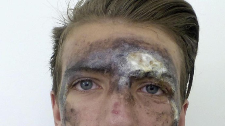
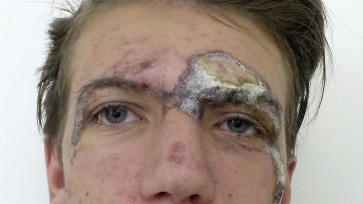
The wound was locally cleaned and disinfected. The patient was then discharged with the prescription of a silver sulfadiazine dressing. On a follow-up visit 3 days later, the patient complained of dark grayish discolorations in the areas of his face where he had applied silver sulfadiazine cream (Fig. 1). Sterile gauze pads with sodium chloride were left on the discolored areas to soak for around 15 min, which allowed for an easy removal of the color in affected areas with healthy skin (Fig. 2). The wounds themselves were not cleaned to avoid interfering with the healing process and causing pain to the patient.
Fig. 1.
Three days after burn.
Fig. 2.
Three days after burn, after cleaning with 0.8% NaCl.
The patient was instructed to stop using the silver sulfadiazine cream and to apply a panthenol-containing cream as needed. He was educated about the importance of using sunscreen SPF 50+ in order to prevent sunlight reacting with any remaining silver ions in the wounds.
Discussion
While previously reported cases described patients who either ingested silver or had longer and more extensive exposure to silver-containing topicals, our patient experienced a strong discoloration after only 3 days due to UV exposure during treatment.
Coombs et al. [11] found a fast absorption of silver through burn wounds, with elevated serum silver levels in 20 of 22 patients included in the study, and hepatic dysfunction in patients with burns affecting more than 10% TBSA. This phenomenon could also be seen in a 17-y-ear-old patient with 30% TBSA mixed depth burns on the lower body half, who was treated with Acticoat silver-coated wound dressing, showing symptoms of hepatotoxicity, and argyria-like symptoms on his face, as well as elevated silver levels in plasma and urine, indicating the absorption of the silver released from the dressing. After removal of the silver-coated dressing, liver enzymes returned to normal, and the patient's facial discoloration disappeared over the next few days [12]. Acticoat wound dressing was also associated with the semi-permanent skin staining described in a 17-year-old burn patient - the skin discoloration in the affected area lasted for 3 years [13].
However, the use of silver was found to be safe to use in moderate burns, as in our patient [11]. The reason for the extreme discoloration in our case might have been the reaction of silver with light, as this patient's burn wounds were in the face, and occurred during summer, so UV exposition was high. Silver is highly reactive with light, a mechanism that is used in photography, resulting in a photoreduction of ionic Ag+ to metallic Ag0. Reduced silver, which exhibits the classic grayish color, subsequently reacts with various ligands (SH), such as enzymes, to Ag2S, and selenium-containing enzymes to form the complex Ag2Se/S [14]. This argyrol complex then deposits in the cell, leading to staining of the affected skin.
Conclusion
Despite being a rather rare adverse effect, local discoloration of the skin after the topical treatment with silver sulfadiazine-containing creams has to be kept in mind. Especially in sun-exposed areas such as the face or the hands, even short-term applications can lead to localized argyria. The risk can be reduced by applying dressings to avoid exposure to light. As silver-induced skin discolorations can remain over years, we recommend limiting the use of silver sulfadiazine-containing creams to non-sun-exposed areas.
Statement of Ethics
The authors have no ethical conflicts to disclose.
Disclosure Statement
The authors report no conflict of interest.
Supplementary Material
Supplementary data
References
- 1.Alexander JW. History of the medical use of silver. Surg Infect (Larchmt) 2009 Jun;10((3)):289–92. doi: 10.1089/sur.2008.9941. [DOI] [PubMed] [Google Scholar]
- 2.Lansdown AB. Silver in health care: antimicrobial effects and safety in use. Curr Probl Dermatol. 2006;33:17–34. doi: 10.1159/000093928. [DOI] [PubMed] [Google Scholar]
- 3.Maitre S, Jaber K, Perrot JL, Guy C, Cambazard F. [Increased serum and urinary levels of silver during treatment with topical silver sulfadiazine] Ann Dermatol Venereol. 2002 Feb;129((2)):217–9. [PubMed] [Google Scholar]
- 4.Cinotti E, Labeille B, Douchet C, Cambazard F, Perrot JL. Dermoscopy, reflectance confocal microscopy, and high-definition optical coherence tomography in the diagnosis of generalized argyria. Journal of the American Academy of Dermatology. 2017;76((2s1)):S66–s68. doi: 10.1016/j.jaad.2016.07.057. doi: Online First: Epub Date]|. [DOI] [PubMed] [Google Scholar]
- 5.Saluja SS, Bowen AR, Hull CM. Resident Rounds: Part III - Case Report: Argyria - A Case of Blue-Gray Skin. J Drugs Dermatol. 2015 Jul;14((7)):760–1. [PubMed] [Google Scholar]
- 6.White JM, Powell AM, Brady K, Russell-Jones R. Severe generalized argyria secondary to ingestion of colloidal silver protein. Clin Exp Dermatol. 2003 May;28((3)):254–6. doi: 10.1046/j.1365-2230.2003.01214.x. [DOI] [PubMed] [Google Scholar]
- 7.Han TY, Chang HS, Lee HK, Son SJ. Successful treatment of argyria using a low-fluence Q-switched 1064-nm Nd:YAG laser. Int J Dermatol. 2011 Jun;50((6)):751–3. doi: 10.1111/j.1365-4632.2010.04796.x. [DOI] [PubMed] [Google Scholar]
- 8.Friedmann DP, Buckley S, Mishra V. Localized Cutaneous Argyria From a Nasal Piercing Successfully Treated With a Picosecond 755-nm Q-Switched Alexandrite Laser. Dermatologic surgery: official publication for American Society for Dermatologic Surgery [et al] 2017;43((8)):1094–1095. doi: 10.1097/DSS.0000000000001162. doi: Online First: Epub Date] [DOI] [PubMed] [Google Scholar]
- 9.Gottesman SP, Goldberg GN. Immediate successful treatment of argyria with a single pass of multiple Q-switched laser wavelengths. JAMA Dermatol. 2013 May;149((5)):623–4. doi: 10.1001/jamadermatol.2013.234. [DOI] [PubMed] [Google Scholar]
- 10.Saager RB, Hassan KM, Kondru C, Durkin AJ, Kelly KM. Quantitative near infrared spectroscopic analysis of Q-Switched Nd:YAG treatment of generalized argyria. Lasers Surg Med. 2013 Jan;45((1)):15–21. doi: 10.1002/lsm.22084. [DOI] [PMC free article] [PubMed] [Google Scholar]
- 11.Coombs CJ, Wan AT, Masterton JP, Conyers RA, Pedersen J, Chia YT. Do burn patients have a silver lining? Burns: journal of the International Society for Burn Injuries. 1992;((3)):179–184. doi: 10.1016/0305-4179(92)90067-5. 18. [DOI] [PubMed] [Google Scholar]
- 12.Trop M, Novak M, Rodl S, Hellbom B, Kroell W, Goessler W. Silver-coated dressing acticoat caused raised liver enzymes and argyria-like symptoms in burn patient. J Trauma. 2006 Mar;60((3)):648–52. doi: 10.1097/01.ta.0000208126.22089.b6. [DOI] [PubMed] [Google Scholar]
- 13.Zweiker D, Horn S, Hoell A, Seitz S, Walter D, Trop M. Semi-permanent skin staining associated with silver-coated wound dressing Acticoat. Ann Burns Fire Disasters. 2014 Dec;27((4)):197–200. [PMC free article] [PubMed] [Google Scholar]
- 14.Dubey P, Matai I, Kumar SU, Sachdev A, Bhushan B, Gopinath P. Perturbation of cellular mechanistic system by silver nanoparticle toxicity: Cytotoxic, genotoxic and epigenetic potentials. Adv Colloid Interface Sci. 2015 Jul;221:4–21. doi: 10.1016/j.cis.2015.02.007. [DOI] [PubMed] [Google Scholar]
Associated Data
This section collects any data citations, data availability statements, or supplementary materials included in this article.
Supplementary Materials
Supplementary data


