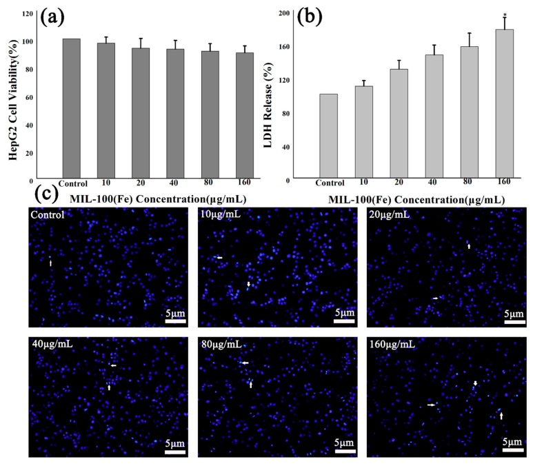
Figure 6.
Effects of MIL-100(Fe) on HepG2 cells’ viability, membrane, and morphology: (a) In vitro cell viabilities of HepG2 cells after being incubated for 48 h with MIL-100(Fe); (b) LDH release of HepG2 cells incubated for 48 h with MIL-100(Fe); (c) nuclear morphology images of HepG2 cells after 48 h exposure to MIL-100(Fe). (* p < 0.05 vs. Control.)

