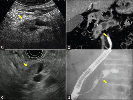
A 64-year-old man was referred to our hospital for workup of a pancreatic mass. Ultrasonography revealed dilatation of the main pancreatic duct (MPD) in the body and tail of the pancreas [Figure 1a]. Abdominal computed tomography demonstrated a mass in the head of the pancreas with gradual enhancement [Figure 1b]. EUS showed a hypoechoic mass in MPD, measuring about 16 mm × 14 mm, with dilatation of MPD in the pancreatic body and tail [Figure 1c]. Endoscopic retrograde pancreatography showed obstruction of MPD in the head of the pancreas [Figure 1d], and we performed biopsy of the lesion. Histological examination of the biopsy specimen suggested combined carcinoma with both ductal and neuroendocrine features.
Figure 1.

(a) Ultrasonography image showing dilatation of the main pancreatic duct in the body and tail of the pancreas. (b) Computed tomography image showing a mass in the head of the pancreas with gradual enhancement. (c) EUS image showing a hypoechoic mass in the main pancreatic duct with dilatation of the main pancreatic duct in the body and tail of the pancreas. (d) Event-related potential image showing obstruction of the main pancreatic duct
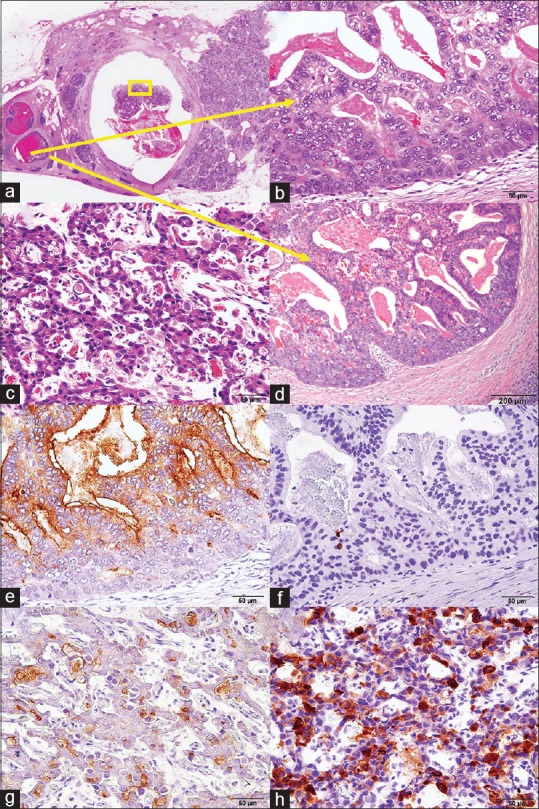
Subtotal stomach-preserving pancreatoduodenectomy was performed. Macroscopically, MPD was occluded by a solid tumor, which measured 16 mm × 8 mm in size and extended 33 mm in length, while we could not find a macroscopic solid lesion in the parenchyma. Microscopically, the tumor consisted of two components: one was a ductal component of cells with irregularly shaped nuclei and conspicuous nucleoli [Figure 2a and b] and the other was a neuroendocrine component of cells with round nuclei, “salt-and-pepper” chromatin, and eosinophilic cytoplasm [Figure 2c]. The transitional zone with minimal invasion into the pancreatic parenchyma was observed in the branch duct adjacent to MPD [Figure 2d]. Immunohistochemically, the former component was positive for cytokeratin-19, CA19-9, and carcinoembryonic antigen (CEA) [Figure 2e], but not for endocrine markers [Figure 2f], while the latter was positive for chromogranin A and synaptophysin [Figure 2g], not for CEA [Figure 2h]. Ki-67 labeling index was over 20% in an endocrine element. Each component comprised more than one-third of the neoplastic tissue; therefore, we finally diagnosed it as mixed ductal-neuroendocrine carcinoma.
Figure 2.

(a-d) Histological findings in the surgically resected specimen. (a) The main pancreatic duct in the head of the pancreas was occluded by a solid tumor. (b) A component in the branch duct was mainly a ductal component. (c) A component in the main pancreatic duct was mainly a neuroendocrine component (yellow square). (d) The transitional zone with minimal invasion into the pancreatic parenchyma was observed in the branch duct adjacent to the main pancreatic duct. (e-h) Immunohistochemically, a ductal component was positive for carcinoembryonic antigen (e), but not for synaptophysin (f), while a neuroendocrine component was positive for synaptophysin neuroendocrine component (g), not for carcinoembryonic antigen (h)
Pancreatic mixed ductal-neuroendocrine carcinoma is a rare tumor whose frequency in pancreatic tumors is < 0.2%[1] and reveals expanding or invasive growth in the pancreatic parenchyma.[2] There has been only one case report of minute mixed ductal-neuroendocrine carcinoma with predominant intraductal growth similar to this case.[3]
In this case, we assume that ductal adenocarcinoma would be initially derived from an intraepithelial lesion in the branch duct adjacent to MPD and that it would partially differentiate into neuroendocrine carcinoma during the growth in MPD. This resulted in a very rare case of unique mixed ductal-neuroendocrine carcinoma with predominant intraductal growth in MPD without a macroscopic intraparenchymal lesion.
Declaration of patient consent
The authors certify that they have obtained all appropriate patient consent forms. In the form the patient has given his consent for his images and other clinical information to be reported in the journal. The patient understands that his name and initial will not be published and due efforts will be made to conceal his identity, but anonymity cannot be guaranteed.
Financial support and sponsorship
Nil.
Conflicts of interest
There are no conflicts of interest.
REFERENCES
- 1.Cubilla AL, Fitzgerald PJ. Cancer of the exocrine pancreas: The pathologic aspects. CA Cancer J Clin. 1985;35:2–18. doi: 10.3322/canjclin.35.1.2. [DOI] [PubMed] [Google Scholar]
- 2.Chetty R, El-Shinnawy I. Intraductal pancreatic neuroendocrine tumor. Endocr Pathol. 2009;20:262–6. doi: 10.1007/s12022-009-9093-z. [DOI] [PubMed] [Google Scholar]
- 3.Terada T, Kawaguchi M, Furukawa K, et al. Minute mixed ductal-endocrine carcinoma of the pancreas with predominant intraductal growth. Pathol Int. 2002;52:740–6. doi: 10.1046/j.1440-1827.2002.01416.x. [DOI] [PubMed] [Google Scholar]


