Abstract
Context:
Hair restoration surgery for androgenetic alopecia (AGA) essentially involves various forms of hair transplantation. There is paucity of studies assessing donor area in Indian men and also no simplified guidelines are available for the safe donor area for follicular unit extraction (FUE). Our study is an attempt to study the donor area in Indian men.
Aims:
To assess the density of follicular units (FUs) in the donor area, that is, both scalp and beard in Indian men, and to propose simplified guidelines for FUE.
Materials and Methods:
The study design was cross-sectional and was carried out for 2 years. All the consenting male patients with male pattern hair loss Hamilton Norwood grading III or more who consulted for a hair restoration surgery were recruited. FU density was assessed in the donor area of scalp by drawing a rectangle with its lower border being a straight line joining two points, which are 27–28mm from the line drawn perpendicular to tragus, passing through external occipital protuberance. Three squares of area 1cm2 were drawn within the rectangle. Average of the FUs and follicles in the three squares was calculated to obtain mean density in the donor area of the scalp. Total number of FU was assessed, considering 25% extraction, total average number of extractable follicles was assessed. Total donor area was divided into three areas (areas 1, 2, and 3) and average number of extractable FU was assessed in each. Donor area in the beard, below the jawline, was divided into a triangle and rectangle. Average number of FU, total number of extractable FU was calculated similarly.
Results:
A total of 580 male patients were recruited in the study. Mean FU density in the scalp and beard was 78.2/cm2 and 49.7/cm2, respectively. The total available number of FUs for extraction in the areas 1, 2, and 3 and beard considering 25% extraction was 2064, 3097, 3612, and 824, respectively. We propose three types of donor areas in the scalp, namely, limited, standard, and extended donor area.
Keywords: Androgenetic alopecia, follicular unit extraction, safe donor zone
INTRODUCTION
Androgenetic alopecia (AGA) is the most common type of hair loss characterized by progressive thinning of the scalp hair and a reduction in hair density and diameter.[1] AGA in men presents with a typical pattern of bitemporal and frontal recession of the hairline or vertex thinning, which gradually extends anteriorly.[2] Hair restoration surgery for AGA essentially involves various forms of hair transplantation. The hair transplantation is based on the principle of donor dominance, that is, hair follicles that are androgen insensitive keep their properties even after transplanting into scalp areas affected by AGA.[3] Follicular unit extraction (FUE) technique for hair restoration has gained popularity in recent times. There is paucity of studies assessing donor area in Indian men and also no simplified guidelines are available for the safe donor area for FUE. Our study is an attempt to study the donor area in Indian men and to devise simplified guidelines for the safe donor area.
AIMS
-
•
To assess the density of follicular units (FUs) in the donor area, that is, both scalp and beard in Indian men
-
•
To propose simplified guidelines for FU extraction from scalp and beard in Indian men
MATERIALS AND METHODS
The study design was cross-sectional and was carried out in a private hair restoration facility in north India for 2 years from February 2016 to January 2018. All the consenting male patients, with male pattern hair loss Hamilton Norwood grading III or more, who consulted for a hair restoration surgery were recruited in the study. Patients with diffuse and unpatterned hair loss were excluded. A thorough clinical examination was carried out in all the patients to rule out any other cause of hair loss. FU density was assessed in the scalp and beard as follows:
Assessing FU density in the donor area of scalp
A straight line was drawn joining two points, which are 27–28mm from the line drawn perpendicular to tragus, passing through external occipital protuberance. A square of area 1cm2 was drawn in the midline, 2cm above the external occipital protuberance. Two squares of the same area were drawn one on each side, 5cm lateral to the first square [Figure 1]. Hairs in all the three squares were trimmed and photographs were taken. Number of FUs and number of follicles were counted in each of the square. Average of the FUs and follicles in the three squares was calculated to obtain mean density in the donor area of the scalp.
Figure 1.
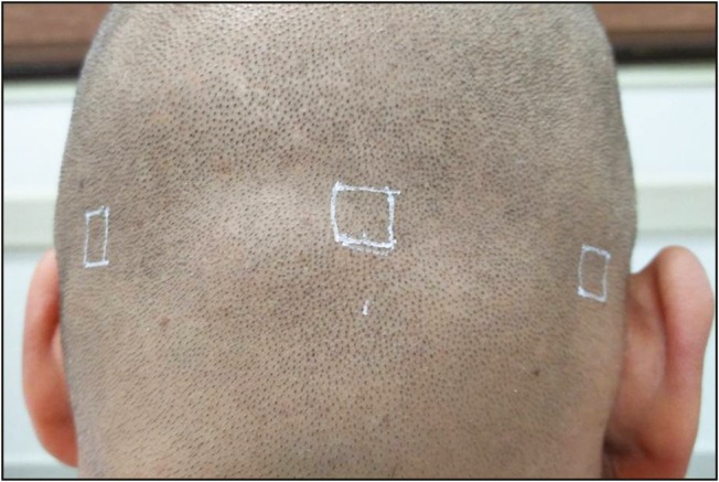
Three squares of 1cm2 area drawn 2cm above external occipital protuberance, 5cm apart from each other
Assessing the number of FUs available for extraction in the scalp
Number of FUs present in the extractable area of the scalp = Total extractable area in the scalp × density of the scalp.
Considering 25% or one-fourth of them can be extracted,
Total number of extractable FUs in the scalp = 0.25 × Total extractable area in the scalp (rectangle) × average density of the scalp
(Extractable area = length × breadth of the rectangle)
In this study, we have calculated extractable number of FUs in the following three areas [Figures 2 and 3]:
Figure 2.
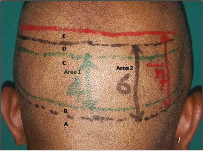
Posterior view of scalp showing areas 1, 2, and 3
Figure 3.
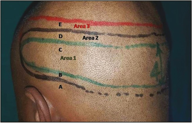
Lateral view of the scalp showing areas 1, 2, and 3
-
•
Area 1: Area between line B and C
-
•
Area 2: Area between line A and D
-
•
Area 3: Area between line A and E
Line A: A straight line was drawn joining two points, which are 27–28mm from the line drawn perpendicular to tragus, passing through external occipital protuberance.
Line B: A line drawn parallel to line A, 1cm above line A.
Line C: A line drawn parallel to line A, 5cm above line A
Line D: A line drawn parallel to line A, 6cm above line A
Line E: A line drawn parallel to line A, 7cm above line A
Total extractable FUs in area 1 = 0.25 × (length of line A × 4) × average density of the scalp
Total extractable FUs in area 2 = 0.25 × (length of line A × 6) × average density of the scalp
Total extractable FUs in area 3 = 0.25 × (length of line A × 7) × average density of the scalp
Assessing FU density in the donor area of beard
We considered beard hair present inferior to the jawline for the extraction. Jawline is the superior border for extractable area in the beard, whereas straight line passing through Adam’s apple was considered inferior border of extractable area in beard. A straight line was drawn joining angles of mandible. A square of area 1cm2 was drawn in the midline, such that the line was passing through the center. Two squares of same area were drawn one on each side, 3.5cm lateral to the first square [Figure 4]. All the three squares were trimmed and photographs were taken. Number of FUs and number of follicles were counted in each of the square. Average of the FUs and follicles in the three squares was calculated to obtain mean density in the donor area of the beard.
Figure 4.
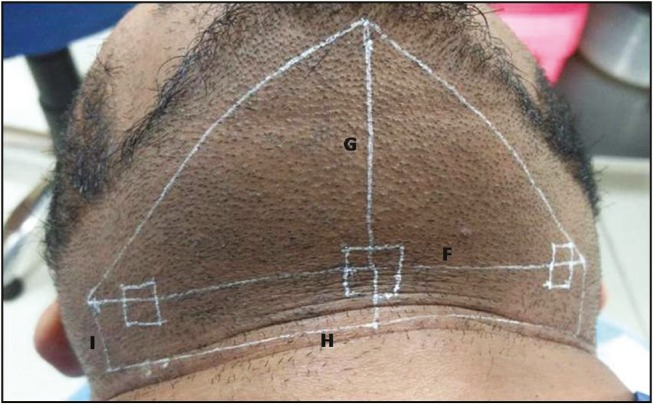
Donor area in the beard, below the jawline, divided into a rectangle and triangle by line F. Three squares of 1cm2 drawn on the line F can also be seen
Assessing the number of FUs available for extraction in the beard
Total extractable area in the beard × average density of FUs in the beard = Total number of FUs present in the extractable area.
Although 25% of them can be extracted without affecting the visual appearance, depending on patient’s preference, percentage of FUs extracted from the beard can be more and can be up to 100%.
Total number of FUs available for extraction = 0.25 × Total extractable area in the beard × average density of FUs in the beard.
Total extractable area in the beard = Area of triangle + area of rectangle [Figure 4]
(Area of triangle = 0.5 × F × G, Area of rectangle = F × I)
where, F: length of line drawn between two angles of mandibles
G: length of line drawn perpendicular to line F, passing through the midline
H: length of straight line drawn parallel to line F up to where the beard hairs exist (if no clear demarcation between beard hairs and neck hairs, line H will be passing through superior border of Adam’s apple).
I: length of perpendicular line drawn between line F and line H
RESULTS
A total of 580 men with AGA who consulted us for hair restoration surgery were recruited in the study. Their age ranged between 24 and 45 years with mean age of 31.7 years. Mean FU density in the scalp and beard was 78.2/cm2 and 49.7/cm2, respectively. Mean number of follicles in each FU in scalp and beard was 1.81 and 1.32, respectively. Mean hair density in scalp and beard was 141.5/cm2 and 65.6/cm2, respectively. Total available number of FU in the donor zones, (area 1, 2, and 3 and beard) extractable number of FUs considering 25% extraction are given in Table 1.
Table 1.
Table showing extractable area, extractable number of FU’s from the scalp and the beard
| Variable | Area 1 (LDA) | Area 2 (SDA) | Area 3 (EDA) | Beard |
|---|---|---|---|---|
| Area (in cm2) | 105.6 | 158.4 | 184.8 | 64.6 |
| Total FU | 8,258 | 12,387 | 14,451 | 3,295 |
| Total extractable FU (considering 25% extraction) | 2,064 | 3,097 | 3,612 | 824 |
| Total extractable FU (in two sittings) | 3,612 | 5,420 | 6,321 |
LDA = limited donor area, SDA = standard donor area, EDA = extended donor area
DISCUSSION
Orentreich[4] was the first American physician to perform hair transplantation for male-patterned baldness. He suggested theory of “donor dominance,” which stated that the transplanted hair keeps the original nature of the donor site even after being transplanted.[4] Donor dominance theory of Orentreich[4] and the definition of a safe donor area are the theoretical foundation, which made modern hair transplantation possible. Exact definition of safe donor area, which is the area that will have no hair loss, could not be defined by anyone. Because no safe donor area guarantees that the hair will be permanent. There only exists a safe donor area anticipated to have no invasion of alopecia. Thus, safe donor area can be defined as an area in which no progression of permanent hair loss occurs.[5]
The donor area described by Rassman and Carson[6] has three significant boundaries. The anterior boundary is vertically superior to that of the external acoustic meatus. The superior boundary is located 2cm above the upper border of the helical rim of the horizontal plane, whereas the inferior border of the donor area is slightly controversial as it may move upward with the passage of time. The most crucial and clinically critical standard for determining a safe donor area is the superior border, which is profoundly related to the maximum extent of vertex alopecia.[7]
The definition of the safe donor area, which is currently being applied globally to surgical practice by most physicians, is an adaptation of theory presented by Unger.[8] He defined alopecia as a continuous progressive condition, by calculating the probability of the worst-case scenario, he proposed the most reasonable standard. His definition was in accordance with the facts that the global mean life expectancy was ≤80 years and >80% of men aged between 70 and 79 years manifested baldness that is less than Norwood type VII.[8]
Cole and Devroye[9] defined the total permanent donor area as 203cm2 and he further divided it into eight major regions (3.5×6cm) and six minor regions (3.5×2cm).[7]
Bernstein and Rassman[10] reported that the safe donor area accounted for approximately 25% of the entire scalp, up to 50% of the hair could be obtained from the donor site and even after harvesting of 50% of hair, the apparent density remained the same.[6,10]
There is no universally acceptable definition for the safe donor zone. This study is an attempt to give a simplified guideline based on the existing knowledge on the safe donor zone and our practical experience. We propose three types of donor areas, namely, limited donor area (area 1), standard donor area (area 2), and extended donor area (area 3) (limited donor area is more permanent than standard area, which is more permanent than extended donor area) [Figure 5]. Mean available number of FUs for extraction from these areas, considering 25% extraction, is given in Table 1. Though 50% hairs can be extracted without apparent decrease in the density, it should not be done in a single sitting as it can lead to extensive microvascular damage, which in turn causes decrease in the quality and thickness of remaining hairs. We recommend 25% extraction in the first sitting and extraction of 25% of the remaining hairs in the second sitting, which should be at least 6 months apart (making a total of 25% + 18.75% [0.25×75] = 43.75%). In beard, the concept of safe donor zone is not delineated. We suggest safe donor zone in the beard, which should be cosmetically acceptable to most patients, that is, below the jawline. In beard, higher proportion of hairs can be extracted depending on the requirement. As we have taken beard hair only below the jawline, cosmesis of the patient is not much affected even after total extraction. If required, hairs can also be taken from above the jawline if patient is comfortable with it. But it is not something we recommend.
Figure 5.
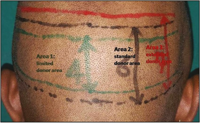
Posterior view of the scalp showing limited, standard, and extended donor areas
Extraction from extended donor area should be carried out only after explaining the patient about the relatively nonpermanent nature of the hairs and that they have to be implanted in a cosmetically less significant zone. In our study, we have seen for the miniaturization of the follicles both downward from vertex and upward from below. We have seen miniaturization in 12% (70) patients, extending either into the extended donor zone or 1cm above it [Figure 6]. Miniaturization was observed in 3% patients, extending into the standard donor zone. If there is miniaturization, area at least 1cm below it has to be avoided. Though we have seen many cases with reverse pattern hair loss, we have not found it extending to external occipital protuberance in any.
Figure 6.
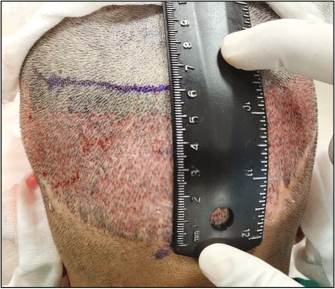
One-year post-follicular unit transfer, now undergoing FUE. A margin of 1cm taken from the point of miniaturization of hair follicles, which was extending into extended donor zone
We used digital photography as a tool for quantification of hairs and FUs in the donor scalp and beard. In our study, we measured hair density at three different regions of the donor scalp and took average of the three. Mean FU density in our study in the donor area of scalp was 78.2/cm2, whereas average hair density was 141.5/cm2.
Jimenez and Ruifernández[11] devised a mathematical model for estimating donor size for FU transplantation. They also used digital photography for quantification of hairs, FUs, and interfollicular distance. They studied hair density by marking a single rectangle of area 0.5cm2 (1×0.5cm) in the occipital scalp and extrapolated the results, considering hair density is maintained constant throughout the donor scalp, and found the mean density of the occipital donor area was 65–85 FUs/cm2.[11]
A review of the literature reveals a significant variation among different authors regarding the counts of hair density, which is probably due to either racial variations or the methodology used to count the hairs per unit area. Limmer[12] finds a range of 120–240 hairs/cm2 and Haber and Stough[13] 144–176 hairs/cm2. Bernstein and Rassman[14] found significant racial variations in the hair density and FU density among Caucasians, Asians, and Africans. The African individual has a lower hair density (average 160 hairs/cm2) than the Asian (average 170 hairs/cm2) and Caucasian (average 200 hairs/cm2).[14] Average hair density in donor area measured by different authors include 150–300/cm2 by Bernstein,[15] 124–200/cm2 by Jimenez and Ruifernández,[11] 190–210/cm2 by Cole and Devroye.[9]
Designing and planning of the recipient area and assessment and management of the donor area are the most important factors in hair transplantation. An estimation of the number of FUs that can be harvested is important for planning a hair transplant session. The placement strategy can be refined by knowing information about the number and type of grafts. Experienced surgeons can predict the harvest requirements at a glance, but for novice surgeons, it is useful to follow some guidelines because an accurate estimation of the donor area is important if surgical results are to be predictable.
Limitations
-
•
There may be little thinning of safe donor zone in the parietal area. It was ignored for the sake of easy calculation.
Financial support and sponsorship
Nil.
Conflicts of interest
There are no conflicts of interest.
REFERENCES
- 1.Khumalo NP, Jessop S, Gumedze F, Ehrlich R. Hairdressing and the prevalence of scalp disease in African adults. Br j Dermatol. 2007;157:981–8. doi: 10.1111/j.1365-2133.2007.08146.x. [DOI] [PubMed] [Google Scholar]
- 2.Hamilton JB. Patterned loss of hair in man; types and incidence. Ann ny Acad Sci. 1951;53:708–28. doi: 10.1111/j.1749-6632.1951.tb31971.x. [DOI] [PubMed] [Google Scholar]
- 3.Kaliyadan F, Nambiar A, Vijayaraghavan S. Androgenetic alopecia: an update. Indian j Dermatol Venereol Leprol. 2013;79:613–25. doi: 10.4103/0378-6323.116730. [DOI] [PubMed] [Google Scholar]
- 4.Orentreich N. Autografts in alopecias and other selected dermatological conditions. Ann ny Acad Sci. 1959;83:463–79. doi: 10.1111/j.1749-6632.1960.tb40920.x. [DOI] [PubMed] [Google Scholar]
- 5.Knudsen RG. The donor area. Facial Plast Surg Clin North Am. 2004;12:233–40. doi: 10.1016/j.fsc.2003.12.003. [DOI] [PubMed] [Google Scholar]
- 6.Rassman WR, Carson S. Micrografting in extensive quantities. The ideal hair restoration procedure. Dermatol Surg. 1995;21:306–11. [PubMed] [Google Scholar]
- 7.Park JH, Na YC, Moh JS, Lee SY, You SH. Predicting the permanent safe donor area for hair transplantation in Koreans with male pattern baldness according to the position of the parietal whorl. Arch Plast Surg. 2014;41:277–84. doi: 10.5999/aps.2014.41.3.277. [DOI] [PMC free article] [PubMed] [Google Scholar]
- 8.Unger W. Delineating the safe donor area for hair transplanting. J Am Acad Cosmet Surg. 1994;4:239–43. [Google Scholar]
- 9.Cole J, Devroye J. A calculated look at the donor area. Hair Transplant Forum Intl. 2001;11:150–4. [Google Scholar]
- 10.Bernstein RM, Rassman WR. Follicular transplantation. Patient evaluation and surgical planning. Dermatol Surg. 1997;23:771–84. discussion 801-5. [PubMed] [Google Scholar]
- 11.Jimenez F, Ruifernández JM. Distribution of human hair in follicular units a mathematical model for estimating the donor size in follicular unit transplantation. Dermatol Surg. 1999;25:294–8. doi: 10.1046/j.1524-4725.1999.08114.x. [DOI] [PubMed] [Google Scholar]
- 12.Limmer BL. The density issue in hair transplantation. Dermatol Surg. 1997;23:747–50. doi: 10.1111/j.1524-4725.1997.tb00408.x. [DOI] [PubMed] [Google Scholar]
- 13.Haber RS, Stough DB. Accurate estimation of graft requirements when using multibladed knives. In: Stough DB, Haber RS, editors. Hair replacement: surgical and medical. St. Louis, MO: Mosby; 1996. pp. 138–42. [Google Scholar]
- 14.Bernstein RM, Rassman WR. The aesthetics of follicular transplantation. Dermatol Surg. 1997;23:785–99. doi: 10.1111/j.1524-4725.1997.tb00419.x. [DOI] [PubMed] [Google Scholar]
- 15.Bernstein R. Accurate follicular unit transplantation. In: Haber R, Stough D, editors. Hair replacement: surgical and medical. St. Louis, MO: Mosby; 1996. pp. 138–42. [Google Scholar]


