

Abstract
Background:
Ulnocarpal impaction is the most common reason to perform ulnar shortening osteotomy. There are 3 osteotomy techniques for ulnar shortening: transverse, step-cut, and oblique cut1-3.
First described by Milch4 in 1941, extra-articular diaphyseal oblique or transverse shortening is the most frequently performed type of shortening. However, it is associated with a nonunion rate of up to 10%, and irritation by implants requiring removal occurs in up to 28% of cases5,6. Intra-articular procedures such as the wafer procedure affect the distal ulnar joint surface, which can lead to stiffness of the distal radioulnar joint (DRUJ) due to scar tissue formation and adhesion of the triangular fibrocartilage complex (TFCC)7. Lapner et al.8 described increased pressure in the DRUJ after the wafer procedure, which may lead to an early onset of osteoarthritis. Complication rates between 8% for open wafer procedures and 21% for arthroscopic wafer procedures have been described9.
Intra-articular shortening has also been described by Slade and Gillon10 in 2007 and Hammert et al.11 in 2012 and was tested in cadavers by Greenberg et al.12 in 2013. This closing wedge technique preserves the distal joint surface of the ulna and also allows for easy correction of the inclination of the hub joint surface of the ulna.
In contrast to the technique of Slade, our described osteotomy is steeper and longer proximally, which allows for fixation with >2 screws13-16. Rapid healing of the metaphyseal bone compared with diaphyseal bone is described, and implant removal is necessary less often14,17,18.
With the described procedure, the interosseous membrane remains untouched, especially the distal oblique bundle, which additionally provides stability of the DRUJ in 40% of patients19.
Description:
A dorso-ulnar approach through the fifth extensor sheath is performed. The ulnocarpal joint and the DRUJ are accessed through an arthrotomy distal and proximal to the TFCC. The foveal attachment of the TFCC and the subsheath of the sixth extensor sheath are visualized. The osteotomy is intra-articular oblique from distal ulnar to proximal radial. Sliding the head of the ulna proximally achieves the desired shortening of up to 5 mm, and the head is fixed using 2, 3, or 4 cannulated headless screws. A slight correction of the axis of the ulnar head is also possible.
Alternatives:
An alternative to this procedure is extra-articular osteotomy using a palmar or dorsal ulnar approach. If necessary, additional ulnocarpal procedures can be performed in an open or arthroscopically assisted manner.
Rationale:
The shortening takes place only in the articular part of the distal aspect of the ulna. This procedure can easily be combined with TFCC repair, synovectomy of the DRUJ, or repair or reconstruction of the lunotriquetral ligament if needed. Shortening of up to 5 mm is possible.
Introductory Statement
The distal ulnar sliding osteotomy is an intra-articular ulnar shortening procedure that allows for shortening of up to 5 mm and also open refixation/repair of the triangular fibrocartilage complex (TFCC).
Indications & Contraindications
Indications
Ulnocarpal impaction syndrome.
Flat shape of the ulnar head (Fig. 1).
Short ulnar styloid.
Need for combined interventions.
Fig. 1.

Ulnocarpal impaction syndrome shown by single-photon emission computed tomography (SPECT).
Contraindications
Long ulnar styloid (risk of ulnar styloid impaction).
Degenerative processes in the distal radioulnar joint (DRUJ).
Need for ulnar shortening of >5 mm.
Additional complex procedures such as the reconstruction of the TFCC described by Adams and Berger20.
Step-by-Step Description of Procedure (Video 1)
Video 1.
Detailed description of the incision and the technique.
Step 1: Incision (Video 1: 00:10)
Perform an S-shaped incision with the arm in full pronation.
Position the patient supine on the surgical table.
Perform radiocarpal/midcarpal arthroscopy to verify the pathology and check for additional lesions—for example, lunotriquetral ligament rupture or chondromalacia of the lunate.
After the arthroscopy, position the arm on an arm-table in full pronation, and perform an S-shaped incision.
Step 2: Exposure of the Ulnar Head (Video 1: 00:26)
Use the access through the fifth extensor sheath and open the DRUJ in an L-shaped manner.
Take care not to damage the dorsal branch of the ulnar nerve.
Open the fifth extensor compartment and elevate an ulnar-based flap in the direction of the sixth extensor compartment. This exposes the tendon of the extensor digiti minimi and the distal part of the extensor carpi ulnaris. Be aware not to extend the incision of the sixth extensor sheath more proximally than the base of the ulnar styloid.
Retract the extensor digiti minimi and extensor carpi ulnaris tendons and expose the dorsal capsule. The anatomical relationship between the TFCC and the capsule requires caution not to damage the radioulnar ligaments (Fig. 2).
Perform an incision in the dorsal capsule parallel to the ulnocarpal joint and distal to the TFCC.
Open the DRUJ in an inverted L-shaped manner on the floor of the fifth extensor sheath. Incise the underlying dorsal capsule of the DRUJ and expose the ulnar head.
Fig. 2.

Illustration of the DRUJ.
Step 3: Osteotomy and Fixation (Video 1: 01:00)
Perform a long oblique osteotomy and use cannulated headless screws for fixation.
Use Hohmann retractors to secure the soft tissue.
Start the osteotomy radial of the foveal attachment of the TFCC (Fig. 3) and make it long enough proximally to allow fixation with at least 2 cannulated headless screws. The osteotomy can also be performed through the ulnar styloid to avoid any damage to the foveal fibers of the TFCC.
Perform the osteotomy with an oscillating saw. Mobilize the radial part of the ulnar head proximally according to your preoperatively measured values. If the shape of the ulnar head is not congruent with the sigmoid notch, the angulation of the ulnar head can be corrected by removal or interposition of a small wedge.
Shorten the proximal tip of the wedge. Use a Weber clamp to secure the reduction. Insert 2, 3, or 4 Kirschner wires parallel and horizontally.
Insert 2, 3, or 4 cannulated headless 2.0 to 2.5-mm screws. Make sure not to penetrate the radial joint surface of the ulnar head.
Use fluoroscopy to check the result (Figs. 4-A, 4-B, and 4-C).
Fig. 3.

Scheme of the osteotomy and positioning of the screws.
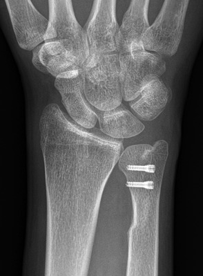
Figs. 4-A, 4-B, and 4-C Case illustration of the ulnar sliding osteotomy.
Fig. 4-A.

Preoperative appearance.
Fig. 4-B.

Six weeks postoperatively.
Fig. 4-C.
Twelve weeks postoperatively.
Step 4: Closure
Close the capsule and fifth extensor compartment, preferably with continuous absorbable sutures.
Close the capsule of the DRUJ with a single absorbable continuous or interrupted suture. In our experience, one of the main stabilizers of the DRUJ besides the subsheath of the extensor carpi ulnaris is the floor of the fifth extensor sheath.
Close the floor with monofilament absorbable double running suture and make sure to bury the knots to avoid disturbing suture material on the floor of the fifth extensor sheath. Check for stability of the DRUJ.
Close the retinaculum above the extensor digiti minimi with an absorbable single running suture, but make sure not to tighten the retinaculum too much as that could induce scarring over the underlying tendon and cause reactive synovitis.
If it is not possible to reposition the tendon, close the tendon sheath proximal and distal to the DRUJ and leave the extensor digiti minimi subcutaneous at the level of the DRUJ.
Step 5: Splinting and Postoperative Treatment
Apply a splint for 6 weeks and start active-assisted movement after confirmation of healing after 6 weeks.
Apply an above-the-elbow or sugar-tong plaster splint in a neutral position for the first night.
On the first or second postoperative day, an anti-pronation/supination splint (Fig. 5) is applied by the occupational therapist. Elbow movement is immediately allowed.
After confirmation of healing on radiographs at 6 weeks, active-assisted movement is allowed and the splint is removed.
Full weight-bearing and an unlimited range of motion should be achieved after 12 weeks.
Fig. 5.

The anti-pronation/supination splint.
Results
Our results regarding osseous union were similar to those reported by Sennwald et al.14, who performed the identical osteotomy. In all of our 10 (unpublished) cases, ulnar shortening was sufficient and osseous union occurred after 4 to 6 weeks. All of the patients returned to work. In 2 cases, the screws had to be removed because of protrusion in the DRUJ, whereas Sennwald et al. reported no complications due to implants14. In another 2 of our cases, the fifth extensor compartment needed to be revised because of chronic synovitis as a complication of the approach.
Pitfalls & Challenges
The DRUJ needs to be opened.
Beware of insufficient shortening (the final result of the procedure should be −1 to −1.5 mm of shortening).
Avoid screw penetration in the DRUJ or the subsheath of the extensor carpi ulnaris.
Nonunion is possible.
Too long an ulnar styloid can lead to ulnar styloid impaction syndrome.
Make sure that the arm is in full pronation during the procedure. The blade needs to be positioned from dorsal radial to palmar ulnar.
Do not damage the foveal insertion of the TFCC.
Footnotes
Published outcomes of this procedure can be found at: J Hand Surg Br. 1995 Apr;20(2):178-84 and J Hand Surg Eur Vol. 2013 Jun;38(5):542-9.
Disclosure: The authors indicated that no external funding was received for any aspect of this work. The Disclosure of Potential Conflicts of Interest forms are provided with the online version of the article (http://links.lww.com/JBJSEST/A239).
References
- 1.Darrow JC, Jr, Linscheid RL, Dobyns JH, Mann JM, 3rd, Wood MB, Beckenbaugh RD. Distal ulnar recession for disorders of the distal radioulnar joint. J Hand Surg Am. 1985. July;10(4):482-91. [DOI] [PubMed] [Google Scholar]
- 2.Darlis NA, Ferraz IC, Kaufmann RW, Sotereanos DG. Step-cut distal ulnar-shortening osteotomy. J Hand Surg Am. 2005. September;30(5):943-8. [DOI] [PubMed] [Google Scholar]
- 3.Chun S, Palmer AK. The ulnar impaction syndrome: follow-up of ulnar shortening osteotomy. J Hand Surg Am. 1993. January;18(1):46-53. [DOI] [PubMed] [Google Scholar]
- 4.Milch H. Cuff resection of the ulna for malunited Colles’ fracture. J Bone Joint Surg Am. 1941. April;23(2):311-3. [Google Scholar]
- 5.Cha SM, Shin HD, Ahn KJ. Prognostic factors affecting union after ulnar shortening osteotomy in ulnar impaction syndrome: a retrospective case-control study. J Bone Joint Surg Am. 2017. April 19;99(8):638-47. [DOI] [PubMed] [Google Scholar]
- 6.Nagy L, Jungwirth-Weinberger A, Campbell D, Pino JG. The AO ulnar shortening osteotomy system indications and surgical technique. J Wrist Surg. 2014. May;3(2):91-7. [DOI] [PMC free article] [PubMed] [Google Scholar]
- 7.Feldon P, Terrono AL, Belsky MR. The “wafer” procedure. Partial distal ulnar resection. Clin Orthop Relat Res. 1992. February;275:124-9. [PubMed] [Google Scholar]
- 8.Lapner PC, Poitras P, Backman D, Giachino AA, Conway AF. The effect of the wafer procedure on pressure in the distal radioulnar joint. J Hand Surg Am. 2004. January;29(1):80-4. [DOI] [PubMed] [Google Scholar]
- 9.Katz DI, Seiler JG, 3rd, Bond TC. The treatment of ulnar impaction syndrome: a systematic review of the literature. J Surg Orthop Adv. 2010. Winter;19(4):218-22. [PubMed] [Google Scholar]
- 10.Slade JF, 3rd, Gillon TJ. Osteochondral shortening osteotomy for the treatment of ulnar impaction syndrome: a new technique. Tech Hand Up Extrem Surg. 2007. March;11(1):74-82. [DOI] [PubMed] [Google Scholar]
- 11.Hammert WC, Williams RB, Greenberg JA. Distal metaphyseal ulnar-shortening osteotomy: surgical technique. J Hand Surg Am. 2012. May;37(5):1071-7. [DOI] [PubMed] [Google Scholar]
- 12.Greenberg JA, Werner FW, Smith JM. Biomechanical analysis of the distal metaphyseal ulnar shortening osteotomy. J Hand Surg Am. 2013. October;38(10):1919-24. Epub 2013 Aug 24. [DOI] [PubMed] [Google Scholar]
- 13.Sennwald GR, Lauterburg M, Zdravkovic V. A new technique of reattachment after traumatic avulsion of the TFCC at its ulnar insertion. J Hand Surg Br. 1995. April;20(2):178-84. [DOI] [PubMed] [Google Scholar]
- 14.Sennwald G, Della Santa D, Beaulieu JY. A comparison of diaphyseal and metaphyseal techniques of ulna shortening. J Hand Surg EurVol. Vol 2013. June;38(5):542-9. Epub 2012 Nov 30. [DOI] [PubMed] [Google Scholar]
- 15.Nakamura T, Nakao Y, Ikegami H, et al. Open repair of the ulnar disruption of the triangular fibrocartilage complex with double three-dimensional mattress suturing technique. Tech Hand Up Extrem Surg. 2004. June;8(2):116-23. [DOI] [PubMed] [Google Scholar]
- 16.Nakamura T, Sato K, Okazaki M, Toyama Y, Ikegami H. Repair of foveal detachment of the triangular fibrocartilage complex: open and arthroscopic transosseous techniques. Hand Clin. 2011. August;27(3):281-90. Epub 2011 Jul 13. [DOI] [PubMed] [Google Scholar]
- 17.Aronson J, Shen X. Experimental healing of distraction osteogenesis comparing metaphyseal with diaphyseal sites. Clin Orthop Relat Res. 1994. April;301:25-30. [PubMed] [Google Scholar]
- 18.Marquez-Lara A, Nuñez FA, Jr, Kiymaz T, Nuñez FA, Sr, Li Z. Metaphyseal versus diaphyseal ulnar shortening osteotomy for treatment of ulnar impaction syndrome: a comparative study. J Hand Surg Am. 2017. June;42(6):477e1-8. Epub 2017 Apr 20. [DOI] [PubMed] [Google Scholar]
- 19.Moritomo H. The function of the distal interosseous membrane and its relevance to the stability of the distal radioulnar joint: an anatomical and biomechanical review. Handchir Mikrochir Plast Chir. 2015. October;47(5):277-80. Epub 2015 May 4. [DOI] [PubMed] [Google Scholar]
- 20.Adams BD, Berger RA. An anatomic reconstruction of the distal radioulnar ligaments for posttraumatic distal radioulnar joint instability. J Hand Surg Am. 2002. March;27(2):243-51. [DOI] [PubMed] [Google Scholar]