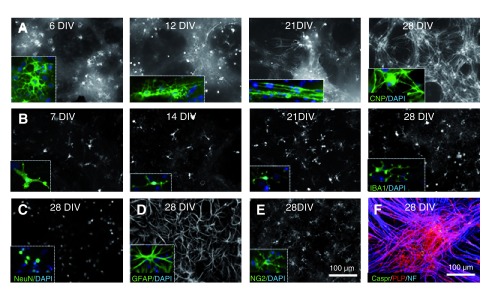
Figure 1. Embryonic day 13 mouse spinal cord cells differentiate over time into the major neural cell types of the nervous system.
A. The morphology of CNP positive cells changes with time in culture. At 6 DIV, multiple cell process extend from the cell body and the cells have a ‘lacy’ appearance, as shown in the inset. By 12 DIV, a small number of myelin-like sheaths are present; often a single single sheath can be observed running in a line through one axis of an otherwise ‘lacy–appearing’ cell. In the inset, two such sheaths can be observed along 3 adjacent ‘lacy-appearing’ cells. By 21 DIV, many cells have extended multiple myelin-like sheaths and by 28 DIV few ‘lacy-appearing’ cells remain and a dense network of myelin sheaths are visible. When stained with antibodies to PLP/DM20, Caspr and phosphorylated neurofilament (NF), many axons are covered by consecutive sheaths with nodes of Ranvier straddled by Caspr positive paranodes (bottom right image). B. Iba1 +ve microglia are present at all stages examined. At 7DIV most microglia appear amoeboid but become ramified and extend multiple processes over time (insets). C. Antibody to NeuN labels neuronal nuclei and sometimes staining extends into the cytoplasm, but rarely enters the cell processes. D. GFAP positive astrocytes are found throughout the culture, ( E) as are NG2 +ve OPCs. F. Combined staining with antibodies to PLP/DM20, Caspr and phosphorylated neurofilament (NF), reveals that many axons are covered by consecutive sheaths with nodes of Ranvier straddled by Caspr positive paranodes. Images A–F were contrast enhanced to ease viewing.

