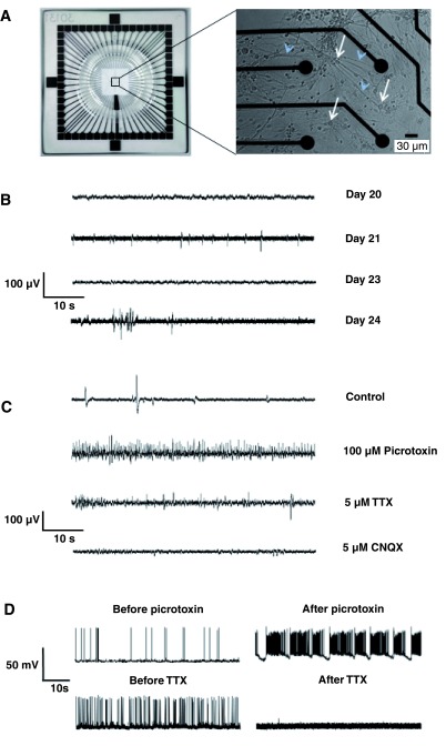
Figure 2. Myelinating cell cultures are spontaneously electrically active.
A. Low power view of the whole MEA (left); all 59 recording electrodes and the ground electrode are concentrated in the center of the array. In the high-power image (right), four electrodes (black circles) can be observed in relation to the cell bodies (white arrows) and processes (including axons; blue arrowheads) of neurons and glia, on an MEA. To facilitate viewing, we selected a region in which cell density is lower than normal. Extracellular activity can be assessed from each of 59 recording electrodes. B. Extracellular recordings from a single electrode between 20 and 24. DIV. Each trace represents the sum of the activity detected at that electrode, which is in direct contact with multiple neuronal and glial cell bodies and processes. ‘Fast’ spike-like activity is evident at DIV 21 and ‘burst-like’ activity develops by DIV 24. C. Additive pharmacology on a DIV 24 culture (sequential administration of drugs without washout) demonstrates in vivo-like neuronal network activity of spinal cord myelinating cultures. The recording shows that addition of picrotoxin, a GABAergic inhibitor, evokes a massive increase in ‘fast’ neuronal activity (second versus first trace). Subsequent addition of TTX (a Na + channel blocker) inhibits much of the ‘fast’ spike-like (presumably axonal) activity. CNQX (a blocker of AMPA/kainate receptors) blocks excitatory synaptic activity. The remaining slow potential changes probably represent local spontaneous depolarizations (at neuronal and glial cell bodies). D. Examples of whole-cell current clamp recordings from single cells, presumably neurons, firing spontaneously at a resting potential of around -50 mV. Top, trace before and 5 minutes after the addition of 1 µM TTX (top). Bottom, trace from a second cell before and 5 minutes after the addition of 100 μM picrotoxin. TTX consistently blocked activity (n = 6 cells), as expected, whilst picrotoxin caused regular burst spiking in every cell tested (n = 18 cells), but the effect on overall spike rate was inconsistent, presumably reflecting the phenotype of the cell.

