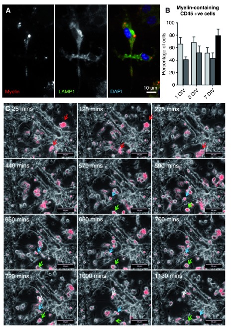
Figure 3. Motile microglia/macrophages take up myelin debris in vitro.
( A) ID4B antibody staining of cultures treated with rhodamine-labelled myelin debris at 27 DIV shows myelin debris (red) within lysosomal associated membrane glycoprotein 1 (green) late endosomes/lysosomes. All cells containing myelin were found to be CD45 +ve (not shown). ( B) After incubation with saturating amounts of myelin debris (data obtained with 0.075 or 0.1 mg myelin protein ml -1 were similar, so values from each were included providing two technical replicates) over three different time periods, the percentage of CD45 positive cells that contained ‘wild type’ (pale grey bars) or Plp1-tg (dark grey bars) myelin debris, which is taken up in a receptor-dependent manner, was between 40–70 % (n = 6; 2 technical replicates for each of 3 independent experiments, per time point); not significantly different from the proportion that contained latex beads (black bar), which are taken up in a receptor-independent manner (n = 3 independent experiments). Bars represent mean +/- s.e.m.. ( C) Time lapse stills over 1130 minutes show myelin debris (red) at time of addition (25 mins – 275mins) and then being phagocytosed by microglia/macrophages (440 mins – 1130 mins). Blue and green arrows highlight the locations of individual cells over time.

