Abstract
BACKGROUND:
Penile cancer accounts for 0.4-0.6% of all malignancy in men in Europe and the United States of America. It also accounts for 10% of all malignancy in men in some Asian, South American, and African countries. P53 protein has the function to regulate apoptosis in the cell cycle. Therefore, the presence of p53 in cells may indicate higher proliferative activity of the cells as a feedback mechanism, indicating disease progression.
AIM:
This study aims to identify the association between p53 expression and survival rate in penile cancer patients.
METHODS:
This study was a retrospective observational analytic study. This study was conducted in Pathology Anatomy Laboratory Faculty of the Medicine University of Sumatera Utara/Haji Adam Malik Hospital/University of Sumatera Utara Hospital to assess p53 expression. This study was conducted from January 2018 to December 2018.
RESULTS:
The total subjects in this study were 33 with the mean age of 50.79 ± 10.62. Based on clinical stage, patients in this study are divided into 11 patients (33.3%) in stage T II and 22 patients (66.7%) in stage T III/T IV. P53 expression was positive in 13 patients (35.3%). There were 19 patients (57.6) alive and 14 patients (42.4%) deceased. Statistical analysis using chi-square showed that there was an association between p53 expression and mortality (p = 0.011). In the Kaplan-Meier Curve for 3-year overall survival based on p53 expression, the survival rate in 36 months in the p53 positive group is 18%, while in p53 negative group, the survival rate was 60%. The survival rate based on p53 status was significantly different (p = 0.025).
CONCLUSION:
There is a significant association between p53 expression and mortality in penile cancer patients. In conclusion, p53 expression in penile cancer cells examined by immunohistochemistry may show prognostic values in the disease progression.
Keywords: P53 expression, Survival rate, Penile cancer, Gene overexpression, Mortality
Introduction
Penile cancer accounts for 0.4-0.6% of all malignancy in men in Europe and the United States of America. It also accounts for 10% of all malignancy in men in some Asian, South American, and African countries [1]. In Indonesia, there were 69 men diagnosed with penile malignancies in Dr Cipto Mangunkusumo Hospital and Dharmais Hospital Jakarta Cancer Center for 11 years period (1994-2005) [2]. Another study in Sanglah Hospital Bali showed that there were 46 penile cancer patients for 8 years period. Meanwhile, in Haji Adam Malik Medan Hospital, the incidence of penile cancer for the last 4 years (2012-2015) was 34 patients [3], [4].
The principal value in the management of penile cancer is the levitation of the tumour with good organ preservation along partial or total penectomy in regards to lowering the recurrence rate. Aside from the treatment of a primary tumour, the involvement of the lymph node remains to be an important factor in enhancing the patient’s prognosis. Penile cancer is an aggressive disease. The success rate of local lesion treatment is only for early stage disease. Besides, the more-progressive and advanced disease with the involvement of regional lymph node or distant metastasis remains a problem in the field of neuro-oncology [4].
The incidence of penile cancer varies within circumcision status, hygiene standard, phimosis, sexual partner, Human papilloma virus (HPV) infection, tobacco exposure, and other factors [1]. Etiologic factors known were chronic irritation from smegma, a product from bacterial activity in desquamated cells that are accumulated in the preputium. The most common histopathology found in penile cancer is Squamous Cell Carcinoma (SCC) [5].
P53 protein is a product from the TP53 gene in the body; this protein has the function to regulate apoptosis in the cell cycle. The presence of p53 in cells may indicate higher proliferative activity of the cells as a feedback mechanism, indicating disease progression. The study showed that the excessive protein expression of p53 was found in penile cancer cells [6]. Furthermore, mutation or deletion of p53 in the cell may precipitate cancer to further progress [7]. Based on this theoretical background, p53 could be used to see prognosis of the disease where ass more expression of the protein is associated with worse clinical parameters [6]. However, the use of any diagnostic or prognostic tool to measure the expressions is not widely implemented [8].
This rationale accelerates the urgency to do a study on the association between p53 expression and survival rate in penile cancer patients.
Material and Methods
This study was a retrospective observational analytic study. This study aimed to analyse the association between p53 expression and survival rate in penile cancer patients. This study was conducted in Pathology Anatomy Laboratory Faculty of Medicine University of Sumatera Utara/ Haji Adam Malik Hospital / University of Sumatera Utara Hospital. This study has the ethical approval issued by Research Ethics Committee Faculty of the Medicine University of Sumatera Utara. This study was conducted from January 2018 to December 2018.
Immunohistochemistry of p53
The protein expression of p53 was observed using Immunohistochemistry examination done on FFPE preparation. Specific antibodies for p53 (mouse monoclonal antibody) obtained from Sigma Aldrich (St Louis, Missouri). Preparation/micro slicing was done for each sample, and slides are provided for each sample for microscopic evaluation.
Microscopic evaluation was performed by an experienced pathologist. The semiquantitative examination was done on random fields per specimen containing a minimum of 500 cells using ImageJ (Research Service Branch, NIH.gov) in 40 times magnification. The expression of the protein would be positive if cells were stained brownish in the cytoplasm or the nuclei with a granular pattern. If the number of positive cells exceeds 60%, the sample is considered positive.
Results
The total subjects in this study were 33 with the mean age of 50.79 ± 10.62. Based on clinical T stage, patients in this study are divided into 11 patients (33.3%) in cT2 and 22 patients (66.7%) in cT3/cT4. There were 22 patients (66.7%) who had cancer invasion to the urethra.
Table 1.
Characteristics of the subjects
| Variable | p-Value | |
|---|---|---|
| Total patients | 33 | |
| Mean Age ±SD | 50.79 ± 10.62 | 0.71** |
| Clinical T stage (cT; %) | 0.72* | |
| cT2 | 11 (33.3) | |
| cT3/cT4 | 22 (66.7) | |
| Urethral invasion (%) | 22 (66.7) | 0.72* |
| Management (%) | ||
| Total penectomy | 18 (54.5) | |
| Partial penectomy | 10 (30.3) | |
| No operation | 5 (15.2) | |
| Chemotherapy (%) | 8 (24.2) | 0.416* |
| p53 expression (%) | ||
| Positive | 13 (39.4) | |
| Negative | 20 (60.6) | |
| Mortality status (%) | ||
| Alive | 19 (57.6) | |
| Deceased | 14 (42.4) |
Fisher’s Exact Test,
Mann-Whitney Test.
Based on the treatment type, there were 18 patients (54.4%) who had total penectomy, 10 patients (30.3%) who had partial penectomy, 5 patients (15.2%) who had no operation, and 8 patients (24.2%) who had completed chemotherapy cycles using TIP (Paclitaxel, ifosfamide, and cisplatin) regimen. P53 expression was positive in 13 patients (35.3%). There were 19 patients (57.6) alive and 14 patients (42.4%) deceased.
Table 2.
Association between p53 expression and mortality
| p53 Expression | Mortality | p-Value | |
|---|---|---|---|
| Alive | Deceased | ||
| Positive | 2 | 11 | 0.011 |
| Negative | 12 | 8 | |
Statistical analysis using chi-square showed that there was an association between p53 expression and mortality (p = 0.011).
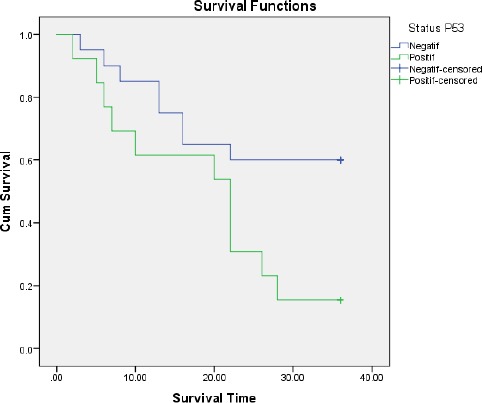
Figure 1.

Kaplan-Meier Curve for 3-year overall survival in Penile Squamous Cell Carcinoma based on p53 status
In the Kaplan-Meier Curve for 3-year overall survival based on p53 expression, the survival rate in 36 months in the p53 positive group is 18%, while in p53 negative group, the survival rate was 60%. The survival rate based on p53 status was significantly different (p = 0.025).
Discussion
In this study, we assessed the role of p53 oncoprotein expression as a prognostic factor in penile cancer. P53 expression was analysed with the survival rate of penile cancer patients. In this study, we assessed the p53 expression as a predictor for mortality in penile cancer patients. Subjects were enrolled in one tertiary hospital which was comprised of the varied stage of cancer.
In this study, p53 positive was found in 13 of 33 patients (39.4%). By previous studies about p53 expression, this study used 20% cut-off point for the nucleus staining. This positive cut-off is higher than the study conducted by Levi et al. (26% or 15 of 58 cases) [9] and almost the same with the study conducted by Lam et al., (40% or 17 of 42 cases) [10]. The difference depends on the antibody reagent used.
Compared to the previous correlation study with other types of cancer, this study did not find a correlation between p53 expression and clinical or histopathological variables. Cordon Carlo et al. who conducted a study about p53 and its correlation with bladder cancer found a positive correlation between p53 expression and vascular invasion [11]. Cabelguenne et al., who studied head and neck tumour [12], and Maeda et al., who studied carcinoma of the stomach, showed that p53 immunoreactivity was not correlated with lymph node metastasis [13]. On the other hand, Unal et al. noted a higher cervical metastasis in tongue cancer case in p53 positive [14].
Lopes et al. showed that the immunoreactivity and staining stage of p53 was significantly associated with lymph node metastasis in N stage penile cancer [15]. Patient with p53 immunoreactivity had 4.8 times risk of having metastasis compared to a patient with the p53 negative. Lymphatic embolism by neoplasm cell and positive p53 was a single predictive factor for lymph node metastasis in multivariate analysis. In this study, metastasis was not assessed because of the lacking number of the patient who had metastasis, some of which could not be followed.
In the study conducted by Marinescu et al., it is found that all (100%) of the poorly differentiated penile cancer had a positive expression of p53 oncoprotein, regardless of the tumour stage. P53 was found in 91.2% of the moderately-differentiated tumour and 72.2% of the well-differentiated tumour [16]. Based on the tumour stage, Marinescu identified a positive association of p53 in all stage II and stage III tumour, and in 43 (84.3%) stage I tumour. In a well-differentiated tumour, the p53 marker is in the nuclei of the peripheral islands of tumour cell and seldom isolated inside the islands, with low or moderate intensity. For moderately- or poorly-differentiated squamous penile cancer, p53 immunohistochemistry staining was found in the nuclei, in the peripheral neoplastic tumour island, with moderate or high intensity. The association between squamous cell carcinoma and carcinoma in situ was statistically different. In addition to that, the association between poorly-differentiated carcinoma and well-differentiated carcinoma was statistically different [16]. In this study, we evaluated the clinical stage of the clinical T stage and found no association between p53 expression and clinical T stage cT2 or cT3 or cT4 penile cancer.
A prospective study conducted by Kamran-Shostari et al. about the clinical significance of p53 and p16 in penile cancer survival rate in North America revealed that p53 could be a predictor for penile cancer carcinoma metastasis to the lymph node. However, this association was only found in patients with the p16 negative. P16 was a predictor in penile carcinoma in association with HPV (Human Papilloma Virus) infection [17]. In another study about penile cancer survival, positive p53 was associated with bad prognosis. Ficarra et al. showed that the survival rate was only 34% in 47 patients with penile cancer. In addition to that, a patient with p53 negative gene expression had a 5-year- and 10-year-survival rate of 64.5% and 54.6%. Meanwhile, the p53 positive gene expression had 30.2% and 26.4%. These differences were significant between two groups (p = 0.009) [18]. This study is by our study which showed that p53 expression is significantly different with 3-year-survival-rate of a penile cancer patient (p = 0.025).
In this study, we found that p53 could be a predictive factor for mortality in penile cancer. However, this study has a limitation in the number of the sample and follow-up. The development of treatment modalities such as prophylaxis lymphadenectomy and chemotherapy have a role that should be considered for further study. Involvement of other than p53 gene, such as p16 should also be considered as a comparator.
In conclusion, there is a significant association (p = 0.025) between p53 expression and mortality in penile cancer patients. In conclusion, p53 expression in penile cancer cells examined by immunohistochemistry may show prognostic values in the disease progression.
Further research is needed to assess p53 expression and its association with treatment response in penile cancer patients. In addition to that, the involvement of other than p53 gene, such as p16 should also be considered as a comparator.
Footnotes
Funding: This research did not receive any financial support
Competing Interests: The authors have declared that no competing interests exist
References
- 1.Pettaway CA, Lance RS, Davis JW. Tumors of the penis. In Campbell-WalshUrology. 2012:901–1000. [Google Scholar]
- 2.Tranggono U, Umbas R. Karakteristik dan terapi penderita keganasan penis di RS Cipto Mangunkusumo dan RS Kanker Dharmais. Indonesian Journal of Cancer. 2008:45–50. [Google Scholar]
- 3.Kusmawan E, Bowolaksono, Widiana R. The Clinical Features of Penile Cancer Patients at Sanglah General Hospital Bali-Indonesia. Bali Medical Journal. 2012;1(1):1–5. [Google Scholar]
- 4.Hakenberg OW, Protzel C. Chemotherapy in penile cancer. Therapeutic Advances in Urology. 2012;4(3):133–8. doi: 10.1177/1756287212441235. https://doi.org/10.1177/1756287212441235 PMid:22654965 PMCid:PMC3361747. [DOI] [PMC free article] [PubMed] [Google Scholar]
- 5.Irawan W, Warli SM. Karakteristik Penderita Kanker Penis di RSUP H Adam Malik Medan. 2015 [Google Scholar]
- 6.Reed SI. In: Cellcycle in Cancer Principles &Practice of Oncology. 7th Ed. DeVita V Jr, Hellman S, Rosenberg S A, editors. Vol. 1. Philadelphia: Lippincott & Wilkins; 2005. pp. 89–94. [Google Scholar]
- 7.Zargar-Shostari K, Spiess P E, Berglund A E, et al. Clinical Significanceof p53 and p16ink4 Status in a Contemporary North American Penile Carcinoma Cohort. Clinicalgenitourinary cancer. 2016;(12):1–6. doi: 10.1016/j.clgc.2015.12.019. [DOI] [PMC free article] [PubMed] [Google Scholar]
- 8.Gunia S, Kakies C, Erbersdobler A, Hakenberg O W, Koch S, May M. Expressionof p53, p21 andcyclin D1 in penile cancer:p53 predictspoor prognosis. JounalofClinicalPathology. 2012;65:232–236. doi: 10.1136/jclinpath-2011-200429. [DOI] [PubMed] [Google Scholar]
- 9.Levi JE, Rahal P, Sarkis Á S, Villa LL. Human papillomavirus DNA and p53 status in penile carcinomas. International journal of cancer. 1998;76(6):779. doi: 10.1002/(sici)1097-0215(19980610)76:6<779::aid-ijc1>3.0.co;2-v. https://doi.org/10.1002/(SICI)1097-0215(19980610)76:6<779::AID-IJC1>3.0.CO;2-V. [DOI] [PubMed] [Google Scholar]
- 10.Lam KY, Chan AC, Chan KW, Leung ML, Srivastava G. Expression of p53 and its relationship with human papillomavirus in penile carcinomas. European Journal of Surgical Oncology (EJSO) 1995;21(6):613–6. doi: 10.1016/s0748-7983(95)95262-4. https://doi.org/10.1016/S0748-7983(95)95262-4. [DOI] [PubMed] [Google Scholar]
- 11.Cordon-Cardo C, Zhang ZF, Dalbagni G, Drobnjak M, Charytonowicz E, Hu SX, Xu HJ, Reuter VE, Benedict WF. Cooperative effects of p53 and pRB alterations in primary superficial bladder tumors. Cancer research. 1997;57(7):1217. PMid:9102201. [PubMed] [Google Scholar]
- 12.Cabelguenne A, Blons H, de Waziers I, Carnot F, Houllier AM, Soussi T, Brasnu D, Beaune P, Laccourreye O, Laurent-Puig P. p53 alterations predict tumor response to neoadjuvant chemotherapy in head and neck squamous cell carcinoma:a prospective series. J Clin Oncol. 2000;18(7):1465–73. doi: 10.1200/JCO.2000.18.7.1465. https://doi.org/10.1200/JCO.2000.18.7.1465 PMid:10735894. [DOI] [PubMed] [Google Scholar]
- 13.Maeda K, Kang SM, Onoda N, Ogawa M, Sawada T, Nakata B, et al. Expression of p53 and vascular endothelial growth factor associated with tumor angiogenesis and prognosis in gastric cancer. Oncology. 2008;55:594. doi: 10.1159/000011918. https://doi.org/10.1159/000011918 PMid:9778629. [DOI] [PubMed] [Google Scholar]
- 14.Unal OF, Ayhan A, Hosal AS. Prognostic value of p53 expression and histopathological parameters in squamous cell carcinoma of oral tongue. J Laryngol Otol. 2009;113:446. doi: 10.1017/s0022215100144184. [DOI] [PubMed] [Google Scholar]
- 15.Lopes A, Bezerra AR, Pinto CA, Serrano SV, et al. p53 as a new prognostic factor for lymph node metastasis in penile carcinoma:analysis of 82 patients treated with amputation and bilateral lymphadenectomy. AUA. 2002;168:81–6. [PubMed] [Google Scholar]
- 16.Marinescu A, Stepan AE, Mărgăritescu C, et al. P53, p16 and Ki67 immunoexpression in cutaneous squamous cell carcinoma and its precursor lesions. Rom J Morphol Embryol. 2016;57(2):691–6. PMid:27833960. [PubMed] [Google Scholar]
- 17.Zargar-Shoshtari K, Spiess PE, Berglund AE, Sharma P, Powsang JM, Giuliano A, Magliocco AM, Dhillon J. Clinical significance of p53 and p16ink4a status in a contemporary North American Penile Carcinoma Cohort. Clinical genitourinary cancer. 2016;14(4):346–51. doi: 10.1016/j.clgc.2015.12.019. https://doi.org/10.1016/j.clgc.2015.12.019 PMid:26794389. [DOI] [PMC free article] [PubMed] [Google Scholar]
- 18.Ficarra V, D'Amico A, Cavalleri S, Zanon G, Mofferdin A, Schiavone D, et al. Surgical treatment of penile carcinoma:our experience from 1976 to 2007. Urol Int. 2009;62:234. doi: 10.1159/000030404. https://doi.org/10.1159/000030404 PMid:10567891. [DOI] [PubMed] [Google Scholar]


