Abstract
NGLY1 deficiency is a rare genetic disease caused by mutations in the NGLY1 gene that encodes N-glycanase 1. The disease phenotype in patient cells is unclear. A human induced pluripotent stem cell (iPSC) line was generated from skin dermal fibroblasts of a patient with NGLY1 deficiency that has compound heterozygous mutations of a p.Q208X variant (c.622C > T) in exon 4 and a p.G310G variant (c.930C > T) in exon 6 of the NGLY1 gene. This iPSC line offers a useful resource to study the disease pathophysiology and a cell-based model for drug development to treat NGLY1 deficiency.
Resource table
| Unique stem cell line identifier | TRNDi002-B |
| Alternative name(s) of stem cell line | HT519B |
| Institution | National Institutes of Health, National Center for Advancing Translational Sciences, Bethesda, Maryland, USA |
| Contact information of distributor | Dr. Wei Zheng, Wei.Zheng@nih.gov |
| Type of cell line | iPSC |
| Origin | Human |
| Additional origin info | Age: 10-year-old Sex: Male Ethnicity: Caucasian |
| Cell Source | Skin fibroblasts |
| Clonality | Clonal |
| Method of reprogramming | Integration-free Sendai viral vectors |
| Genetic modification | NO |
| Type of modification | N/A |
| Associated disease | NGLY1 Deficiency |
| Gene/locus | NGLY1Q208X; NGLY1G310G |
| Method of modification | N/A |
| Name of transgene or resistance | N/A |
| Inducible/constitutive system | N/A |
| Date archived/stock date | 04-27-2018 |
| Cell line repository/bank | Human Pluripotent Stem Cell Registry https://hpscreg.eu/cell-line/TRNDi002-B |
| Ethical approval | NIGMS Informed Consent Form was obtained from patient at time of sample submission. Confidentiality Certificate: CC-GM-15-004 |
Resource utility
This hiPSC line is a useful tool for studies of disease phenotype, disease pathophysiology, and use as a cell-based disease model for drug development to treat patients with NGLY 1 deficiency.
Resource details
NGLY1 deficiency or NGLY1-congenital disorder of deglycosylation is a rare autosomal recessive disorder caused by mutations in the NGLY1 gene, which encodes N-glycanase 1. It cleaves N-linked glycans from glycoproteins. Deficiency in N-glycanase 1 results in malfunctions in deglycosylation of N-glycosylated proteins. Because protein glycosylation and deglycosylation play an important role in post-translational modification of proteins, deficiency of this enzyme can cause protein misfolding and aggregation in the endoplasmic reticulum and cytosol, as well as proteasome dysfunction due to defective processing of Nrf1. The typical features of NGLY1 deficiency include developmental delay or intellectual disability of varying degrees, lack of tear production, elevated liver transaminases, and a complex movement disorder (Lam et al., 1993; Suzuki, 2016; Tomlin et al., 2017).
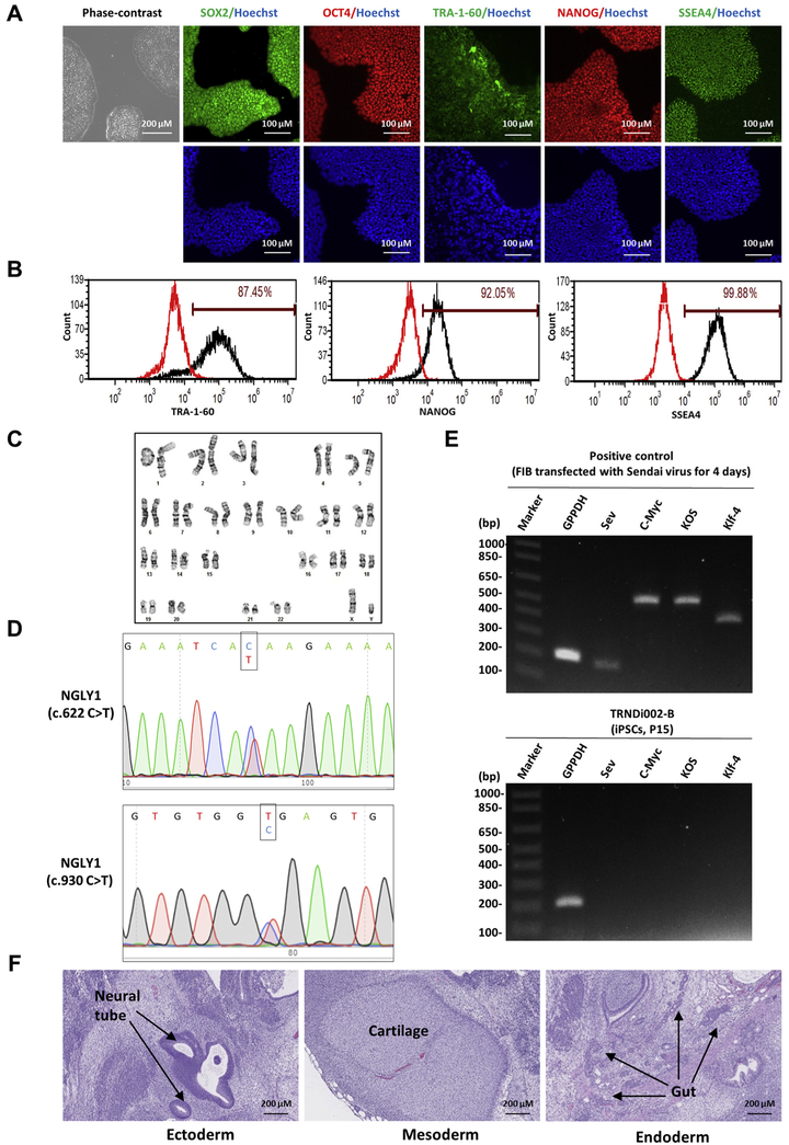
In this study, a human induced pluripotent stem cell line was established from skin fibroblasts of a 10-year-old male patient (GM25344, Coriell Institute) carrying compound heterozygous mutations of a p. Q208X variant (c.622C > T) in exon 4 and a p.G310G variant (c.930C > T) in exon 6 of the NGLY1 gene (Table 1). This subject is also found to be heterozygous for a c.4060 A > T mutation (Thr1354Ser) in the CACNA1S gene based on the information released by Coriell Institute, which might represent one of the risk factors for increased susceptibility to malignant hyperthermia (MH) (Beam et al., 2017). To generate the iPS cells, the non-integrating CytoTune-Sendai viral vector kit (A16517, Thermo Fisher Scientific) containing OCT3/4, KLF4, SOX2 and C-MYC pluripotency transcription factors was employed to transduce the patient fibroblasts using the method described previously (Beers et al., 2015). The iPSC line named TRNDi002-B was generated from the patient fibroblasts. The mutations (c.622C > T and C.930C > T) of the NGLY1 gene described by Coriell Institute were confirmed in the TRNDi002-B iPSC line by Sanger sequencing of the PCR product harboring the single nucleotide variation (SNV) (Fig. 1D). The patient iPS cells exhibited a classical embryonic stem cell morphology (Fig. 1A) and normal karyotype (46, XY), as confirmed by the G-banded karyotyping at passage 11 (Fig. 1C), expressed the major pluripotent protein markers of NANOG, SOX2, OCT4, SSEA4 and TRA-1-60 (Fig. 1A, B) evidenced by both immunofluorescence staining and flow cytometry analysis. Sendai virus vector (SeV) clearance was detected with reverse transcription polymerase chain reaction (RT-PCR) using SeV-specific primers, the vector disappeared by passage 15 (Fig. 1E). This iPSC line was not contaminated with mycoplasma (Supplementary Fig. S1) and were authenticated using STR DNA profiling analysis, which demonstrated matching genotypes at all 18 loci examined (information available with the authors). Furthermore, the pluripotency of this iPS cell line was confirmed by the teratoma formation experiment that exhibited its ability to differentiate into cells of all three germ layers (Ectoderm, neural tube; Mesoderm, cartilage; Endoderm, gut) in vivo (Fig. 1F).
Table 1.
Characterization and validation.
| Classification | Test | Result | Data |
|---|---|---|---|
| Morphology | Photography | Normal | Fig. 1 Panel A |
| Phenotype | Immunocytochemistry | SOX2, OCT4, NANOG, SSEA-4, TRA-1-60 | Fig. 1 Panel A |
| Flow cytometry | TRA-1-60 (87.5%); NANOG (92.1%); SSEA-4 (99.9%) | Fig. 1 Panel B | |
| Genotype | Karyotype (G-banding) and resolution | 46XY Resolution: 350–400 | Fig. 1 Panel C |
| Identity | Microsatellite PCR (mPCR) OR | Not performed | N/A |
| STR analysis | 18 sites tested, all sites matched | Available with the authors | |
| Mutation analysis (IF APPLICABLE) | Sequencing | Compound heterozygous mutation of NGLY1 | Fig. 1 Panel D |
| Southern Blot OR WGS | N/A | N/A | |
| Microbiology and virology | Mycoplasma | Mycoplasma testing by luminescence. Negative | Supplementary Fig. S1 |
| Differentiation potential | Teratoma formation | Teratoma with three germlayers formation.
Ectoderm (neural tube); Mesoderm (cartilage); Endoderm (gut) |
Fig. 1 Panel F |
| Donor screening (OPTIONAL) | HIV 1 + 2 Hepatitis B, Hepatitis C | N/A | N/A |
| Genotype additional info | Blood group genotyping | N/A | N/A |
| (OPTIONAL) | HLA tissue typing | N/A | N/A |
Fig. 1. Characterization of TRNDi002-B iPSC line.
A) Left: Phase contrast imaging of TRNDi002-B colonies grown on Matrigel at passage 10. Right: Representative immunofluorescent micrographs of iPSCs positive for stem cell markers: SOX2, OCT4, TRA-1-60, NANOG, and SSEA4. Nucleus is labelled with Hoechst (in blue). B) Flow cytometry analysis of pluripotency protein markers: TRA-1-60, NANOG and SSEA4. C) Cytogenetic analysis showing a normal karyotype (46, XY). D) Detection of compound heterozygous of a p.Q208X variant (c.622C > T) in exon 4 and a p.G310G (c.930C > T) in exon 6 of the NGLY1 gene. E) RT-PCR verification of the clearance of Sendai virus from the reprogrammed cells. Sendai virus vector transduced fibroblasts was used as positive control. F) Pathological analysis of a teratoma from TRNDi002-B iPSC, showing a normal ectodermal, endodermal and mesodermal differentiation.
Materials and methods
Cell culture
Patient skin fibroblasts were purchased from Coriell Cell Repositories (GM25344) and cultured in DMEM supplemented with 10% fetal bovine serum, 100 units/ml penicillin and 100 μg/ml streptomycin in a humidified incubator with 5% CO2 at 37 °C. Human iPS cells were cultured in StemFlex medium (Thermo Fisher) on Matrigel (Corning, 354277)-coated plates at 37 °C in humidified air with 5% CO2 and 5% O2. The cells were passaged with 0.5 mM Ethylenediaminetetraacetic acid (EDTA) at generally 1:6 ratio when they reach 80% confluency.
Reprogramming of human skin fibroblasts
Patient fibroblasts were reprogrammed into iPS cells using the non-integrating Sendai virus technology following the method described previously (Beers et al., 2015).
Genome analysis
The genome analysis of variants in NGLY1 was conducted through Applied StemCell (Milpitas, California, USA). Briefly, genomic DNA was extracted from hiPSC line TRNDi002-B using QuickExtract™ DNA Extraction Solution (Lucigen) followed by PCR amplification using MyTaq™ Red Mix (Bioline, Taunton, MA). Amplifications were carried out on T00 Thermal Cycler from Bio-Rad (#1861096) using the following program: 95 °C, 2 mins; 35 cycles of [95 °C, 15 s; 60 °C, 15 s; 72 °C, various length depending on size of amplicon], 72 °C 5 mins; 4 °C, indefinite. Genotyping of the compound heterozygous for a p. Q208X variant (c.622C > T) in exon 4 and a p.G310G variant (c.930C > T) in exon 6 of the NGLY1 gene were performed using Sanger sequencing analysis. The specific primers for gene amplification and sequencing are listed in Table 2.
Table 2.
Reagents details.
| Antibodies used for
immunocytochemistry/flow-cytometry | |||
|---|---|---|---|
| Antibody | Dilution | Company Cat # and RRID | |
|
| |||
| Pluripotency markers | Mouse anti-SOX2 | 1:50 | R & D systems, Cat# MAB2018, RRID: AB_358009 |
| Pluripotency markers | Rabbit anti-NANOG | 1:400 | Cell signaling, Cat# 4903, RRID: AB_10559205 |
| Pluripotency markers | Rabbit anti-OCT4 | 1:400 | Thermo Fisher, Cat# A13998, RRID: AB_2534182 |
| Pluripotency markers | Mouse anti-SSEA4 | 1:1000 | Cell signaling, Cat# 4755, RRID: AB_1264259 |
| Pluripotency markers | Mouse anti-TRA-1-60- Alexa Fluor 488 | 1:10 | BD Biosciences, Cat# 560173, RRID: AB_1645379 |
| Secondary antibodies | Donkey anti-Mouse IgG (Alexa Fluor 488) | 1:400 | Thermo Fischer, Cat# A21202, RRID: AB_141607 |
| Secondary antibodies | Donkey anti-Rabbit IgG (Alexa Fluor 594) | 1:400 | Thermo Fischer, Cat# A21207, RRID: AB_141637 |
| Flow cytometry antibodies | Anti-Tra-1-60-DyLight 488 | 1:50 | Thermo Fischer, Cat# MA1–023-D488X, RRID: AB_2536700 |
| Flow cytometry antibodies | Anti-Nanog-Alexa Fluor 488 | 1:50 | Millipore, Cat# FCABS352A4, RRID: AB_10807973 |
| Flow cytometry antibodies | anti-SSEA-4-Alexa Fluor 488 | 1:50 | Thermo Fischer, Cat# 53–8843-41, RRID: AB_10597752 |
| Flow cytometry antibodies | Mouse-IgM-DyLight 488 | 1:50 | Thermo Fischer, Cat# MA1–194-D488, RRID: AB_2536969 |
| Flow cytometry antibodies | Rabbit IgG-Alexa Fluor 488 | 1:50 | Cell Signaling, Cat# 4340S, RRID: AB_10694568 |
| Flow cytometry antibodies | Mouse IgG3-FITC | 1:50 | Thermo Fischer, Cat# 11–4742-42, RRID: AB_2043894 |
|
| |||
| Primers | |||
| Target | Forward/reverse primer (5′−3′) | ||
|
| |||
| Sev specific primers (RT-PCR) | Sev/181 bp | GGA TCA CTA GGT GAT ATC GAG C/ACC AGA CAA GAG TTT AAG AGA TAT GTA TC | |
| Sev specific primers (RT-PCR) | KOS/528 bp | ATG CAC CGC TAC GAC GTG AGC GC/ACC TTG ACA ATC CTG ATG TGG | |
| Sev specific primers (RT-PCR) | Klf4/410 bp | TTC CTG CAT GCC AGA GGA GCC C/AAT GTA TCG AAG GTG CTC AA | |
| Sev specific primers (RT-PCR) | C-Myc/523 bp | TAA CTG ACT AGC AGG CTT GTC G/TCC ACA TAC AGT CCT GGA TGA TGA TG | |
| House-keeping gene (RT-PCR) | GAPDH/197 bp | GGA GCG AGA TCC CTC CAA AAT/GGC TGT TGT CAT ACT TCT CAT GG | |
| Targeted mutation analysis (PCR) | NGLY1 (c.622C > T)/577bp | CCA TGC AGT TCA AAC CCA TGT TCT TC/GAT GCA TAG ATC AGA GCT CTG TAT TGA TCC | |
| Targeted mutation analysis (PCR) | NGLY1 (c.930C > T)/558bp | CTG CAA AGC CCT ACT ACC CTC/ATT AAA GGC CGG GCG CAG | |
Immunocytochemistry
For immunofluorescence staining, patient iPSCs were fixed in 4% paraformaldehyde for 15 mins, rinsed with DPBS, and permeabilized with 0.3% Triton X-100 in DPBS for 15 mins. The cells were incubated with the Image-iT™ FX signal enhancer (ThermoFisher Scientific) for 40 mins at room temperature in a humidified environment and then followed by incubation individually with primary antibodies including SOX2, OCT4, NANOG, SSEA4 and TRA-1-60, diluted in the Image-iT™ FX signal enhancer blocking buffer, for overnight at 4 °C. After washing with DPBS, a corresponding secondary antibody conjugated with Alexa Fluor 488 or Alex Fluor 594 was added to the cells and incubated for 1 h at room temperature (Antibodies used are listed in Table 2). Cells were then stained with Hoechst 33342 for 15 mins after a wash and imaged using an INCell Analyzer 2200 imaging system (GE Healthcare) with 20× objective lens and Texas Red, FITC and DAPI filter sets.
Flow cytometry analysis
The iPSCs were harvested using TrypLE Express Enzyme (ThermoFisher Scientific). Cells were fixed with 4% paraformaldehyde for 10 mins at room temperature and then washed with DPBS. Before fluorescence-activated cell sorting analysis, cells were permeabilized with 0.2% Tween-20 in DPBS for 10 mins at room temperature and stained with fluorophore conjugated antibodies for 1 h at 4 °C on a shaker (Antibodies used are listed in Table 2). Cells were then analyzed on a BD Accuri™ C6 FlowCytometry system (BD Biosciences).
G-banding karyotype
The G-banding karyotype analysis was conducted at WiCell Research Institute (Madison, WI, USA). A total of 20 randomly selected metaphases were analyzed by G-banding for each cell line.
Short tandem repeat (STR) analysis
Patient fibroblasts and derived iPSC lines were sent to the Johns Hopkins University Genetic Resources Core Facility for STR DNA profile analysis using a Promega PowerPlex 18D Kit. The PCR product was electrophoresed on an ABI Prism® 3730x1 Genetic Analyzer and data was analyzed using GeneMapper® v 4.0 software (Applied Biosystems).
Mycoplasma detection
Mycoplasma testing was performed and analyzed using the Lonza MycoAlert kit following the instructions from the company. Ratio B/A > 1.2 indicates mycoplasma positive; 0.9–1.2 Result indicates ambiguous; < 0.9 indicates mycoplasma negative.
Sendai virus detection
Total RNA was extracted using RNeasy Plus Mini Kit (Qiagen). Human fibroblasts (GM05659, Coriell Institute) after transfection with Sendai virus for 4 days was used as the positive control. 1 μg of RNA was reverse transcribed into cDNA with Superscript™ III First-Strand Synthesis SuperMix kit and PCR was performed using Platinum II Hot- Start PCR Master Mix (ThermoFisher Scientific) and the amplifications were carried out using the following program: 94 °C, 2 mins; 30 cycles of 94 °C, 15 s, 60 °C, 15 s and 68 °C, 15 s on Mastercycler pro S (Eppendorf) with the primers listed in Table 2. The products were then loaded to the E-Gel® 1.2% with SYBR Safe™ gel, and imaged by G: Box Chemi-XX6 gel doc system (Syngene, Frederick, MD).
Teratoma formation assay
Patient iPSCs cultured in 6-well plates were dissociated with DPBS containing 0.5 mM EDTA, and approximately 1 × 10⁷ dissociated cells were resuspended in 400 μl culture medium supplied with 25mM HEPES (pH 7.4) and stored on ice. Then, 50% volume (200 μl) of cold Matrigel (Corning, 354277) was added and mixed with the cells. The mixture was injected subcutaneously into NSG mice (JAX No. 005557) at 150 μl per injection site. Visible tumors were removed 6–8 weeks post injection, and were immediately fixed in 10% Neutral Buffered Formalin. The fixed tumors were embedded in paraffin and stained with hematoxylin and eosin.
Supplementary data to this article can be found online at https://doi.org/10.1016/j.scr.2018.101362.
Supplementary Material
Acknowledgement
We would like to thank Dr. Zu-xi Yu of the pathology Core of National Heart, Lung and Blood Institute, National Institutes of Health for sectioning and staining the teratoma. We also would like to thank Tobie Lee and Kelli Wilson of the Research Services Section at National Center for Advancing Translational Sciences for coordinating the STR DNA analysis and mycoplasma testing service. We would like to acknowledge Beth Aselage from Retrophin and Carrie Ostrea from NGLY1.org for helpful discussions. This work was supported by the Therapeutics for Rare and Neglected Diseases Program, Division of Preclinical Innovation, National Center for Advancing Translational Sciences, National Institutes of Health, under a CRADA collaboration between NCATS, NGLY1.org, and Retrophin.
References
- Beam TA, Loudermilk EF, Kisor DF, 2017. Pharmacogenetics and pathophysiology of CACNA1S mutations in malignant hyperthermia. Physiol. Genomics 49, 81–87. [DOI] [PubMed] [Google Scholar]
- Beers J, Linask KL, Chen JA, Siniscalchi LI, Lin Y, Zheng W, Rao M, Chen G, 2015. A cost-effective and efficient reprogramming platform for large-scale production of integration-free human induced pluripotent stem cells in chemically defined culture. Sci. Rep. 5, 11319. [DOI] [PMC free article] [PubMed] [Google Scholar]
- Lam C, Wolfe L, Need A, Shashi V, Enns G, 1993. NGLY1-related congenital disorder of deglycosylation. In: Adam MP, Ardinger HH, Pagon RA, Wallace SE, Bean LJH, Stephens K, Amemiya A (Eds.), GeneReviews((R)), Seattle (WA). [PubMed] [Google Scholar]
- Suzuki T, 2016. Catabolism of N-glycoproteins in mammalian cells: molecular mechanisms and genetic disorders related to the processes. Mol. Asp. Med. 51, 89–103. [DOI] [PubMed] [Google Scholar]
- Tomlin FM, Gerling-Driessen UIM, Liu YC, Flynn RA, Vangala JR, Lentz CS, Clauder-Muenster S, Jakob P, Mueller WF, Ordonez-Rueda D, Paulsen M, Matsui N, Foley D, Rafalko A, Suzuki T, Bogyo M, Steinmetz LM, Radhakrishnan SK, Bertozzi CR, 2017. Inhibition of NGLY1 inactivates the transcription factor Nrf1 and potentiates proteasome inhibitor cytotoxicity. ACS Cent. Sci. 3, 1143–1155. [DOI] [PMC free article] [PubMed] [Google Scholar]
Associated Data
This section collects any data citations, data availability statements, or supplementary materials included in this article.


