Figure 2.

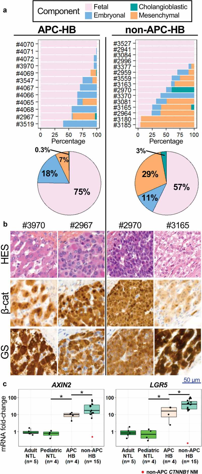
Pathological comparison of APC and non-APC HB.
(a) Histological component distribution assessed by pathological reviewing of APC and non-APC HB. (b) Representative HES and IHC staining of β-catenin (β-cat) and GS within the tumors of APC and non-APC HB patients (high magnification). Lymphocytes without staining are seen in APC-HB. (c) Levels of AXIN2 and LGR5 mRNAs assessed by qRT-PCR in APC and non-APC HB as well as in adult and pediatric non-tumor liver (NTL), expressed as a fold-change compared to adult NTL. The non-APC and non-CTNNB1-mutated HB sample is marked in red. * P < .05 (Wilcoxon test, without taking into account the non-APC CTNNB1 NM tumor).
