Introduction
Symptomatic migraine or migraine‐like headache due to a brainstem lesion is rarely reported, and limited reports of brainstem lesions with migraine‐like headache onset are all directed to the pons or midbrain [1, 2]. We describe a new onset of recurrent unilateral headache whose clinical features were similar to that of migraine with aura in a patient who had a lateral medullary infarction, i.e., Wallenberg syndrome.
Case Report
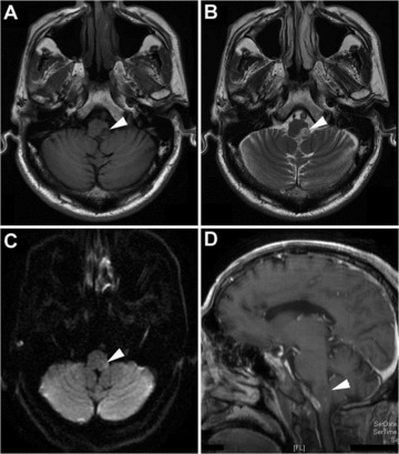
A 60‐year‐old man presented with acute onset of nausea, vomiting, vertigo and fluctuating colorful stripes around his visual field 15 days earlier, unsteadiness, swallowing disturbance, and sensory loss of the right limb and the left face developed subsequently. His past medical history was remarkable for hypertension, which was not regularly controlled as well as for smoking for 30 years. The neurological examination revealed a left Horner syndrome, moderate dysarthria, mild dysphagia (bucking while drinking a large mouthful of water), evident soft palate paresis and disappeared gag reflex on the left, hypoalgesia and thermoanesthesia of left face and right two limbs, and moderate left limb ataxia. Other neurological and physical examinations were normal. At the onset of nausea, vomiting and vertigo, computed tomography scan did not disclose any significant abnormalities. After admission, brain magnetic resonance imaging (performed the following day, i.e., sixteen days after the symptom onset, revealed a low signal on T1‐weighted imaging, high signal on T2‐weighted and diffusion‐weighted imaging in the dorsal and lateral aspect of the left medulla (Figure 1). The patient was diagnosed as lateral medullary infarction, i.e., Wallenberg syndrome and managed with aspirin and atorvastatin. His condition steadily improved and he had nearly no motor or sensory defect induced by stroke three months after discharge.
Figure 1.

MR imaging obtained 16 days after symptom onset. MR image through the medulla oblongata at presentation shows a patchy hypointense (A), hyperintense (B and C), and nonenhancing (D) lesion over the left posterolateral aspect of the medulla oblongata (arrow heads) on T1‐weighted (A), T2‐weighted (B), diffusion‐weighted (C), and enhancing (D) imaging, respectively.
One week after admission, i.e., 3 weeks after the stroke onset, he complained of paroxysmal headache attacks. A typical attack would begin with a visual aura, which was in a shape of fluctuating colorful stripes around his visual field. The visual aura would last 1–5 min and would be followed by a right‐sided headache with nausea, photophobia, and aggravation by head movement. Every episode of headache would last half day or would disappear over a sleep. The headache would attack once a day or once every other day. With respect to the treatment, ibuprofen, rizatriptan and pregabalin he had ever taken failed to prevent the recurrence of headaches but attenuated the pain severity mildly. The recurrent headaches persisted for three weeks and disappeared spontaneously. In the next 3 months, he had five times of "cluster"‐like headaches with a similar headache pattern. Gradually, the headache would attack almost every day with the visual aura rarely appeared and finally disappeared. But the headache severity became milder after he accepted treatment with sodium valproate 1000 mg daily). Before this event, he had neither personal nor familial history of headaches.
Discussion
This patient had severe, recurrent and right side‐locked headaches with visual auras associated with a left side Wallenberg syndrome. According to current criteria of headache classification, the International Classification of Headache Disorders (ICHD‐II) criteria [3], this case belongs to the headache attributed to ischemic stroke (cerebral infarction). Whereas, the migraine with aura‐like clinical features, e.g., chronic recurrence and the precedence of visual aura, are different from the regular headache induced by cerebral infraction, but more suggestive of "symptomatic migraine with aura," though the headache characteristics do not meet ICHD‐2 criteria for a specific primary headache disorder [3].
The underlying pathophysiology of symptomatic migraine caused by lesions in the midbrain or pons has been postulated as a loss of inhibition descending from certain nucleus, e.g., locus coeruleus, periaqueductal gray (PAG) and nucleus cuneiformis (NCF) in midbrain. This results in hyperexcitability of trigeminovascular neurons, i.e., facilitation of the trigeminal nociception, thought to underlie migraine headache [4, 5]. In current case, the lesion causing migraine with aura‐like headache was neither in midbrain nor in pons but in medulla where the trigeminal nucleus is located. The sensory symptom of Wallenberg syndrome and its later complete recovery also indicated that the trigeminal sensory nucleus was partially damaged. The partial damage of trigeminal nucleus would cause a structure change, probably inducing a facilitation of the ipsilateral trigeminal nociception and the relevant ipsilateral headache. But the contralateral headache and the preceding visual aura involving visual cortex in current case seemly make this possible mechanism illogical. In fact, up to date, no study has ever referred the pathogenesis of migraine to the medulla, and no symptomatic migraine or migraine‐like headache has ever been reported to be caused by lesion in medulla. Thus, the pathophysiology of the migraine with aura‐like headache generated from a Wallenberg syndrome in current case is largely unknown.
In migraineurs, deficient habituation of cortical response to repeated sensory stimuli has been detected during the interictal period. Current hypotheses attribute this deficient habituation to insufficient intracortical inhibition or to low level of sensory cortical pre‐activation ultimately due to the hypofunction of the brainstem [6]. In our patient with Wallenberg syndrome, ascending neural activity coming from ipsilateral trigeminal nucleus caudalis and contralateral spinal cord posterior horn was impaired. This may lead to the deficits of cortical excitatory habituation, i.e., cortical hyperexcitability, the physiological basis of cortical spreading depression (CSD), which can activate central trigeminovascular neurons in the spinal trigeminal nucleus [7] inducing headache attack. Though the occipital area from which the visual aura originated is not related to the parietal lobe, the central area processing neural activity ascending from ipsilateral trigeminal nucleus caudalis and contralateral spinal cord posterior horn, a recent study has demonstrated that these two areas are closely connected as a peripheral pain stimulation can alter the excitability of parietal lobe as well as of occipital lobe [8]. And now it is well accepted that CSD is the physiological basis of visual aura. Thus, the migraine with aura‐like headaches in current patient might be due to the impaired ascending preactivation. This is supported by the recurrence of headaches, though not of classic or common migraine, in patients with traumatic transections of the cervical spinal cord with the ascending neural activity coming from bilateral spinal cord posterior horn impaired [9], as nonmigraine headache, e.g., medication‐overuse headache, is also associated with cortical habituation [10]. The deficits of the cortical habituation due to impaired ascending preactivation seemly also explain why the headache was bilateral in patients with traumatic transections of the cervical spinal cord [9] but unilateral in our patient with a lateral medullary infarction. Given the preactivation and CSD mechanisms correct, weeks’ lagging time might be needed to build up the cortical hyperexcitability inducing CSD.
In conclusion, this case provides evidence for the first time that the medulla lesion can cause the initiation of migraine with aura‐like headaches. Deficits of the cortical habituation induced by the decreasing of ascending preactivation due to the lesion of the medulla might be involved in the pathogenesis of the headaches.
Informed consent is available from the patient.
Disclosure
This article consists of original, unpublished work, which is not submitted elsewhere.
Conflict of Interest
The authors declare no conflict of interest.
References
- 1. Friedman D. Unilateral headache associated with a pontine infarction. Cephalalgia 2010;30:1524–1526. [DOI] [PubMed] [Google Scholar]
- 2. Afridi S, Goadsby PJ. New onset migraine with a brain stem cavernous angioma. J Neurol Neurosurg Psychiatry 2003;74:680–682. [DOI] [PMC free article] [PubMed] [Google Scholar]
- 3. Headache Classification Subcommittee of the International Headache Society. The International Classification of Headache Disorders: 2nd edition. Cephalalgia 2004;24(Suppl 1):9–160. [DOI] [PubMed] [Google Scholar]
- 4. Sprenger T, Goadsby PJ. Migraine pathogenesis and state of pharmacological treatment options. BMC Med 2009;7:71. [DOI] [PMC free article] [PubMed] [Google Scholar]
- 5. Katsarava Z, Egelhof T, Kaube H, Diener HC, Limmroth V. Symptomatic migraine and sensitization of trigeminal nociception associated with contralateral pontine cavernoma. Pain 2003;105:381–384. [DOI] [PubMed] [Google Scholar]
- 6. Coppola G, Pierelli F, Schoenen J. Is the cerebral cortex hyperexcitable or hyperresponsive in migraine? Cephalalgia 2007;27:1427–1439. [DOI] [PubMed] [Google Scholar]
- 7. Zhang X, Levy D, Kainz V, Noseda R, Jakubowski M, Burstein R. Activation of central trigeminovascular neurons by cortical spreading depression. Ann Neurol 2011;69:855–865. [DOI] [PMC free article] [PubMed] [Google Scholar]
- 8. Coppola G, Serrao M, Curra A, Di Lorenzo C, Vatrika M, Parisi V, Pierelli F. Tonic pain abolishes cortical habituation of visual evoked potentials in healthy subjects. J Pain 2010;11:291–296. [DOI] [PubMed] [Google Scholar]
- 9. Spierings EL, Foo DK, Young RR. Headaches in patients with traumatic lesions of the cervical spinal cord. Headache 1992;32:45–49. [DOI] [PubMed] [Google Scholar]
- 10. Coppola G, Curra A, Di Lorenzo C, et al Abnormal cortical responses to somatosensory stimulation in medication‐overuse headache. BMC Neurol 2010;10:126. [DOI] [PMC free article] [PubMed] [Google Scholar]


