Abstract
Abstract. The aim of this investigation is to determine whether rapamycin treatment has any effect on endothelial progenitor cells (EPCs). Total mononuclear cells (MNCs) were isolated from peripheral blood by Ficoll density gradient centrifugation, and then the cells were plated on fibronectin‐coated culture dishes. After 7 days in culture, attached cells were stimulated with rapamycin (in a series of final concentrations: 0.1, 1.0, 2.0 and 5.0 g/ml) for 6, 12, 24 and 48 h. EPCs were characterized as adherent cells, double positive for DiLDL uptake and lectin binding by direct fluorescence staining. EPC proliferation and migration were determined using the MTT assay and a modified version of the Boyden chamber assay, respectively. An EPC adhesion assay was performed by replating the cells on fibronectin‐coated dishes; adherent cells were then counted. Tube formation activity was assayed by using a tube formation assay kit and endothelial nitric oxide synthase (eNOS) was assayed by Western blot analysis. Incubation of isolated human MNCs with rapamycin decreased the number of EPCs present; rapamycin also decreased EPCs proliferative, migratory, adhesive, tube formation capacity and eNOS production in a concentration‐ and time‐dependent manner. Rapamycin was found to decrease the number, proliferative, migratory, adhesive and tube formation capacities of the EPCs, and also was found to decreases eNOS in the EPCs.
INTRODUCTION
Vascular endothelial progenitor cells (EPCs) are the precursors of mature endothelial cells. Increasing evidence suggests that circulating progenitor cells contribute to post‐natal neovascularization. These cells home to sites of ischaemia, adopt an endothelial phenotype and contribute to new blood vessel formation; however, the identity of the circulating cells that contribute to neovascularization is not entirely clear. Bone marrow‐derived haematopoietic progenitor cells can give rise to EPCs and can contribute to endothelial recovery and new capillary formation after ischaemia.
Rapamycin (sirolimus, RAPA) is a bacterial macrolide that forms a complex with FK‐binding protein (FKBP‐12) that in turn binds to the mammalian target of rapamycin (mTOR) with high affinity (Dumont et al. 1990; Sehgal 1998). In a recent investigation, RAPA has been shown to inhibit tumour growth by an anti‐angiogenic effect in an experimental mouse model (Guba et al. 2002).
At the molecular level, some factors such as statins and vascular endothelial growth factor (VEGF), can improve proliferation of EPCs by activating the phosphatidyl‐inositol‐3‐kinase (PI‐3K)–Akt–endothelial nitric oxide synthase (eNOS) system, suggesting that the PI‐3K–Akt–eNOS signalling pathway may be the important signalling transduction pathway in EPCs (Urbich & Dimmeler 2005). To further elucidate RAPA's effect as an anti‐angiogenic agent and the mechanism of the eNOS role in these events, our group has investigated the effects of RAPA on number, activity and eNOS production of EPCs from peripheral blood.
MATERIALS AND METHODS
Isolation and culture of EPCs
EPCs were cultured according to previously described techniques (Kalka et al. 2000; Hill et al. 2003; Chen et al. 2004). Briefly, total mononuclear cells (MNCs) were isolated from blood of human study subjects by Ficoll density gradient centrifugation. Cells were plated on culture dishes coated with human fibronectin (Chemicon, Pittsburgh, PA, USA) and were maintained in Medium 199 (Sigma, St. Louis, MO, USA) supplemented with 20% foetal‐calf serum, penicillin (100 U/ml) and streptomycin (100 g/ml). After 4 days in culture, non‐adherent cells were removed by washing with PBS, new media was applied and the culture was maintained for a total of 7 days. Attached cells were stimulated with rapamycin (Sigma, to make a series of final concentrations: 0.1, 1.0, 2.0 and 5.0 µg/ml) for 6, 12, 24 and 48 h.
Cell staining
Direct fluorescent staining was used to detect dual binding of FITC‐labelled Ulex europaeus agglutinin (UEA‐1; Sigma) and 1,1‐dioctadecyl‐3,3,3,3‐tetramethylindocarbocyanine (DiI)‐labelled acetylated low‐density lipoprotein (acLDL; Molecular Probe Data Base, Genova, Italy). Cells were first incubated with acLDL at 37 °C and later were fixed with 2% paraformaldehyde for 10 min. After washing, cells were reacted with UEA‐1 (10 g/ml) for 1 h. After staining, samples were viewed using an inverted fluorescence microscope (Leica Microsystems AG, Wetzlar, Germany) and were further demonstrated by laser scanning confocal microscopy (LSCM, Leica). Cells demonstrating double positive fluorescence were identified as differentiating EPCs (Kalka et al. 2000; Vasa et al. 2001; Chen et al. 2004). In some cases two or in other cases three independent investigators evaluated the number of EPCs per well by counting 15 randomly selected high‐power fields (200).
Migration assay
EPC migration was evaluated by using a modified Boyden chamber assay (Jiangsu Qilin Medical Equipment Factory, Shuzhou, China). In brief, Isolated EPCs were detached using 0.25% trypsin, harvested by centrifugation, resuspended in 500 l M199 and were counted; then 2 × 10⁴ EPCs were placed in the upper chamber of a modified Boyden vessel. M199 and human recombinant VEGF (50 ng/ml) were placed in the lower compartment. After 24 h incubation at 37 °C, the lower side of the filter was washed with PBS and cells were fixed in 2% paraformaldeyde. For quantification, cells were stained with Giemsa solution. Cells migrating into the lower chamber were counted manually in three random microscopic fields (200) (Vasa et al. 2001; Chen et al. 2004).
Cell adhesion assay
EPCs were washed with PBS and were gently detached with 0.25% trypsin. After centrifugation and resuspension in M199, 5% FBS, identical cell numbers were replated onto fibronectin‐coated culture dishes and were incubated for 30 min at 37 °C. Adherent cells were counted blind by independent investigators (Walter et al. 2002; Chen et al. 2004).
EPC proliferation assay
The effect of rapamycin on EPC proliferation was determined by the MTT assay. After being cultured for 7 days, EPCs were digested with 0.25% trypsin and then were cultured in serum‐free medium in 96‐well culture plates (200 l per well). EPCs were supplemented with 10 l MTT (5 g/l) and were incubated for another 6 h. The supernatant was then discarded by aspiration and the EPC preparation was shaken with 200 l DMSO for 10 min, before the OD value was measured at 490 nm (Chen et al. 2004).
Tube formation assay
A tube formation assay was performed with a tube formation assay kit (Chemicon). The protocol was according to the manufacturer's instructions. Briefly, ECMatrix‘ solution was thawed on ice overnight, was mixed with 10×ECMatrix‘ diluent and was placed in a 96‐well tissue culture plate at 37 °C for 1 h to allow the matrix solution to solidify. EPCs were harvested as described previously and were replated (10 000 cells per well) on top of the solidified matrix solution. Cells were incubated at 37 °C for 24 h. Tube formation was inspected under an inverted light microscope at 200× magnification. Tube formation was defined as a structure exhibiting a length four times its width (Reyes et al. 2002; Tepper et al. 2002; Chen et al. 2004). Five independent fields were assessed for each well, and the average number of tubes per 200 fields was determined.
Western blot analysis for eNOS
The cell monolayers were washed three times with PBS and were lysed with RIPA buffer containing phenylmethylsulphonyl fluoride and aprotinin as protease inhibitors. The cell lysates (30 g total protein) were denatured and subjected to 7.5% sodium dodecyl sulphate–polyacrylamide gel electrophoresis (SDS–PAGE). The proteins were transferred to nitrocellulose membranes by electroblotting. Membranes were soaked in a blocking solution containing PBS with 5% non‐fat dried milk and 0.05% Tween 20 for 1 h at room temperature. The membranes were then incubated with anti‐eNOS monoclonal antibodies (Santa Cruz Biotechnology, Santa Cruz, CA, USA) for 2 h and then with peroxidase conjugated secondary antibodies for 2 h at room temperature. The bands corresponding to eNOS were detected using the appropriate chemiluminescence reagent (Amersham Pharmacia Biotech, Piscataway, NJ, USA).
Statistical analysis
Data were expressed as mean ± SD. We used one‐way anova and independent‐samples t‐test to analyse the differences of variables. Differences were considered to be significant if P value < 0.05. All statistical analyses were performed with spss 11.5.
RESULTS
Characteristics of human EPCs
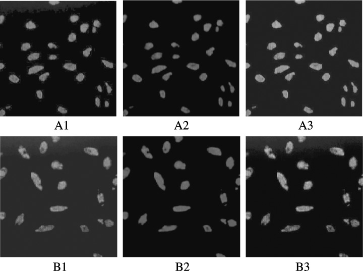
Total MNCs isolated and cultured for 7 days resulted in spindle‐shaped, endothelial cell‐like morphology. EPCs were characterized as adherent cells, double positive for DiLDL uptake and lectin binding, under a laser scanning confocal microscope (Fig. 1). We and other investigators have previously demonstrated that EPCs isolated in this fashion also exhibit many other endothelial characteristics, including expression of CD31, vWF and vascular endothelial growth factor receptor 2 (VEGFR‐2) (Kalka et al. 2000; Vasa et al. 2001; Tepper et al. 2002; Chen et al. 2004).
Figure 1.

Characteristics of human EPCs. MNCs were cultured for 7 days, and adherent cells lectin binding (green, exciting wavelength 477 nm) and DiLDL uptake (red, exciting wavelength 543 nm) were assessed under a laser scanning confocal microscope. Double positive cells appearing yellow in the overlay were identified as differentiating EPCs (×400). Group rapamycin 5.0 g/ml (B1, B2, B3) significantly decreased the number of EPCs compared with control group (A1, A2, A3).
Effect of rapamycin on EPC numbers
Incubation of isolated human MNCs with rapamycin decreased the number of EPCs in a concentration‐ (Table 1) and time‐dependent (Table 2) manner. The low concentration group (0.1 µg/ml) were of no significant difference compared to the control group.
Table 1.
Effects of rapamycin on EPC number, proliferation, migration, adhesion and tube formation capacity (n = 6)
| Group | Number (cells × 200) | Proliferation (490 nm light absorbance) | Migration (migratory cells × 200) | Adhesion (adherent cells × 200) | Tube formation (tubes × 200) |
|---|---|---|---|---|---|
| Control | 59.5 ± 6.2 | 0.503 ± 0.051 | 13.3 ± 3.1 | 31.4 ± 5.5 | 23.3 ± 3.5 |
| 0.1 µg/ml | 54.1 ± 4.5 | 0.472 ± 0.045 | 11.9 ± 2.1 | 28.4 ± 3.1 | 21.3 ± 2.6 |
| 1.0 µg/ml | 52.2 ± 5.5 a | 0.430 ± 0.040 b | 9.7 ± 2.1 a | 26.0 ± 3.3 a | 17.7 ± 3.2 b |
| 2.0 µg/ml | 40.0 ± 5.0 b | 0.308 ± 0.030 b | 7.5 ± 2.0 b | 20.7 ± 2.7 b | 13.0 ± 3.1 b |
| 5.0 µg/ml | 24.6 ± 4.1 b | 0.212 ± 0.024 b | 6.3 ± 2.4 b | 13.1 ± 3.4 b | 8.2 ± 2.6 b |
Compared to control group, a P < 0.05; b P < 0.01.
Table 2.
Effect of 5.0 g/ml rapamycin on EPC number (200 cells) in a time‐course experiment (n = 6)
| Group | 0 h | 6 h | 12 h | 24 h | 48 h |
|---|---|---|---|---|---|
| Control | 59.5 ± 6.2 | 60.0 ± 4.4 | 60.8 ± 4.8 | 59.9 ± 6.1 | 61.2 ± 7.6 |
| 5.0 µg/ml | 58.6 ± 5.0 | 50.5 ± 5.7 a | 43.8 ± 5.5 b | 31.8 ± 4.8 b | 24.6 ± 4.1 b |
Compared to control group, a P < 0.05; b P < 0.01.
Effects of rapamycin on the proliferative capacity of isolated EPCs
Incubation of isolated human MNCs with rapamycin decreased EPCs’ proliferative capacity in a concentration‐ (Table 1) and time‐dependent (Table 3) manner. The low concentration group (0.1 g/ml) were of no significant difference compared to the control group.
Table 3.
Effect of 5.0 g/ml rapamycin on EPC proliferative capacity (490 nm light absorbance) in time‐course experiment (n = 6)
| Group | 0 h | 6 h | 12 h | 24 h | 48 h |
|---|---|---|---|---|---|
| Control | 0.503 ± 0.050 | 0.507 ± 0.062 | 0.504 ± 0.068 | 0.513 ± 0.068 | 0.511 ± 0.068 |
| 5.0 µg/ml | 0.519 ± 0.066 | 0.405 ± 0.044 a | 0.362 ± 0.041 a | 0.280 ± 0.038 a | 0.212 ± 0.024 a |
Compared to control group, a P < 0.01.
Effects of rapamycin on the migratory capacity of isolated EPCs
Incubation of isolated human MNCs with rapamycin decreased EPCs’ migratory capacity in a concentration‐ (Table 1) and time‐dependent (Table 4) manner. The low concentration group (0.1 g/ml) were of no significant difference compared to the control group.
Table 4.
Effect of 5.0 g/ml rapamycin on EPC migratory capacity (migratory cells 200) in time‐course experiment (n = 6)
| Group | 0 h | 6 h | 12 h | 24 h | 48 h |
|---|---|---|---|---|---|
| Control | 13.3 ± 3.1 | 13.5 ± 3.1 | 14.0 ± 3.1 | 14.0 ± 2.7 | 13.9 ± 2.6 |
| 5.0 µg/ml | 13.2 ± 2.7 | 10.3 ± 3.2 | 8.8 ± 3.5 a | 7.6 ± 3.1 b | 6.3 ± 2.4 b |
Compared to control group, a P < 0.05; b P < 0.01.
Effect of rapamycin on the adhesive capacity of isolated EPCs
Incubation of isolated human MNCs with rapamycin decreased EPCs adhesive capacity in a concentration‐ (Table 1) and time‐dependent (Table 5) manner. The low concentration group (0.1 g/ml) were of no significant difference compared to the control group.
Table 5.
Effect of 5.0 g/ml rapamycin on EPC adhesive capacity (adherent cells, 200) in the time‐course experiment (n = 6)
| Group | 0 h | 6 h | 12 h | 24 h | 48 h |
|---|---|---|---|---|---|
| Control | 31.4 ± 5.5 | 30.3 ± 4.7 | 31.6 ± 6.9 | 30.1 ± 6.4 | 30.7 ± 5.2 |
| 5.0 µg/ml | 31.3 ± 7.0 | 23.7 ± 5.1 a | 20.8 ± 4.9 a | 14.4 ± 5.1 b | 13.1 ± 3.4 b |
Compared to control group, a P < 0.05; b P < 0.01.
Effects of rapamycin on EPC tube formation
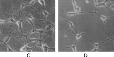
Incubation of isolated human MNCs with rapamycin decreased EPCs’ tube formation capacity (Fig. 2) in a concentration‐dependent manner (Table 1). The low concentration group (0.1 g/ml) were of no significant difference compared to the control group.
Figure 2.

Effects of rapamycin on EPCs’ tube formation. After 24 h, the tubes formed in group 5.0 g/ml rapamycin (D) were seen less than the control group (C) significantly.
Effects of rapamycin on EPCs eNOS
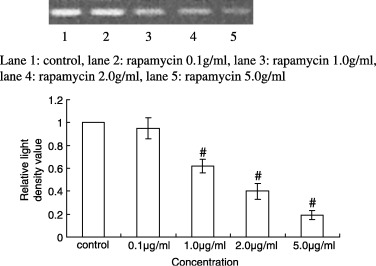
Western blot analysis showed that the expression of eNOS (the relative light density values of control group and rapamycin 0.1, 1.0, 2.0 and 5.0 g/ml were 1.00, 0.95 ± 0.09, 0.62 ± 0.06, 0.40 ± 0.07 and 0.19 ± 0.04, respectively) had decreased significantly in the cells treated with rapamycin in a concentration‐dependent manner (Fig. 3), strongly suggesting that the inhibitory effect of rapamycin is mediatedthrough decreasing eNOS.
Figure 3.

Western blot analysis of eNOS of EPCs by a variety of concentrations of rapamycin. The expression of eNOS was significantly decreased in the cells treated with rapamycin in a concentration‐dependent manner. Compared to control group, #P < 0.01.
DISCUSSION
There is strong evidence that EPCs play a significant role in neovascularization and re‐endothelialization, particularly during ischaemic conditions. More recently, in animals and in human subjects, two groups have documented that EPCs contribute up to 25% of endothelial cells in newly formed vessels (Murayama et al. 2002; Suzuki et al. 2003). Increasing the number of circulating EPCs by transplantation of haematopoietic stem cells or by injection of in vitro differentiated EPCs has been shown to improve neovascularization of ischaemic hindlimbs (Murohara et al. 2000), to accelerate blood flow in diabetic mice (Schatteman et al. 2000) and to improve cardiac function (Kocher et al. 2001). Moreover, Vasa et al. (2001) have recently reported that patients with coronary heart disease (CHD) have reduced levels and functional impairment of EPCs, which correlated with risk factors for CHD. In addition, we have observed previously that hyperhomocysteine, a major risk factor of cardiovascular diseases, induced the reduction of EPCs with decreased functional activity in vitro (Chen et al. 2004). Therefore, the stimulation of mobilization and/or differentiation of EPCs may provide a useful novel therapeutic strategy to improve post‐natal neovascularization and re‐endothelialization in patients with CHD.
RAPA is an important agent used in drug‐eluting stents; it can reduce re‐stenosis significantly. But it also has some serious side‐effects, such as inhibiting re‐endothelialization. In the past, RAPA has been shown to possess anti‐angiogenic effects in mouse models (Guba et al. 2002) and that it could reduce metastatic tumour growth and neo‐angiogenesis (Luan et al. 2003). mTOR is present in freshly isolated CD133+ cells, and in proliferating and differentiating EPCs (Butzal et al. 2004). In our study, we investigated the effects of rapamycin on EPCs. We found it decreased their number, proliferation, migratory, adhesive and tube formation capacities in a concentration‐ and time‐dependent manner. This inhibition was of a similar magnitude as that found for haematopoietic cell lines, especially for the T‐cell line. Because T cells represent the main target of the immunosuppressive effect of RAPA and are believed to be exquisitely sensitive to this agent, it is important to acknowledge that its anti‐angiogenic effects occur in the same dose range.
Besides the inhibition of proliferation and induction of apoptosis, RAPA has pronounced effects on the differentiation of EPCs, resulting in generation of markedly reduced numbers of mature endothelial cells (Butzal et al. 2004). RAPA reduced the expression of endothelial antigens on non‐adherent differentiating EPCs. This may be an effect of gene regulation of both antigens, but more likely it represents a block in differentiation down the endothelial pathway. The population of CD133+ cells contains cells with haemangioblast potential, which may give them the opportunity for endothelial or haematopoietic differentiation (Gehling et al. 2000; Salven et al. 2003; Loges et al. 2004). RAPA may influence the fate of these stem cells and redirect them away from the endothelial pathway. Fukuda et al. (2005) have recently reported that the potent inhibitory effects of RAPA on circulating smooth muscle progenitor cells may mediate the clinical efficacy of the RAPA‐eluting stent; RAPA potentially may affect re‐endothelialization after stent implantation. These results are similar to ours.
RAPA is believed to possess at least two modes of action, exerting its anti‐angiogenic effect. First, it has been shown to reduce secretion of VEGF by tumour cells (Guba et al. 2002). This may be caused by down‐regulation of hypoxia‐inducible factor 1 alpha (HIF‐1α), which is upstream of VEGF expression (Zhong et al. 2000). Because sufficient amounts of exogenous VEGF were present in our medium during proliferation and differentiation of EPCs, this mechanism cannot be responsible for our findings. Second, VEGF receptor signalling may be perturbed by RAPA (Ilan et al. 1998). This mechanism might prevent cell proliferation and differentiation of circulating EPCs and result in a lack of neo‐angiogenesis from bone marrow‐derived cells. Walter et al. (2004) have shown that eNOS mRNA stability and eNOS activity through a PI‐3K/Akt is a common signal transduction pathway in endothelium and EPCs. We also found that rapamycin decreased the eNOS production of EPCs in a concentration‐dependent manner. Thus, we deduce that decreasing eNOS is one of the mechanisms of the inhibitory effect of RAPA on EPCs.
REFERENCES
- Butzal M, Loges S, Schweizer M, Fischer U, Gehling UM, Hossfeld DK, Fiedler W (2004) Rapamycin inhibits proliferation and differentiation of human endothelial progenitor cells in vitro . Exp. Cell Res. 66, 65. [DOI] [PubMed] [Google Scholar]
- Chen JZ, Zhu JH, Wang XX, Zhu JH, Xie XD, Sun J, Shang YP, Guo XG, Dai HM, Hu SJ (2004) Effects of homocysteine on number and activity of endothelial progenitor cells from peripheral blood. J. Mol. Cell Cardiol. 36, 233. [DOI] [PubMed] [Google Scholar]
- Dumont FJ, Staruch MJ, Koprak SL, Melino MR, Sigal NH (1990) Distinct mechanisms of suppression of murine T cell activation by the related macrolides FK‐506 and rapamycin. J. Immunol. 144, 251. [PubMed] [Google Scholar]
- Fukuda D, Sata M, Tanaka K, Nagai R (2005) Potent inhibitory effect of sirolimus on circulating vascular progenitor cells. Circulation 111, 926. [DOI] [PubMed] [Google Scholar]
- Gehling UM, Ergun S, Schumacher U, Wagener C, Pantel K, Otte M, Schuch G, Schafhausen P, Mende T, Kilic N, Kluge K, Schafer B, Hossfeld DK, Fiedler W (2000) In vitro differentiation of endothelial cells from AC133‐positive progenitor cells. Blood 95, 3106. [PubMed] [Google Scholar]
- Guba M, Von Breitenbuch P, Steinbauer M, Koehl G, Flegel S, Hornung M, Bruns CJ, Zuelke C, Farkas S, Anthuber M, Jauch KW, Geissler EK (2002) Rapamycin inhibits primary and metastatic tumor growth by antiangiogenesis: involvement of vascular endothelial growth factor. Nat. Med. 8, 128. [DOI] [PubMed] [Google Scholar]
- Hill JM, Zalos G, Halcox JP, Schenke WH, Waclawiw MA, Quyyumi AA, Finkel T (2003) Circulating endothelial progenitor cells, vascular function, and cardiovascular risk. N. Engl. J. Med. 348, 593. [DOI] [PubMed] [Google Scholar]
- Ilan N, Mahooti S, Madri JA (1998) Distinct signal transduction pathways are utilized during the tube formation and survival phases of in vitro angiogenesis. J. Cell Sci. 111, 3621. [DOI] [PubMed] [Google Scholar]
- Kalka C, Masuda H, Takahashi T, Kalka‐Moll WM, Silver M, Kearney M, Li T, Isner JM, Asahara T (2000) Transplantation of ex vivo expanded endothelial progenitor cells for therapeutic neovascularization. Proc. Natl Acad. Sci. USA 97, 3422. [DOI] [PMC free article] [PubMed] [Google Scholar]
- Kocher AA, Schuster MD, Szabolcs MJ, Takuma S, Burkhoff D, Wang J, Homma S, Edwards NM, Itescu S (2001) Neovascularization of ischemic myocardium by human bone‐marrow‐derived angioblasts prevents cardiomyocyte apoptosis, reduces remodeling and improves cardiac function. Nat. Med. 7, 430. [DOI] [PubMed] [Google Scholar]
- Loges S, Fehse B, Brockmann MA, Lamszus K, Butzal M, Guckenbiehl M, Schuch G, Ergun S, Fischer U, Zander AR, Hossfeld DK, Fiedler W, Gehling UM (2004) Identification of the adult human hemangioblast. Stem Cells Dev. 13, 229. [DOI] [PubMed] [Google Scholar]
- Luan FL, Ding R, Sharma VK, Chon WJ, Lagman M, Suthanthiran M (2003) Rapamycin is an effective inhibitor of human renal cancer metastasis. Kidney Int. 63, 917. [DOI] [PubMed] [Google Scholar]
- Murayama T, Tepper OM, Silver M, Ma H, Losordo DW, Isner JM, Asahara T, Kalka C (2002) Determination of bone marrow‐derived endothelial progenitor cell significance in angiogenic growth factor‐induced neovascularization. Exp. Hematol. 30, 967. [DOI] [PubMed] [Google Scholar]
- Murohara T, Ikeda H, Duan J, Shintani S, Sasaki K, Eguchi H, Onitsuka I, Matsui K, Imaizumi T (2000) Transplanted cord blood‐derived endothelial precursor cells augment postnatal neovascularization. J. Clin. Invest. 105, 1527. [DOI] [PMC free article] [PubMed] [Google Scholar]
- Reyes M, Dudek A, Jahagirdar B, Koodie L, Marker PH, Verfaillie CM (2002) Origin of endothelial progenitors in human postnatal bone marrow. J. Clin. Invest. 109, 337. [DOI] [PMC free article] [PubMed] [Google Scholar]
- Salven P, Mustjoki S, Alitalo R, Alitalo K, Rafii S (2003) VEGFR‐3 and CD133 identify a population of CD34+ lymphatic/vascular endothelial precursor cells. Blood 101, 168. [DOI] [PubMed] [Google Scholar]
- Schatteman GC, Hanlon HD, Jiao C, Dodds SG, Christy BA (2000) Blood‐derived angioblasts accelerate blood‐flow restoration in diabetic mice. J. Clin. Invest. 106, 571. [DOI] [PMC free article] [PubMed] [Google Scholar]
- Sehgal SN (1998) Rapamune (RAPA, rapamycin, sirolimus): mechanism of action immunosuppressive effect results from blockade of signal transduction and inhibition of cell cycle progression. Clin. Biochem. 31, 335. [DOI] [PubMed] [Google Scholar]
- Suzuki T, Nishida M, Futami S, Fukino K, Amaki T, Aizawa K, Chiba S, Hirai H, Maekawa K, Nagai R (2003) Neoendothelialization after peripheral blood stem cell transplantation in humans. A case report of a Tokaimura nuclear accident victim. Cardiovasc. Res. 58, 487. [DOI] [PubMed] [Google Scholar]
- Tepper OM, Galiano RD, Capla JM, Kalka C, Gagne PJ, Jacobowitz GR, Levine JP, Gurtner GC (2002) Human endothelial progenitor cells from type II diabetics exhibit impaired proliferation, adhesion, and incorporation into vascular structures. Circulation 106, 2781. [DOI] [PubMed] [Google Scholar]
- Urbich C, Dimmeler S (2005) Risk factors for coronary artery disease, circulating endothelial progenitor cells, and the role of HMG‐CoA reductase inhibitors. Kidney Int. 67, 1672. [DOI] [PubMed] [Google Scholar]
- Vasa M, Fichtlscherer S, Aicher A, Adler K, Urbich C, Martin H, Zeiher AM, Dimmeler S (2001) Number and migratory activity of circulating endothelial progenitor cells inversely correlate with risk factors for coronary artery disease. Cir. Res. 89, E1. [DOI] [PubMed] [Google Scholar]
- Walter DH, Dimmeler S, Zeiher AM (2004) Effects of statins on endothelium and endothelial progenitor cell recruitment. Semin. Vasc. Med. 4, 385. [DOI] [PubMed] [Google Scholar]
- Walter DH, Rittig K, Bahlmann FH, Kirchmair R, Silver M, Murayama T, Nishimura H, Losordo DW, Asahara T, Isner JM (2002) Statin therapy accelerate reendothelialization: a novel effect involving mobilization and incorporation of bone marrow‐derived endothelial progenitor cells. Circulation 105, 3017. [DOI] [PubMed] [Google Scholar]
- Zhong H, Chiles K, Feldser D, Laughner E, Hanrahan C, Georgescu MM, Simons JW, Semenza GL (2000) Modulation of hypoxia‐inducible factor 1alpha expression by the epidermal growth factor/phosphatidylinositol 3‐kinase/PTEN/AKT/FRAP pathway in human prostate cancer cells: implications for tumor angiogenesis and therapeutics. Cancer Res. 60, 1541. [PubMed] [Google Scholar]


