Opportunistic invaders in all biomes
The Microsporidia are a diverse intracellular parasite phylum infecting everything from single-celled protists to humans in all major global biomes. Its 200 or more known genera are grouped into at least five major clades, with vast numbers of taxa in currently unknown hosts remaining undiscovered [1]. One clade that contains perhaps the most intriguing genera within the phylum, the Enterocytozoon group Microsporidia (EGM), includes parasites that infect the cells (and sometimes the nuclei) of invertebrates and fish hosts from aquatic habitats, terrestrial birds and mammals, and human patients with underlying immune-suppressive conditions such as HIV/AIDS [1]. The EGM was not known prior to the HIV/AIDS pandemic in the 1980s but now contains the most prevalent human microsporidian pathogen, Enterocytozoon bieneusi [2]. It is also associated with recent emergent diseases in domestic and companion animals [3] and outbreaks in intensive rearing operations for marine shrimp [4], fish [5], and other wildlife [6]. Certain members of the group exhibit life cycles that passage between invertebrate and vertebrate hosts [7]. It has been proposed that emergence of disease caused by members of this group over the past 5 decades may be indicative of inherent stressors acting upon those biomes in which their hosts exist. Under these suboptimal conditions, opportunistic infections with enhanced potential to cross host taxonomic boundaries have led to increased prevalence and host range in the EGM [1].
An enigmatic clade
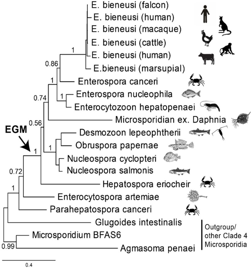
The short subunit (SSU) rRNA gene phylogeny in Fig 1 shows the relationship of E. bieneusi to its closest known relative, Enterospora canceri, a parasite of marine crabs, and all other characterised members of the EGM. Except for E. bieneusi, all members of the EGM are parasites of fish and aquatic invertebrates (including Enterocytospora and Parahepatospora, which do not branch robustly with the rest of EGM and so are not included in its circumscription here). The combined ecological and phylogenetic evidence suggests an aquatic origin of E. bieneusi, which unlike all other members of the clade (so far as is known), is the only EGM lineage to parasitise terrestrial warm-blooded vertebrates. However, it is unknown whether E. bieneusi has aquatic hosts. Notably, unlike other subbranches within microsporidian clade 4, the EGM has rarely been detected in environmental DNA (eDNA) studies [8], suggesting that their life cycles do not include small hosts or host-free stages that are usually sampled from water, sediments, and soils by such studies. Two exceptions to this are the Daphnia-derived lineages shown on Fig 1, but these also apparently have not yet been detected in eDNA studies. Other lineages such as Desmozoon/Paranucleospora are known to cycle between vertebrate and invertebrate hosts and could potentially be detected in eDNA studies [8]. It is possible that broadly targeted microsporidian-specific primers bias against detection of EGM; the use of clade-specific primers will mitigate against this. The question of when and how E. bieneusi effected the transition to terrestrial homeotherms can be addressed by further sampling of putative hosts and eDNA probing of relevant environments. It is important to distinguish between evolutionary and ecological transitions. In evolutionary terms, other currently unknown lineages may be discovered with a closer sister relationship to E. bieneusi than E. canceri. The host affiliation(s) and habitat of these lineages, if they exist, will be informative about evolutionary intermediates between the marine invertebrate–infecting E. canceri and E. bieneusi. An ecological approach would determine whether E. bieneusi is restricted to vertebrate hosts in nonmarine habitats, whether it infects marine mammals, and from a different perspective, whether E. canceri can also infect vertebrates or nonmarine animals. Parasite life cycles can also be investigated with eDNA methods, screening both potential hosts and environmental samples for insight into alternative hosts, vectors, and reservoirs. Freshwater and terrestrial habitats have been undersampled for Microsporidia by these approaches compared with marine systems.
Fig 1. Phylogeny and host range of known members of the EGM.

Except for E. bieneusi infecting humans, terrestrial mammals, and birds, all other members of the EGM infect aquatic hosts. Erection of the genus Enterocytozoon coincided with emergence of its type species E. bieneusi during the HIV/AIDS pandemic in the 1980s. Subsequent description of proposed cogeneric EGM genera (Nucleospora, Paranucleospora/Desmozoon, Enterospora, Hepatospora, Obruspora) and closely related genera (e.g., Enterocytospora and Parahepatospora) have occurred over the past 3 decades. In most cases, emergence has been reported in domesticated terrestrial and aquatic food animals. However, certain EGM and related taxa have also been described infecting wild-animal hosts (e.g., foxes, primates, crabs), some of which reside in invasive populations outside of their native ranges. In at least one case (Paranucleospora/Desmozoon), the parasite is known to cycle between an invertebrate (copepod crustacean) and fish (salmon) host, raising the prospect of the capacity for trophic transfer in other members of the EGM. The Bayesian phylogenetic analysis was conducted using MrBayes version 3.2.5 [27]. The evolutionary model applied included a GTR substitution matrix, a four-category autocorrelated gamma correction, and the covarion model for 4 million generations, 1 million of which were discarded as burn-in. EGM, Enterocytozoon group Microsporidia; GTR, generalized time reversible.
Deep obligates
Microsporidia are often used as examples of extreme metabolic streamlining in the eukaryotes. No other known eukaryotic group has a smaller number of protein-coding genes or a higher level of metabolic dependency on a host. This streamlining likely occurred in the ancestor of the Microsporidia after the divergence of the main group of microsporidia away from early-branching ‘intermediate’ Microsporidia such as Nucleophaga, Paramicrosporidium, and Mitosporidium [9]. Large numbers of genes were lost early in microsporidian evolution, followed by lineage-specific duplication of transporters and expansion of gene families according to the needs of the parasites and their relationships with their host [10]. Perhaps the most archetypal metabolic losses in the phylum are those of the tricarboxylic acid and electron transport chain and the ability to produce ATP in the mitochondria. This was potentially facilitated by the acquisition of ATP transporter by lateral gene transfer to uptake ATP and other nucleotides from the host [11]; however, in the spore stage, there is no access to host ATP, so here, glycolysis is the key pathway for intrinsic energy generation. The EGM has taken microsporidian reductionism a step further and no longer encodes the genes for glycolysis, making them utterly reliant on their hosts for energy [12]. Multiple genomes from the EGM have now been sequenced, and none of them encodes a functional glycolytic pathway, though curiously, each lineage retains genes for different parts of the pathway [13]. Nor do they have the genes for the pentose phosphate pathway or trehalose metabolism, and the capacity to metabolise fatty acids is severely reduced compared with other Microsporidia. What is different about EGM compared with other Microsporidia that may have allowed this metabolic degeneration? They are distinctive in their relationship to the host, either closely abutting the host nucleus or even exclusively living within it [14]. It might be intuitive to think that development in the nucleus eliminates access to the host mitochondria and thus a source of ATP (many Microsporidia gather host mitochondria around their cell); however, ATP levels are not thought to differ between nucleus and cytoplasm, suggesting neither advantage nor disadvantage to nuclear localisation with regard to energy acquisition [15]. However, several other microbes that are also energy parasites live within host nuclei [16]. Regardless of which selection pressures led to the loss of the glycolytic pathway in the EGM, it still leaves this group with the problem of not being able to generate/acquire energy in the spore stage, meaning that they do not have immediate access to ATP for the potentially energetic process of spore germination. To overcome this, future research will tell us whether they are able to either store ATP in the dormant spore, acquire ATP from the host as an endocytosed spore prior to germination, or encode a yet-unknown mechanism of generation of ATP from a storage product.
Infection, disease, and host outcomes
Without exception, infection by members of the EGM is confined to the gastrointestinal tract and directly associated organs of their hosts. In humans, E. bieneusi infects the enterocytes of the superficial lining, rarely disseminating to fibrovascular tissues of the laminar propria [17], leading to self-limiting or persistent diarrhoea and wasting in immune-competent and immune-compromised patients, respectively [18]. Similarly, for fish, Enterospora nucleophila infects enterocytes and rodlet cells of the intestine, causing lethargy, stunting, and cachexia, resulting in emaciation [19]. In shrimp, Enterocytozoon hepatopenaei infects the epithelial cells of the hepatopancreas (a digestive organ associated with the gut), where it associates with a slow-growth syndrome [4], whereas in crabs, E. canceri infects the same cell type but resides almost exclusively within the nucleoplasm of host cells [14]. In all cases, systemic infection of other organ and tissue systems is not observed, suggesting that autoinfection (cell-to-cell within the gut) and transmission (via faeces) underpin the high prevalence observed in susceptible human and animal populations, as well as the role of food and water in spread [1]. Recent studies that challenge the concept that human enterocytes are nonphagocytic [20] coupled with those demonstrating potential for phagocytic uptake of some Microsporidia prior to their translocation across the cytoplasm in vacuoles [21] may indicate that EGM can be translocated by this mechanism into host cells. If so, this simple strategy, leading to infection of the primary contact layer within the host gut, could indicate almost sole reliance on the metabolic conditions therein to support their own spore germination and replication [13]. A key question relates to why exploitation of these apparently easy targets primarily occurs when these diverse hosts animals are immunocompromised.
One health sentinels
The universal emergence of EGM is suggestive of multifactorial intestinal barrier dysfunction across susceptible host groups. Epithelial cells of the intestine form both metabolic and physical barriers to invasion by microbes and maintain an immunoregulatory function by influencing stasis of gut mucosal cells [22]. Direct disruption in this function can occur via diverse physical and psychological stressors [23] and, during ageing [24], lead to a multitude of disease outcomes in the host [25]. The infection of gut epithelial cells by EGM may then occur because of underlying immune, metabolic, or microbiological disruption (as proposed for co-incidence of E. bieneusi with immune-compromised human hosts [1]). In addition, infection with EGM may predispose hosts to direct and indirect effects of coinfection with other pathogens (e.g., infection with E. hepatopenaei makes shrimp more susceptible to effects of the bacterial agent of acute hepatopancreatic necrosis disease [AHPND] [26]). We have previously proposed that, for these reasons, opportunistic microsporidian parasites, epitomised by EGM, are living sentinels of host immune competence that traverse both host taxonomy and the biomes in which these hosts reside [1]. We predict that an increasing prevalence of immunosuppression in hosts from diverse systems—and driven by shifting demographic, environmental, pathophysiological, and psychological forces—will underpin further emergence of EGM in human and animal hosts. Understanding this emergence in the context of wider intestinal barrier dysfunction will be an inevitable focus of future research.
Funding Statement
This work was supported by Defra project FB002 and British Council Newton Fund Institutional Links Project C7104 and Newton Fund Prize project C7724 (to GDS, DB) and by Cefas and the University of Exeter under the auspices of the Centre for Sustainable Aquaculture Futures (to BAPW, DB, and GDS). The funders had no role in study design, data collection and analysis, decision to publish, or preparation of the manuscript.
References
- 1.Stentiford GD, Becnel J, Weiss L, Keeling P, Didier E, Williams B, et al. Microsporidia–Emergent Pathogens in the Global Food Chain. Trends Parasitol. 2016; 32: 336–348. 10.1016/j.pt.2015.12.004 [DOI] [PMC free article] [PubMed] [Google Scholar]
- 2.Desportes I, Le Charpentier Y, Galian A, Bernard F, Cochand-Priollet B, Lavergne A, et al. Occurrence of a new microsporidan: Enterocytozoon bieneusi n.g., n. sp., in the enterocytes of a human patient with AIDS. J Protozool. 1985; 32: 250–254. [DOI] [PubMed] [Google Scholar]
- 3.Santin M, Fayer R. A longitudinal study of Enterocytozoon bieneusi in dairy cattle. Parasitol. Res. 2009; 105: 141–144. 10.1007/s00436-009-1374-4 [DOI] [PubMed] [Google Scholar]
- 4.Tourtip S, Wongtripop S, Sritunyalucksana K, Stentiford GD, Bateman KS, Sriurairatana S, et al. Enterocytozoon hepatopenaei sp. nov. (Microspora: Enterocytozoonidae), a parasite of the black tiger shrimp Penaeus monodon (Decapoda: Penaeidae): fine structure and phylogenetic relationships. J Invertebr Pathol. 2009;102: 21–29. 10.1016/j.jip.2009.06.004 [DOI] [PubMed] [Google Scholar]
- 5.Palenzuela O, Redondo MJ, Cali A, Takvorian PM, Alonso-Naveiro M, Alvarez-Pellitero P, et al. A new intranuclear microsporidium, Enterospora nucleophila n. sp., causing an emaciative syndrome in a piscine host (Sparus aurata), prompts the redescription of the family Enterocytozoonidae. Internat J Parasitol. 2014; 44: 189–203. [DOI] [PubMed] [Google Scholar]
- 6.Fayer R, Santin-Duran M. Epidemiology of Microsporidia in Human Infections In: Weiss L, Becnel JJ, editors. Microsporidia: Pathogens of Opportunity. 1st ed. Oxford: John Wiley & Sons; 2014. p. 135–164. [Google Scholar]
- 7.Nylund S, Nylund A, Watanabe K, Arnesen CE, Karlsbakk E. Paranucleospora theridion n. gen., n. sp. (Microsporidia, Enterocytozoonidae) with a life cycle in the salmon louse (Lepeophtheirus salmonis, Copepoda) and Atlantic salmon (Salmo salar). J Eukaryot Microbiol. 2010; 57: 95–114. 10.1111/j.1550-7408.2009.00451.x [DOI] [PubMed] [Google Scholar]
- 8.Williams BAP, Hamilton KM, Jones MD, Bass D. Group-specific environmental sequencing reveals high levels of ecological heterogeneity across the microsporidian radiation. Environ Microbiol Rep. 2018; 10: 328–336. 10.1111/1758-2229.12642 [DOI] [PMC free article] [PubMed] [Google Scholar]
- 9.Quandt CA, Beaudet D, Corsaro D, Walochnik J, Michel R., Corradi N et al. The genome of an intranuclear parasite, Paramicrosporidium saccamoebae, reveals alternative adaptations to obligate intracellular parasitism. Elife. 2017; 6: e29594 10.7554/eLife.29594 [DOI] [PMC free article] [PubMed] [Google Scholar]
- 10.Nakjang S, Williams TA, Heinz E, Watson AK, Foster PG, Sendra KM, et al. Reduction and expansion in microsporidian genome evolution: new insights from comparative genomics. Genom Biol Evolut. 2013; 5: 2285–303. [DOI] [PMC free article] [PubMed] [Google Scholar]
- 11.Dean P, Hirt RP, Embley TM. Microsporidia: Why make nucleotides if you can steal them? PLoS Pathog. 2016; 12 (11): e1005870 10.1371/journal.ppat.1005870 [DOI] [PMC free article] [PubMed] [Google Scholar]
- 12.Keeling PJ, Corradi N, Morrison HG, Haag KL, Ebert D, Weiss LM et al. The reduced genome of the parasitic microsporidian Enterocytozoon bieneusi lacks genes for core carbon metabolism. Genom Biol Evolut. 2010; 2: 304–9. [DOI] [PMC free article] [PubMed] [Google Scholar]
- 13.Wiredu Boakye D, Jaroenlak P, Prachumwat A, Williams TA, Bateman KS, Itsathitphaisarn O, et al. Decay of the glycolytic pathway and adaptation to intranuclear parasitism within Enterocytozoonidae microsporidia. Environ Microbiol. 2017; 19(5): 2077–2089. 10.1111/1462-2920.13734 [DOI] [PubMed] [Google Scholar]
- 14.Stentiford GD, Bateman KS, Feist SW. Enterospora canceri n.gen., n.sp., an intranuclear microsporidian infecting European edible crab (Cancer pagurus). Dis Aquat Org. 2007; 75: 61–72. 10.3354/dao075061 [DOI] [PubMed] [Google Scholar]
- 15.Imamura H, Nhat KP, Togawa H, Saito K, Iino R, Kato-Yamada Y, et al. Visualization of ATP levels inside single living cells with fluorescence resonance energy transfer-based genetically encoded indicators. Proc Nat Acad Sci U S A. 2009; 106: 15651–6. [DOI] [PMC free article] [PubMed] [Google Scholar]
- 16.Schulz F, Horn M. Intranuclear bacteria: inside the cellular control center of eukaryotes. Trends Cell Biol. 2015; 25: 339–46. 10.1016/j.tcb.2015.01.002 [DOI] [PubMed] [Google Scholar]
- 17.Orenstein JM, Tenner M, Kotler DP. Localization of infection by the microsporidian Enterocytozoon bieneusi in the gastrointestinal tract of AIDS patients with diarrhea. AIDS. 1992; 6: 195–197. [DOI] [PubMed] [Google Scholar]
- 18.Weiss LM. Clinical syndromes associated with microsporidiosis In: Weiss LM, Becnel JJ, editors. Microsporidia: Pathogens of Opportunity. 1st ed. Oxford: John Wiley & Sons; 2014. p. 371–401. [Google Scholar]
- 19.Palenzuela O, Redondo MJ, Cali A, Takvorian PM, Alonso-Naveiro M, Alvarez-Pellitero P, et al. A new intranuclear microsporidium, Enterospora nucleophila n. sp., causing an emaciative syndrome in a piscine host (Sparus aurata), prompts the redescription of the family Enterocytozoonidae. Internat J Parasitol. 2014; 44: 189–203. [DOI] [PubMed] [Google Scholar]
- 20.Dean P, Quitard S, Bulmer DM, Roe AJ, Kenny B. Cultured enterocytes internalise bacteria across their basolateral surface for, pathogen-inhibitable, trafficking to the apical compartment. Scientif Rep. 2015: 5: 17359. [DOI] [PMC free article] [PubMed] [Google Scholar]
- 21.Scheid P. Mechanism of intrusion of a microspordian-like organism into the nucleus of host amoebae (Vannella sp.) isolated from a keratitis patient. Parasitol Res. 2007; 101: 1097–102. 10.1007/s00436-007-0590-z [DOI] [PubMed] [Google Scholar]
- 22.Peterson LW, Artis D. Intestinal epithelial cells: regulators of barrier function and immune homeostasis. Nat Rev Immunol. 2014; 14: 141–153. 10.1038/nri3608 [DOI] [PubMed] [Google Scholar]
- 23.Söderholm JD, Perdue MH. Stress and the gastrointestinal tract. II. Stress and intestinal barrier function. Am J Physiol Gastrointest Liver Physiol. 2001; 280: G7–G13. 10.1152/ajpgi.2001.280.1.G7 [DOI] [PubMed] [Google Scholar]
- 24.Hu DJK, Jasper H. Epithelia: understanding the cell biology of intestinal barrier dysfunction. Curr Biol. 2017; 27: R185–R187. 10.1016/j.cub.2017.01.035 [DOI] [PubMed] [Google Scholar]
- 25.König J, Wells J, Cani PD, García-Ródenas CL, MacDonald T, Mercenier A, et al. Human intestinal barrier function in health and disease. Clin Translat Grastroenterol. 2016; 7: e196. [DOI] [PMC free article] [PubMed] [Google Scholar]
- 26.Aranguren LF, Han JE, Tang KFJ. Enterocytozoon hepatopenaei (EHP) is a risk factor for acute hepatopancreatic necrosis disease (AHPND) and septic hepatopancreatic necrosis (SHPN) in the Pacific white shrimp Penaeus vannamei. Aquacult. 2017; 471: 37–42. [Google Scholar]
- 27.Ronquist F, Teslenko M, Van Der Mark P, Ayres DL, Darling A, Hoehna S, et al. Mrbayes 3.2: Efficient bayesian phylogenetic inference and model choice across a large model space. Systemat Biol. 2012; 61: 539–542. [DOI] [PMC free article] [PubMed] [Google Scholar]


