Abstract
Background and aims Current endoscopic methods of biliary decompression in malignant pancreatic neoplasms are often limited by anatomical and technical challenges. In this case series, we report our experience with endoscopic ultrasound (EUS)-guided placement of an electrocautery-enhanced, lumen-apposing self-expandable metallic stent (LAMS) via transmural gallbladder drainage.
Methods This is a retrospective case series of nine patients (five male, mean age 63.1 years) who underwent EUS-guided LAMS placement for malignant, obstructive jaundice in the pancreatic head. All nine cases were performed by an experienced interventional endoscopist at a single, tertiary medical center. We review the technical and clinical success rates as well as the incidence of procedural adverse events across the nine patients.
Results LAMS placement was technically successful in all cases and there were no procedural adverse events. Seven of nine (77.78 %) patients showed clinical and laboratory improvement immediately following the procedure. One case required re-intervention with interventional radiology guided biliary drain placement. The mean fluoroscopy time was 1.02 minutes.
Conclusions EUS-guided LAMS placement for transmural gallbladder drainage in malignant obstruction appears to be a safe and effective technique, allowing patients to proceed to surgery, chemotherapy, or hospice care.
Introduction
Malignant neoplasms of the pancreatic head often present with clinical and technical challenges. Not infrequently, patients require upfront chemotherapy, either in preparation for surgery or for palliative intent. Therefore, patients often require biliary drainage through established modalities such as endoscopic retrograde cholangiopancreatography (ERCP). However, ERCP may occasionally fail due to anatomical and technical challenges, for example, in cases of inaccessible ampulla 1 . Traditionally, alternative minimally invasive methods for biliary drainage include percutaneous transhepatic cholangiogram (PTC) placed by interventional radiology (IR). Though effective, percutaneous biliary drainage has been associated with adverse events such as biliary peritonitis, pneumothorax, tube dislodgement, patient discomfort, need for ongoing drain care, as well as restrictions on bathing and swimming 2 .
Recently, endoscopic ultrasound (EUS)-guided biliary drainage techniques have been found to be effective in biliary decompression after unsuccessful ERCP 3 . However, techniques such as EUS rendezvous and antegrade stenting are still limited by the need to advance a guidewire through a distal bile duct that may be completely occluded by an obstructive mass. EUS transluminal drainage including choledochoduodenostomy (EUS-CDS) and hepaticogastrostomy (EUS-HGS) are multi-stage procedures and result in the creation of a permanent fistula – despite ongoing technique refinements, EUS-CDS as well as EUS-HGS using conventional techniques carry a risk of stent dislodgement and bile peritonitis. Novel single-stage EUS-guided techniques may avoid these adverse events and provide a more effective means of biliary decompression.
Currently, there are limited data on the efficacy and outcomes of EUS-guided transmural gallbladder drainage, specifically for biliary drainage in the setting of malignant distal biliary obstruction. Recent studies have found that EUS-guided placement of a lumen-apposing self-expandable metallic stent (LAMS) is technically feasible, and a safe and effective alternative in the treatment of acute cholecystitis in patients unsuitable for surgery or IR drain placement 4 5 . We report our experience with LAMS placement for retrograde biliary drainage via the gallbladder (either cholecystoduodenostomy or cholecystogastrostomy) in a case series of patients presenting with malignant obstruction requiring biliary decompression.
Methods
Supplementary Video 1 Table-top depiction of stent deployment.
Supplementary Video 2 Stent deployment method. Step 1: Unlock locking catheter, tent mucosa. Step 2: Apply electrocautery, enter gallbladder (stage 1). Step 3: Lock locking catheter. Step 4: Unlock locking catheter and deployment hub (stage 2) to deploy stent without “jumping forward”. Step 5: Retract catheter, tent internal flange, lock catheter lock (stage 3). Step 6: Unlock deployment hub, finish stent deployment inside scope (stage 4). Step 7: Unlock locking catheter, simultaneously back away EUS scope while pushing stent out of the channel.
This is a retrospective case series from October 2016 to December 2017. Nine patients underwent EUS-guided LAMS placement with transgastric/transduodenal gallbladder drainage for malignant obstructive jaundice in the pancreatic head. The patients were initially referred from both inpatient and outpatient settings for endoscopic attempt at biliary drainage. All nine patients had native pancreaticobiliary anatomy (i. e. no prior history of cholecystectomy nor biliary surgeries). At the time of initial consent for traditional ERCP, patients were also consented for the possible off-label use of a LAMS for biliary decompression. All procedures were performed by the same interventional endoscopist (K.K.) at Kaiser Permanente Los Angeles Medical Center, a tertiary care referral center for the Southern California region of the Kaiser Foundation Health Plan.
The Olympus UCT-140 curvilinear echoendoscope (Olympus Corp., Center Valley, Pennsylvania, United States) and electrocautery-enhanced AXIOS stent (Boston Scientific Co., Natick, Massachusetts, United States) were used in all procedures. The AXIOS stent is an electrocautery-enhanced LAMS, which consists of a fully covered, nitinol, braided stent with a “dumbbell” configuration and bilateral anchor flanges that appose two lumens and minimize the chance of stent migration ( Fig. 1 ).
Fig. 1.
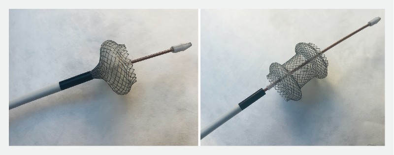
Representative images of the 9F electrocautery-enhanced lumen apposing metal stent.
Cystic duct patency, which is a key requirement of this technique, was confirmed doubly. First, during pre-procedure planning, there was meticulous review of cross-sectional imaging (including magnetic resonance cholangiopancreatography (MRCP) which was available in 8/9 patients) demonstrating the presence of a distended, hydropic gallbladder, intrahepatic biliary dilation, as well as obstructing mass isolated only to the pancreatic head. Second, when the decision was made to proceed with EUS-guided LAMS placement, the gallbladder and cystic duct takeoff were interrogated in detail with EUS to confirm the absence of obvious stones or obstructing masses. To reduce the potential of bile leak or otherwise technical failure during device exchange, the senior author decided against pre-puncture and contrast instillation with a fine needle aspiration (FNA) needle before LAMS placement. To maximize patient safety, all procedures were performed under general endotracheal anesthesia (GETA). All lumen apposing stents were placed via the freehand technique.
The method of stent placement was modified depending on the degree of gallbladder distension. If the ideal short axis diameter for all cases (linear distance between the transducer and the distal gallbladder wall) was greater than 3.5 cm, then stent deployment would proceed in standard fashion. However, if the short axis diameter were less than 3.5 cm, the stent deployment method would be modified to allow partial pre-deployment and prevent excess deployment against the distal gallbladder wall ( Fig. 2 – 5 and Supplementary Videos 1 and 2 ). Due to the nature of the disease (pancreatic adenocarcinoma), in all cases, the LAMS were either left in place for long-term biliary drainage, or removed as part of the surgical explant.
Fig. 2.
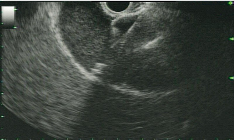
Initial EUS-guided stent deployment into gallbladder.
Fig. 3.
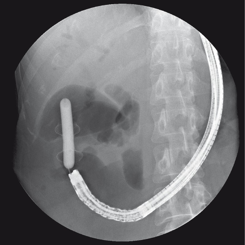
Dilation of lumen apposing metal stent (LAMS) to 15 mm.
Fig. 4.
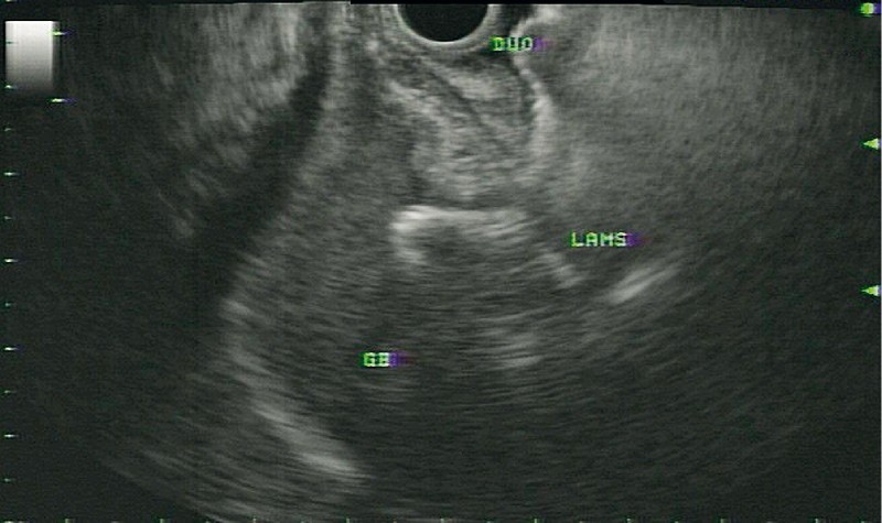
EUS image of lumen apposing metal stent (LAMS) from gallbladder to duodenum.
Fig. 5.
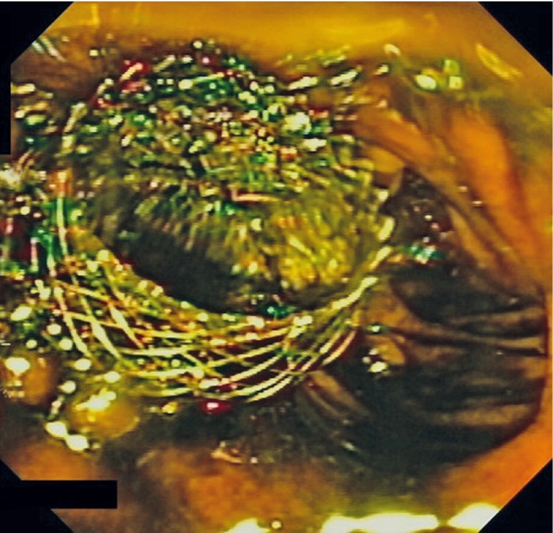
Endoscopic view of lumen apposing metal stent (LAMS) in duodenum.
The primary outcomes of interest included technical and clinical success, and the incidence of procedural adverse events. Secondary outcomes included fluoroscopy time and whether EUS rendezvous was previously attempted. Technical success was defined as successful stent placement in accessing and draining the gallbladder. Clinical success was defined as symptom and post-procedural liver chemistry improvement. The patients were followed for post-procedure adverse events including stent migration, perforation, and stent occlusion requiring re-intervention within a 30-day period. All patients were censored at time of last follow-up within the integrated health system or at study end (January 15, 2018).
The data were analyzed using descriptive statistics to calculate mean values for the primary outcomes. This study was approved by the Institutional Review Board of Kaiser Permanente Southern California (reference number 023563).
Results
Clinical characteristics
A total of nine patients (four women, five men, mean age 63.1 years) underwent LAMS placement for gallbladder drainage in obstructive jaundice ( Table 1 ). All patients were found to have tissue-proven pancreatic ductal adenocarcinoma. During the study period, six of nine (66.7 %) patients underwent chemotherapy and two of nine (22.2 %) patients underwent surgical resection during the study period. One patient had undergone a loop gastrojejunostomy for gastric outlet obstruction due to duodenal extension of pancreatic cancer before index endoscopic procedure and another patient underwent pancreaticoduodenectomy after the end of our study period. Three of nine (33.3 %) patients elected to go on hospice care with no cancer directed therapy.
Table 1. Baseline characteristics of patients in study.
| Characteristic | Number |
| Number of patients | 9 |
| Sex (M/F) | 5/4 |
| Mean age (range), years | 63.1 (41 – 80) |
| Reason for referral – obstructive jaundice | 9 (100 %) |
| Pathology with pancreatic ductal adenocarcinoma | 9 (100 %) |
| Duodenal obstruction | 2 (22.2 %) |
| Chemotherapy | 6 (66.7 %) |
| Surgery post biliary decompression | 2 (22.2 %) during study period |
| Hospice | 3 (33.3 %) |
| Deaths due to underlying disease progression | 4 (44.4 %) |
Endoscopic features
In four of nine (44.44 %) patients, ERCP with EUS rendezvous was initially attempted (transduodenal antegrade wire advancement to the ampulla), but was unsuccessful due to suboptimal scope position, abnormal duodenal anatomy due to obstructing pancreatic head mass, or inability to advance a guidewire antegrade through the ampullary orifice into the duodenum. In the remaining patients (five of nine, 55.6 %), traditional ERCP with EUS rendezvous was not attempted, and instead LAMS placement was the primary modality of choice due to the presence of a markedly dilated biliary system with hydropic gallbladder ( Fig. 6 ) and anticipated difficulties with traditional methods (two referrals for repeated ERCP failures, two for duodenal obstruction, and one for attempted but unsuccessful standard ERCP by the senior author). In one patient, there was a stricture at the juncture of the duodenal bulb and second portion that first required placement of a Wallflex duodenal stent (22 mm × 60 mm, Boston Scientific Co). In another case, there was an incomplete obstruction of the duodenal sweep that prevented adequate duodenoscope advancement past the duodenal bulb and subsequently required Wallflex duodenal stent placement at a repeat endoscopy 6 months later. In these cases, single stage biliary decompression was preferred to decrease the risk of iatrogenic duodenal stent migration and the technical difficulty of traditional ERCP.
Fig. 6.
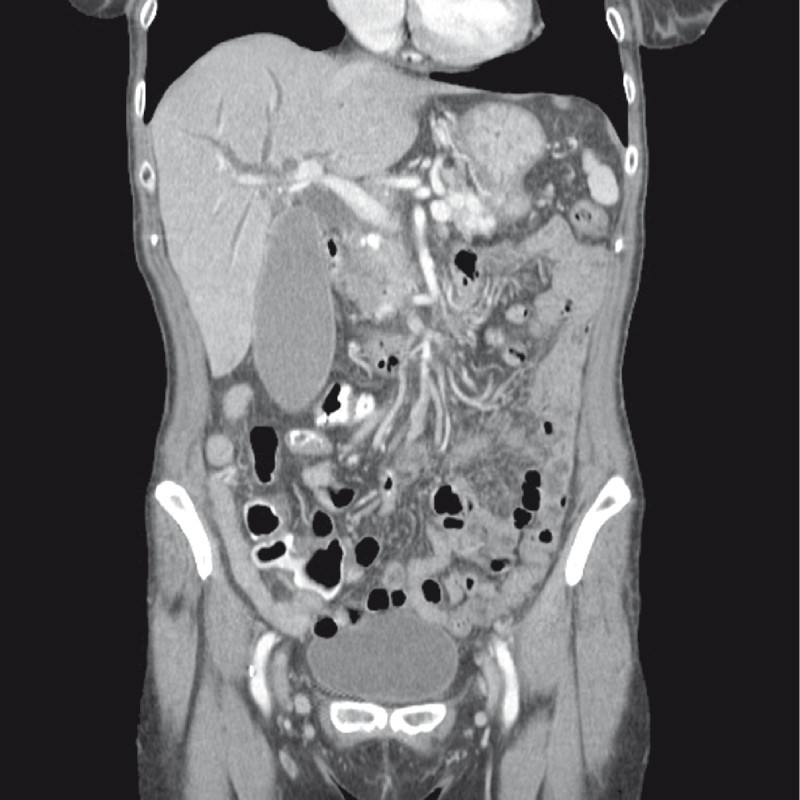
A representative patient with a pancreatic head mass resulting in a hydropic gallbladder.
Technical efficacy and clinical outcomes
Placement of the LAMS was technically successful in all nine patients. Either a transduodenal or transgastric approach was used depending on where the largest window of gallbladder access was identified. A 15 mm × 10 mm LAMS was placed in most of the patients (six of nine, 66.7 %); in three of nine (33.3 %) patients, a double pigtail stent was placed coaxially if there were concerns about either tissue overgrowth or stent embedment that would prevent future stent removal if needed ( Table 2 ). The LAMS was placed from the duodenal bulb to gallbladder in five of nine (55.6 %) patients and from the gastric antrum to gallbladder in four of nine (44.4 %) patients. Mean fluoroscopy time was 1.02 minutes.
Table 2. Interventions and outcomes for each patient in the study.
| Age/sex | LAMS, mm | Coaxial stent (double pigtail plastic stent through LAMS) | Access route | Duodenal obstruction | EUS rendezvous attempted | Fluoroscopy time, min | Complications | Technical success | Clinical success |
| 73 F | 15 × 10 | No | Transgastric | No | No | 0.5 | None | Yes | Lost to follow-up |
| 57 F | 15 × 10 | Yes | Transgastric | Partial obstruction | No | 0.17 | None | Yes | Yes |
| 68 F | 10 × 10 | No | Transduodenal | No | Yes | 0.97 | None | Yes | Yes |
| 48 M | 15 × 10 | No | Transduodenal | No | No | 0.15 | None | Yes | Yes |
| 57 M | 15 × 10 | No | Transgastric | Prior loop gastrojejunostomy | No | 1.78 | None | Yes | Yes |
| 41 F | 10 × 10 | No | Transduodenal | No | Yes | 1.63 | None | Yes | Required IR drain |
| 76 M | 15 × 10 | Yes | Transduodenal | No | Yes | 1.55 | None | Yes | Yes |
| 80 M | 10 × 10 | No | Transduodenal | No | Yes | 0.18 | None | Yes | Yes |
| 68 M | 15 × 10 | Yes | Transgastric | Duodenal stricture requiring stent | No | 2.25 | None | Yes | Yes |
EUS, endoscopic ultrasound; IR, interventional radiology; LAMS, lumen-apposing self-expandable metallic stent.
There were no procedural adverse events. Seven of nine (77.8 %) patients showed clinical improvement following the procedure with improvement in symptoms and immediate improvement in liver enzymes on repeat blood work done within a week of the procedure. One patient presented with a persistently elevated bilirubin level and had ongoing abdominal pain despite LAMS placement, requiring an IR percutaneous transhepatic cholangiogram and external biliary drainage catheter placement within 2 weeks of LAMS placement. The final patient elected to proceed with hospice care and did not have follow-up lab evaluation. Another patient improved clinically after the initial LAMS placement for biliary decompression, but developed recurrent biliary obstruction with progressive disease 7 months after the index procedure, necessitating repeat endoscopic intervention with choledochoduodenostomy. She again demonstrated improvement in symptoms and liver enzymes after the repeat procedure.
There was no need for any endoscopic re-intervention due to stent malfunction in any of the cases. To date, no stents required endoscopic removal due to malfunction. Of note, two stents were uneventfully removed as part of the surgical explant specimen in two patients who underwent a pancreaticoduodenectomy. The average time of follow-up was 130.7 days with a mortality rate of four of nine (44.44 %) due to underlying disease progression.
Discussion
In this retrospective case series, we found that EUS-guided LAMS placement for transmural gallbladder drainage in malignant obstruction was technically successful and improved clinical outcomes, either as a temporizing measure to facilitate neoadjuvant chemotherapy or as a definitive, palliative measure. Of the six patients who sought full cancer therapy post biliary decompression, five were able to proceed with planned chemotherapy with LAMS placement alone. Only in one case was there a need to undergo re-intervention with IR percutaneous transhepatic drain placement within 2 weeks of LAMS placement. However, recurrent biliary obstruction may also have been related to the aggressive nature of that patient’s pancreatic ductal adenocarcinoma.
This procedure was technically successful in all nine cases, including four of nine cases where EUS rendezvous was initially attempted but unsuccessful. There were no procedural adverse events or stent malfunctions that required re-intervention. In one case of eventual tumor progression leading to recurrent obstructive jaundice, repeat endoscopic intervention with EUS-guided choledochoduodenostomy using LAMS placement also proved to be technically and clinically successful. Additionally, the mean fluoroscopy time of 1.02 minutes is significantly lower than published fluoroscopy times in ERCP cases with failed biliary cannulation 6 . This reduction in fluoroscopy time is an important safety consideration to the interventional endoscopist as well as the nursing team, since radiation exposure is cumulative over a lifetime [ALARA (As Low As Reasonably Achievable) principle].
Current methods of EUS-guided biliary drainage include rendezvous, transduodenal choledochoduodenostomy, and antegrade techniques. EUS rendezvous techniques have a reported overall success rate of 81 % with a complication rate of 10 % 7 . EUS transluminal biliary drainage via CDS or HGS has higher success rates, up to 84 – 94 % depending on the access route 3 8 . However, techniques such as EUS-CDS or HGS have been associated with up to 23 % risk of adverse events such as bile peritonitis, pneumoperitoneum, hemobilia, cholangitis, or stent migration 9 10 . There is also a higher risk of stent migration with the use of traditional plastic stents or covered self-expandable metallic stents 3 . Bile leakage in these cases may be a result of the prolonged multi-staged procedure including bile duct puncture, fistula creation and dilation, and stent deployment.
For these reasons, we believe that, in the properly selected patient, cholecystoduodenostomy through a single-stage, electrocautery enhanced EUS-guided LAMS may be a useful adjunct to alternative endoscopic methods of non-ampullary biliary drainage (choledochoduodenostomy and hepaticogastrostomy) and offer key efficiency and safety advantages. First, EUS-guided LAMS placement can be done as a single-stage procedure by utilizing the cautery-enhanced tip of the LAMS, which allows transmural gallbladder penetration and stent deployment to be completed in one step, without the need for multi-step fistulous tract dilation. This allows for a shorter procedure that not only decreases fluoroscopy time, but also minimizes the risk of bile leak leading to peritonitis. Second, because the LAMS design facilitates rapid lumen apposition, it may be a technically feasible option in patients with malignant ascites. Third, the gallbladder and gastrointestinal lumen are both capacious enough to accommodate a 24-mm inner and outer flange, thereby reducing the risk of variable biliary obstruction from stent-duct size mismatch and possible cholangitis. Fourth, in the absence of cholecystitis, up to 75 % of hepatic bile flow typically enters the gallbladder, and thus, we believe this endoscopic drainage technique can more closely mimic normal enterohepatic bile circulation 11 . In fact, this may explain why patients with malignant distal biliary obstruction often present with markedly hydropic gallbladders, the diameter of which is typically much larger than the bile duct upstream from the obstruction.
Although a cholecystoduodenostomy/cholecystogastrostomy may result in a slightly more challenging pancreaticoduodenectomy dissection due to adhesions, the same can occur with a choledochoduodenostomy as well as hepaticogastrostomy. With proper pre-procedure planning, the LAMS can be removed as part of the en bloc surgical explant without additional intervention on the part of the surgeon, as was seen in two of nine patients in this case series. In fact, this method of transluminal endoscopic drainage may be preferable as the stent is fully contained within the explanted specimen, thereby reducing the likelihood of surgeon injury that may result from inadvertently transecting the indwelling metal stent. Finally, deploying a fully covered LAMS preserves all options, including stent removal at a later date if a patient is deemed inoperable despite neoadjuvant chemotherapy.
Although historically there was limited data on the efficacy of EUS-guided transmural gallbladder drainage in malignant obstruction, there is increasing awareness of this technique’s utility in biliary decompression for malignant obstruction. A retrospective review of 12 patients with obstructive jaundice demonstrated the technical and clinical success of EUS-guided gallbladder drainage using an electrocautery-unenhanced LAMS in cases of failed ERCP due to malignant distal biliary obstruction 12 . In that series, the technical and clinical success rates were 100 % and 92 %, respectively; the 16.7 % adverse event rate (2/12) was similar to previously published data on EUS-CDS/EUS-HGS. We believe that the electrocautery enhancement to the LAMS delivery system is crucial to an efficient and safe procedure, to reduce the likelihood of bile leakage and peritonitis that may occur during multi-stage delivery using first-generation devices. The first prospective multicenter study using an electrocautery-enhanced LAMS for EUS-CDS reported 100 % technical and 95 % clinical success rates with 15.8 % procedure-related adverse event rate 13 . We suspect the adverse event rate in that trial (3/29 cases) may have been attributable either to smaller LAMS diameters (6 mm and 8 mm, neither of which are currently available in the United States), or potentially to the angle of deployment resulting in variable stent obstruction, due to the relatively smaller bile duct diameter compared with a hydropic gallbladder. It is worth noting that the LAMS design (which has a “dumbbell” shape) requires an oversized inner and outer flange to maintain lumen apposition. For example, in the prospective multicenter study, even the 6 mm LAMS (not currently available in the United States) has a 14 mm flange diameter; the 10 and 15 mm LAMS in use in the United States have up to a 24 mm flange diameter 13 . Nevertheless, this study demonstrated that EUS-CDS using the LAMS was superior to EUS-CDS with conventional tubular stents; previously reported technical success rate and procedure-related adverse event rate were 84.3 % and 32.6 %, respectively 8 13 . Additionally, for acute cholecystitis, there is increasing literature to support the view that EUS-guided transmural gallbladder drainage with LAMS placement resulted in shorter hospital stays, lower pain scores, need for fewer interventions, and decreased adverse events compared to percutaneous transhepatic gallbladder drainage 5 13 14 15 16 17 .
There were some limitations to this retrospective case series, including small sample size and limited follow-up time. Additionally, due to the heterogeneity of patients and retrospective design, we were not able to perform case matching with similar patients who underwent alternative endoscopic methods of biliary decompression (e. g. traditional ERCP, EUS-CDS).
In conclusion, EUS-guided LAMS placement for transmural gallbladder drainage in malignant obstruction appears to be a safe and effective technique. Patients were able to reach desired treatment end points with this method of biliary decompression, whether in pursuing further chemotherapy and surgery or transitioning to hospice care. In the properly selected patient, this method may be a reasonable alternative when patients are poor candidates for percutaneous transhepatic drain placement or previously described EUS-guided techniques are suboptimal. Future comparative effectiveness studies are needed to define the role of transmural gallbladder LAMS placement, in comparison to EUS-CDS or EUS-HGS, for biliary decompression in malignant obstruction.
Acknowledgments
The authors would like to acknowledge Victoria O’Connor, MD, for her surgical perspective.
Footnotes
Competing interests None
References
- 1.Kahaleh M, Hernandez A J, Tokar J et al. Interventional EUS-guided cholangiography: evaluation of a technique in evolution. Gastrointest Endosc. 2006;64:52–59. doi: 10.1016/j.gie.2006.01.063. [DOI] [PubMed] [Google Scholar]
- 2.Oh H C, Lee S K, Lee T Y et al. Analysis of percutaneous transhepatic cholangioscopy-related complications and the risk factors for those complications. Endoscopy. 2007;39:731–736. doi: 10.1055/s-2007-966577. [DOI] [PubMed] [Google Scholar]
- 3.Iwashita T, Doi S, Yasuda I. Endoscopic ultrasound-guided biliary drainage: a review. Clin J Gastroenterol. 2014;7:94–102. doi: 10.1007/s12328-014-0467-5. [DOI] [PMC free article] [PubMed] [Google Scholar]
- 4.Patil R, Ona M A, Papafragkakis C et al. Endoscopic ultrasound-guided placement of the lumen-apposing self-expandable metallic stent for gallbladder drainage: a promising technique. Ann Gastroenterol. 2016;29:162–167. doi: 10.20524/aog.2016.0001. [DOI] [PMC free article] [PubMed] [Google Scholar]
- 5.Irani S, Ngamruengphong S, Teoh A et al. Similar efficacies of endoscopic ultrasound gallbladder drainage with a lumen-apposing metal stent versus percutaneous transhepatic gallbladder drainage for acute cholecystitis. Clin Gastroenterol Hepatol. 2017;15:738–745. doi: 10.1016/j.cgh.2016.12.021. [DOI] [PubMed] [Google Scholar]
- 6.Romagnuolo J, Cotton P B. Recording ERCP fluoroscopy metrics using a multinational quality network: establishing benchmarks and examining time-related improvements. Am J Gastroenterol. 2013;108:1224–1230. doi: 10.1038/ajg.2012.388. [DOI] [PubMed] [Google Scholar]
- 7.Tsuchiya T, Itoi T, Sofuni A et al. Endoscopic ultrasonography-guided rendezvous technique. Dig Endosc. 2016;28:96–101. doi: 10.1111/den.12611. [DOI] [PubMed] [Google Scholar]
- 8.Gupta K, Perez-Miranda M, Kahaleh M et al. Endoscopic ultrasound-assisted bile duct access and drainage. J Clin Gastroenterol. 2014;48:80–87. doi: 10.1097/MCG.0b013e31828c6822. [DOI] [PubMed] [Google Scholar]
- 9.Ogura T, Higuchi K. Technical tips of endoscopic ultrasound-guided choledochoduodenostomy. World J Gastroenterol. 2015;21:820–828. doi: 10.3748/wjg.v21.i3.820. [DOI] [PMC free article] [PubMed] [Google Scholar]
- 10.Ogura T, Higuchi K. Technical tips for endoscopic ultrasound-guided hepaticogastrostomy. World J Gastroenterol. 2016;22:3945–3951. doi: 10.3748/wjg.v22.i15.3945. [DOI] [PMC free article] [PubMed] [Google Scholar]
- 11.Turumin J L, Shanturov V A, Turumina H E. The role of the gallbladder in humans. Rev Gastroenterol Mex. 2013;78:177–187. doi: 10.1016/j.rgmx.2013.02.003. [DOI] [PubMed] [Google Scholar]
- 12.Imai H, Kitano M, Omoto S et al. EUS-guided gallbladder drainage for rescue treatment of malignant distal biliary obstruction after unsuccessful ERCP. Gastrointest Endosc. 2016;84:147–151. doi: 10.1016/j.gie.2015.12.024. [DOI] [PubMed] [Google Scholar]
- 13.Tsuchiya T, Yuen A, Teoh B et al. Long-term outcomes of EUS-guided choledochoduodenostomy using a lumen-apposing metal stent for malignant distal biliary obstruction: a prospective multicenter study. Gastrointest Endosc. 2018;87:1138–1146. doi: 10.1016/j.gie.2017.08.017. [DOI] [PubMed] [Google Scholar]
- 14.Dollhopf M, Larghi A, Will U et al. EUS-guided gallbladder drainage in patients with acute cholecystitis and high surgical risk using an electrocautery-enhanced lumen-apposing metal stent device. Gastrointest Endosc. 2017;86:636–643. doi: 10.1016/j.gie.2017.02.027. [DOI] [PubMed] [Google Scholar]
- 15.Teoh A YB, Serna C, Penas I et al. Endoscopic ultrasound-guided gallbladder drainage reduces adverse events compared with percutaneous cholecystostomy in patients who are unfit for cholecystectomy. Endoscopy. 2017;49:130–138. doi: 10.1055/s-0042-119036. [DOI] [PubMed] [Google Scholar]
- 16.Tyberg A, Saumoy M, Sequeiros E V et al. EUS-guided versus percutaneous gallbladder drainage: isn’t it time to convert? J Clin Gastroenterol. 2018;52:79–84. doi: 10.1097/MCG.0000000000000786. [DOI] [PubMed] [Google Scholar]
- 17.Saumoy M, Novikov A, Kahaleh M. Long-term outcomes after EUS-guided gallbladder drainage. Endosc Ultrasound. 2018;7:97–101. doi: 10.4103/eus.eus_9_18. [DOI] [PMC free article] [PubMed] [Google Scholar]


