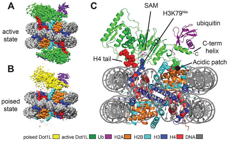
Figure 1: Structures Dot1L bound to H2B-ubiquitin nucleosome.

A, Cryo-EM reconstruction of the 2-to-1 active state complex. B, Cryo-EM reconstruction of the poised state complex. C, Atomic model of the active state complex between Dot1L and the H2B-Ub nucleosome. The H4 tail, H3K79Nle and the acidic patch are depicted as spheres and the SAM cofactor is depicted in stick representation. See also Figures S1–S4.
