Abstract
Background:
Though still thought to be rare, in recent years, vasospasm as a result of primary intraventricular hemorrhage (IVH) has been increasingly recognized in patients with spontaneous primary intraventricular hemorrhage, of various etiologies. Unlike vasospasm in aneurysmal subarachnoid hemorrhage (SAH), which has a well-defined time frame of 3–21 days, such a window is poorly defined for primary spontaneous intraventricular hemorrhage from other vascular etiologies.
Case Description:
We report on two cases of prolonged delayed proximal intracranial cerebral vasospasm occurring 29 and 22 days after the initial presentation.
Conclusion:
To our knowledge, this is the first report of such delayed vasospasm in spontaneous primary intraventricular hemorrhage secondary to a dural arteriovenous fistula and cavernous malformation. Our two cases of vasospasm in patients with nontraumatic nonaneurysmal SAH with IVH presented outside the expected time period of 21 days. It is important to recognize that symptomatic vasospasm secondary to intraventricular hemorrhage is a rare but devastating complication that can have serious deleterious consequences if gone unrecognized and untreated.
Keywords: Delayed cerebral ischemia, delayed cerebral vasospasm, intraventricular hemorrhage
BACKGROUND
Though still thought to be rare, in recent years, vasospasm of vascular origin with intraventricular hemorrhage (IVH) has been increasingly recognized in patients with spontaneous primary intraventricular hemorrhage, of various etiologies.[1,2,3,4,5,6,7,8] We report on two cases of prolonged delayed proximal intracranial cerebral vasospasm occurring 29 and 22 days after the initial presentation. To our knowledge, this is the first report of such delayed vasospasm in spontaneous primary intraventricular hemorrhage secondary to a ruptured dural arteriovenous fistula and cavernous malformation. Our two cases of vasospasm in patients with nontraumatic nonaneurysmal subarachnoid hemorrhage with IVH also presented outside the usual expected time period of 21 days.
CASE DESCRIPTION
Patient 1
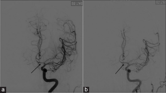
A 47-year-old man was presented with acute onset of nausea, light-headedness, palpitations, and fatigue. His mental status deteriorated but his neurological exam was otherwise nonfocal. Computed tomography (CT) [Figure 1] revealed a right parietal intraparenchymal hemorrhage with IVH. A ventriculostomy was placed for hydrocephalus. Catheter angiography showed a Cognard type IV dural arteriovenous fistula (dAVF) [Figure 2], which was embolized the next day and cured [Figure 3]. On post-bleed day (PBD) 12, he became stuporous. An MRI revealed acute bilateral basal ganglia infarcts and a magnetic resonance angiography for severe multivessel vasospasm. He improved with induced hypertension; however, his symptoms worsened and he was taken for angiogram. At angiogram, verapamil was delivered to the right internal carotid and vertebral arteries; furthermore, both A1 segments of the anterior cerebral arteries underwent balloon angioplasty. On PBD 29, he became aphasic and angiogram now showed severe left internal carotid artery vasospasm [Figures 4 and 5], which was in a different vascular distribution than his prior vasospasm episode. Induced hypertension was initiated with no improvement. Balloon angioplasty was then performed with improvement in exam. He underwent workup for vasculitides that were negative given the severity of the vasospasm; however, afterward, it did not recur. On discharge on PBD 52, he was conversant and ambulatory without assistance. He made a full recovery 3 months after discharge.
Figure 1.
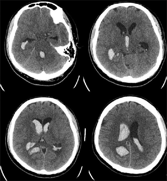
Case #1 Presenting axial non-contrast CT scan showing large amount of IVH
Figure 2.
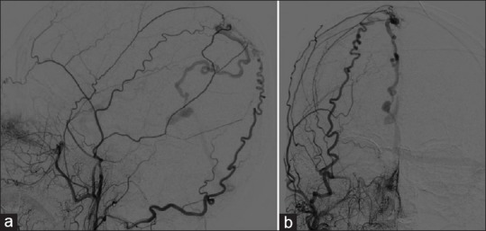
Case #1 Catheter angiography showed a Cognard type IV dAVF [(a) Lateral and (b) A/P Views]
Figure 3.
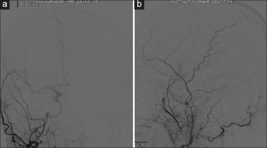
Case #1 Catheter angiography showing cured dAVF [(a) A/P and (b) Lateral Views]
Figure 4.
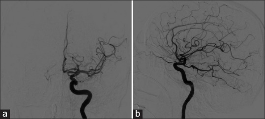
Case #1 Catheter angiography of initial left ICA injection [(a) A/P view and (b) Lateral View]
Figure 5.
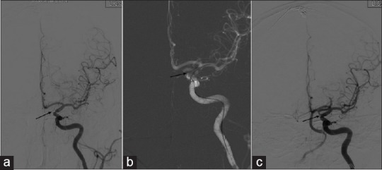
(a) Catheter angiography from PBD 29 showing severe supraclinoid left ICA vasospasm. (b) Balloon inflation angioplasty of vasospasm segment. (c) Improvement of vasospasm s/p balloon angioplasty of left ICA
Patient 2
The patient is a 66-year-old right-handed man with a remote history of idiopathic intraventricular hemorrhage 6 years prior to presentation at our clinic. He was presented to the ER with severe headaches and was found to again have an intraventricular hemorrhage on CT scan [Figure 6]. Angiogram [Figure 7] was again negative and an MRI [Figure 8] showed what appeared to be a cavernous malformation with of the third ventricle with surrounding hemorrhage and layering in the atria of the occipital horns bilaterally. Subsequently, the hemorrhage eventually recirculated into the subarachnoid space and he developed hydrocephalus requiring external ventricular drainage on PBD 9. Two days later, he underwent a right transcortical approach for removal of the cavernous malformation. His hospital course was complicated by cerebral vasospasm on PBD 22, which failed medical management and required interventional treatment by angioplasty of the left vertebral artery, bilateral A1, right M1, and a verapamil injection to the right ICA [Figure 9]. Upon discharge, he was dysphasic, with poor memory and balance; however, he improved to functional independence over the subsequent 2 months.
Figure 6.
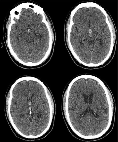
Case #2. Presenting axial non-contrast CT scan showing primary 3rd ventricular IVH
Figure 7.
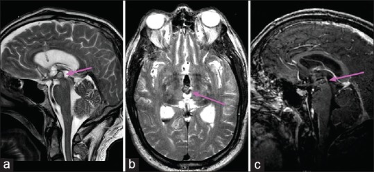
Case #2 MRI showing typical appearance of cavernoma in the 3rd ventricle. [(a) Sagittal T2, (b) Axial T2, (c) Sagittal T1 with contrast]
Figure 8.
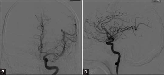
Case #1 (a) Initial Catheter angiography AP projection of left ICA. (b) Initial Catheter angiography lateral projection of left ICA
Figure 9.
Case #2 (a) Catheter angiography from PBD 22 showing a dominant left A1 segment with vasospasm. (b) Improvement of vasospasm s/p balloon angioplasty of left A1 segment
CONCLUSION
We described above two rare cases of patients with IVH with symptomatic vasospasm occurring outside the anticipated window for vasospasm. The proposed mechanism for delayed vasospasm is thought to be recirculation of IVH into the subarachnoid space as opposed to vasospasm due to the direct presence of blood in the subarachnoid space. Since the process of recirculation takes time to occur, the actual time for vasospasm presentation is directly related to the time it takes for blood to enter the subarachnoid space. When accounting for this time delay, the vasospasm actually occurs within the normal timeframe. We speculate that due to this process of recirculation, the window of concern for vasospasm is both delayed and prolonged, as was seen in our two patients. The understanding of resorption and clearance of IVH is poorly described, but time of clearance would presumably be longer and may contribute to the delayed effect on the intracranial arteries in the subarachnoid space. Additionally, large volumes of blood would also have prolonged clearance time and thus prolong the exposure of blood, blood breakdown products, and inflammatory cytokines to the blood vessels leading to delayed vasospasm. It is important to recognize that symptomatic vasospasm secondary to intraventricular hemorrhage is a rare but devastating complication that can have serious deleterious consequences if remained unrecognized and untreated.
Declaration of patient consent
The authors certify that they have obtained all appropriate patient consent forms. In the form the patient(s) has/have given his/her/their consent for his/her/their images and other clinical information to be reported in the journal. The patients understand that their names and initials will not be published and due efforts will be made to conceal their identity, but anonymity cannot be guaranteed.
Financial support and sponsorship
Nil.
Conflicts of interest
There are no conflicts of interest.
Footnotes
Contributor Information
Rose Fluss, Email: fluss@mail.einstein.yu.edu.
Avra Laarakker, Email: avracadavra@gmail.com.
Jonathan Nakhla, Email: jonathan.nakhla@gmail.com.
Allan Brook, Email: abrook@montefiore.org.
David Joseph Altschul, Email: daltschu@montefiore.org.
REFERENCES
- 1.Dull C, Torbey MT. Cerebral vasospasm associated with intraventricular hemorrhage. Neurocrit Care. 2005;3:150–2. doi: 10.1385/NCC:3:2:150. [DOI] [PubMed] [Google Scholar]
- 2.Gerard E, Frontera JA, Wright CB. Vasospasm and cerebral infarction following isolated intraventricular hemorrhage. Neurocrit Care. 2007;7:257–9. doi: 10.1007/s12028-007-0057-1. [DOI] [PubMed] [Google Scholar]
- 3.Kobayashi M, Takayama H, Mihara B, Kawase T. Severe vasospasm caused by repeated intraventricular haemorrhage from small arteriovenous malformation. Acta Neurochir (Wien) 2002;144:405–6. doi: 10.1007/s007010200059. [DOI] [PubMed] [Google Scholar]
- 4.Kothbauer K, Schroth G, Seiler RW, Do D-D. Severe symptomatic vasospasm after rupture of an arteriovenous malformation. Am J Neuroradiol. 1995;16:1073–5. [PMC free article] [PubMed] [Google Scholar]
- 5.Macdonald RL. Delayed neurological deterioration after subarachnoid haemorrhage. Nat Rev Neurol. 2014;10:44–58. doi: 10.1038/nrneurol.2013.246. [DOI] [PubMed] [Google Scholar]
- 6.Park BS, Won YS, Choi CS, Kim BM. Severe symptomatic vasospasm following intraventricular hemorrhage from arteriovenous fistula. J Korean Neurosurg Soc. 2009;45:300–2. doi: 10.3340/jkns.2009.45.5.300. [DOI] [PMC free article] [PubMed] [Google Scholar]
- 7.Yanaka K, Hyodo A, Tsuchida Y, Yoshii Y, Nose T. Symptomatic cerebral vasospasm after intraventricular hemorrhage from ruptured arteriovenous malformation. Surg Neurol. 1992;38:63–7. doi: 10.1016/0090-3019(92)90214-8. [DOI] [PubMed] [Google Scholar]
- 8.Yokobori S, Watanabe A, Nakae R, Onda H, Fuse A, Kushimoto S, et al. Cerebral vasospasms after intraventricular hemorrhage from an arteriovenous malformation-case report. Neurol Med Chir (Tokyo) 2010;50:320–3. doi: 10.2176/nmc.50.320. [DOI] [PubMed] [Google Scholar]


