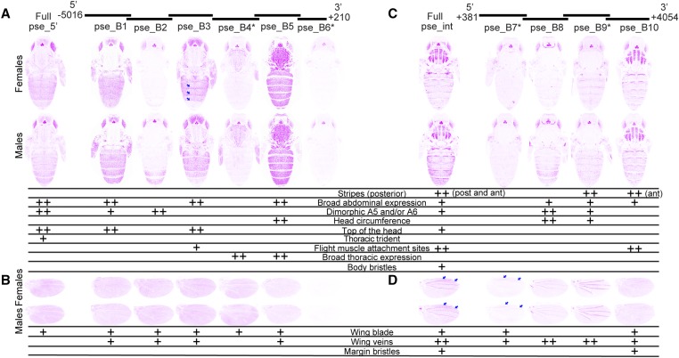
Figure 4.
Enhancer activity of D. pseudoobscura yellow fragments. Below the schematic of overlapping sequence regions from the 5′ intergenic and intronic regions of the D. pseudoobscura yellow gene tested for enhancer activity are images of transgenic pupae that show expression from the GFP reporter genes in magenta. Expression driven by fragments from the 5′ intergenic region (A and B) and the intron (C and D) is shown in the body (A and C) and wings (B and D), for both female (top row) and male (bottom row) flies. Summary tables below each pair of pupal bodies (A and C) and wings (B and D) show our interpretation of these images: “++” indicates strong fluorescence observed in the body region, whereas “+” indicates weaker fluorescence. Note that in addition to the stripes seen at the posterior edge (post) of abdominal segments in many cases, the full D. pseudoobscura intron (“Full pse_int”) as well as fragment pse_B10 drove elevated expression in stripes at the anterior edge of each abdominal segment (indicated by the “ant” notation in the table). Blue arrows highlight elevated expression along the posterior edge of abdominal segments in females driven by the pse_B3 fragment, and between the L1 and L2, as well as L2 and L3, veins in the wing blade driven by full pse_int and the pse_B7 fragment. The asterisks next to the pse_B4, pse_B6, pse_B7, and pse_B9 fragments indicate that activities of these elements are shown for flies heterozygous for the reporter gene. All other images show GFP expression in flies homozygous for the reporter gene. The magenta color used in this figure makes it easier to see low expression levels; a copy of this figure with GFP expression shown in the more traditional green is provided as Figure S3. The wing blade expression driven by pse_B5 was equivocal and went through a secondary inspection before reaching a consensus among authors.

