Abstract
Purpose:
To compare the delivered radiation dose between a low-dose hip CT (Computed Tomography) scan protocol and traditional hip CT scan protocols (referred to as: traditional CT).
Methods:
This was a retrospective comparative cohort study. A group of patients who underwent hip preservation surgery (including arthroscopy, surgical hip dislocation, or periacetabular osteotomy procedures) at our institution between 2016-2017 were identified. Patients were excluded if they had a BMI>35 kg/m2, previous surgery, or absence of a radiation dose report. The low-dose group included patients undergoing hip CT at our institution utilizing a standardized protocol of 100 kVp, 100 mAs and limited scanning field. The traditional CT group included patients who had hip CT scans performed at outside institutions. The total effective dose (Ehip), effective dose per millimeter-body-length-scanned (Emm), and the patients’ age, BMI were compared by univariate analysis. The correlation of Ehip to BMI was assessed.
Results:
Forty-one consecutive patients were included in the low-dose group, and 18 consecutive patients were included in the traditional CT group. Low-dose CT resulted in 90% reduction in radiation exposure compared to traditional CT (Ehip=0.97 ± 0.28 vs. 9.68 ± 6.67mSv, p<0.0001). Age (28±11 vs. 26±10 years, p=0.42), sex (83% vs. 76% female, p=0.74), and BMI (24±3 vs. 24±3 kg/m2, p=0.75) were not different between the two groups. Ehip had poor but significant correlation to BMI in the low-dose CT group (R2=0.14, slope=0.03, p=0.02), and did not correlate to BMI in the traditional CT group (R2=0.13, p=0.14).
Conclusions:
Low-dose hip CT protocol for the purpose of hip preservation surgical planning resulted in 90% reduction in radiation exposure compared to traditional CT.
Level of Evidence:
Diagnostic Study, Level II
Keywords: computed tomography, low-dose CT, radiation exposure, hip preservation, periacetabular osteotomy, hip arthroscopy, image gently
Introduction
The numbers of adolescents and young adult patients undergoing hip preservation surgery continues to grow. Thorough bony correction of the osseous pathomorphology remains critical for achieving optimal outcomes and minimizing the need for revision procedures. 1-5 Plain radiographs have limitations with regards to three-dimensional characterization of hip morphology. This includes precise localization and characterization of cam topography and assessment of acetabular morphology, given the influence of changes in pelvic tilt and rotation.
While some surgeons solely utilize plain radiographs and intraoperative dynamic and fluoroscopic assessment of bony morphology, a growing number of hip surgeons are utilizing computed tomography (CT) in addition to these other modalities to allow a more comprehensive assessment of the bony patho-morphology preoperatively. A CT scan provides detailed visual and quantitative information, allowing clinicians to better conceptualize the morphology of the pelvis, acetabulum and the femur with modern three-dimensional (3D) reconstruction. Potentially false-positive findings on plain films such as cross-over signs or posterior wall signs can also be further evaluated, as well as correction for altered pelvic tilt or rotation present on plain radiographs. 6 However, this increased information from CT does result in increased radiation exposure, which has been a valid concern amongst hip preservation surgeons treating adolescents and young adults.
Recent technological advances as well as refined protocols have permitted the ability to significantly reduce radiation dose when performing these scans. Low-dose CT imaging protocols have been successfully applied to both pediatric and adult populations for various diagnostic and surgical purposes.7-11,12-18. while the application of low-dose hip CT scans specifically for hip preservation surgical planning has not been well established. The purpose of the current study was to compare the delivered radiation dose between a low-dose hip CT scan protocol and traditional hip CT scan protocols (referred to as: traditional CT). We hypothesized that a low-dose hip CT scan protocol utilized at our institution results in lower effective dose than traditional CT in patients undergoing hip preservation surgery.
Materials and Methods
Study Design
The present study was a retrospective comparative diagnostic study of consecutive patients that was approved by our institutional IRB. Patients who underwent hip preservation surgery including arthroscopic and open procedures (surgical hip dislocation, periacetabular osteotomy) between year 2016-2017 in our institution were identified. Patients were excluded from the study if they had previous surgery, or if radiation dosage summaries were unavailable. Patients whose body mass index (BMI) was greater than 35 kg/m2 were also excluded from both groups to minimize the potential effect of obesity on increased radiation dose. The enrolled patients had hip CT scans for preoperative surgical planning including characterization of the cam deformity, femoral version, acetabular version and coverage. They were assigned to two study groups: the low-dose group and the traditional CT group.
The low-dose group included patients who had low-dose CT for preoperative planning at a single institution. The traditional CT group included patients who had their hip CT scans done outside of our institution prior to referral to our institution to undergo the hip preservation surgery. A standardized low-dose CT protocol was developed and optimized for patients with a BMI < 35 kg/m2, and has been in place for several years prior to the study period. The standardized low-dose protocol utilized a kVp of 100 and a mAs of 100, in addition to a limited relevant field of volume scanning the pelvis from the inferior sacroiliac joint to the lesser trochanter of proximal femur. The traditional CT scans were performed according to the protocols in each outside institution, which may have also included attempts to minimize radiation exposure. Three-dimensional reformats were generally utilized in both groups but do not affect the radiation exposure. The low-dose CT protocol included cuts thru the distal femur to assess femoral version and allows assessment of bilateral hips with a single scan.
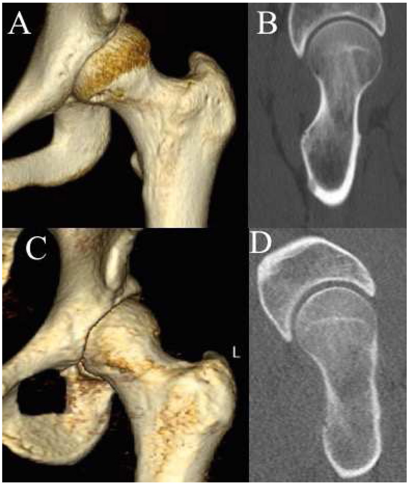
Radiation dosing for femoral version cuts, as well as scout hip CT images, were not routinely available in the traditional CT group. Given this difference between groups, the radiation exposure from cuts thru the distal femur and scout CT were not included for the primary comparison in this study. All scans in the current study were deemed adequate for clinical use. Subjectively, the traditional CT images appear crisper with less quantum noise as compared to the low-dose CT scans, but this did not affect the surgeon’s ability to identify the bony morphology and deformities using the low-dose CT scans (Figure 3).
Figure 3.
Representative CT three-dimensional reformats and two-dimensional axial oblique images in two patients with a 28 times greater radiation exposure with traditional CT compared to low-dose CT. Traditional CT (A,B) – 22 year old female, BMI 21.1, kVp 120, mAs 383, radiation exposure of 11.3 mSv. Low-dose CT (C,D) – 21 year old female, BMI 20.9, kVp 100, mAs 100, radiation exposure of 0.4 mSv
Data Acquisition and Determination of The Radiation Dose
CT scan radiation dosage and scan parameters were documented for both groups. The total slices (m) and slice thickness (u) for the hip CT scan were recorded. The values of kVp, mAs, and the dose-length product (DLP, in the unit of milligray•cm, mGy•cm) for the hip scan (DLPhip) were obtained from the official radiation dose summary report attached to each CT scan. The parameters of the scout topogram, including DLPscout, were recorded as well if they were available. The total scan length (lhip), for the hip was approximated by:
The DLP reflected the “absorbed dose”, which represented the radiation energy in the unit of mGy•cm. Even with the same amount of radiation energy, different types of radiation can cause different effects on living tissues. Therefore, the DLP (absorbed dose) was then converted to the “equivalent dose” in the unit of milliSievert (mSv), by applying the radiation weighing factor (WR) of 1.0 for diagnostic imaging such as X-rays and CT scans. The “equivalent dose” was then converted into “effective dose”, in the same unit of mSv, to account for the effect on health for specific anatomical areas of the human body, as described below.
The total effective dose of the hip CT scan, Ehip, and the effective dose of the scout topogram, Escout, both in the unit of mSv, were determined according to the tissue weighting factors and organ dose estimates by Monte Carlo simulations of the human pelvis being scanned, per the International Commission on Radiological Protection (ICRP) Publications 103. 19 That is, the conversion factor (κ) of 0.015 mSv of effective dose per 1 mSv of the equivalent dose, for hip CT scans. Consequently, the effective dose for unit millimeter length scanned, Emm, was determined by normalizing Ehip to lhip. Namely:
Statistical Analyses
An a priori power analysis was performed to determine estimated requirements for sample size. Traditional CTs were less commonly performed so an estimated 2:1 ratio between the 2 groups was utilized. In order for 80% power and alpha level of 0.05, in order to detect a 50% decrease in radiation exposure (6±3 vs. 3±3 mSv) a total of 52 patients (35 low-dose and 17 traditional CTs). The study period was chosen to meet these requirements resulting in a total of 59 patients.
The difference in the radiation dose between the low-dose CT and traditional CT was assessed by comparing the Ehip and Emm between the two groups, respectively, using two-tailed unpaired student t-test. The age, sex, and BMI were compared between the low-dose CT and traditional CT groups, respectively, using two-tailed unpaired student t-test and Chi-square or Fisher’s Exact (FET) tests . The association of each patient’s BMI with the received total effective dose of the hip CT scan, Ehip, was assessed by linear correlation test. P values less than 0.05 were considered statistically significant.
Results
The present study analyzed a total of 59 patients, of which 41 were included in the low-dose group and the other 18 were in the traditional CT group. The two groups were not different in terms of age (28±11 vs. 26±10 years, p=0.42), sex (83.3% vs. 75.6% female, p=0.74 FET) or BMI (24±3 vs. 24±3 kg/m2, p=0.75). (Table 1)
Table 1.
Demographics and the hip CT scan parameters of the patient group receiving low-dose CT protocol or the traditional CT protocol. BMI: body mass index; SD: standard deviation; n/a: non-applicable.
| Low-dose CT n=41 |
Traditional CT n=18 |
p value | |
|---|---|---|---|
| Demographics | |||
| Age at CT scan (years) | 28±11 (14–66) | 26±10 (12–44) | 0.45 |
| Sex (% Male: % Female) | 24% : 76% | 17% : 83% | 0.74* |
| BMI (kg/m2) | 23.8±3.4 (17.7–31.3) | 23.5±3.1 (18.6–31.8) | 0.75 |
| Body Weight (kg) | 69.7±14.5 (46.4–101.4) | 65.5±12.8 (48.2–106.4) | 0.29 |
| Body Height (cm) | 170.6±9.4 (152.0–193.0) | 166.6±8.5 (155.0–183.0) | 0.13 |
| Hip CT scan parameters | |||
| kVp | 97.6±6.6 (80.0–100.0) | 130.2±25.8 (100.0–223.0) | p<0.001 |
| mAs | 99.0±7.7 (80.0–120.0) | 269.3±127.8 (74.0–500.0) | p<0.001 |
| cut thickness (mm) | All 0.6 mm thickness | 1.8±1.3 (0.5–5.0) | n/a |
Data are shown as mean±SD (range);
: Fisher’s Exact test
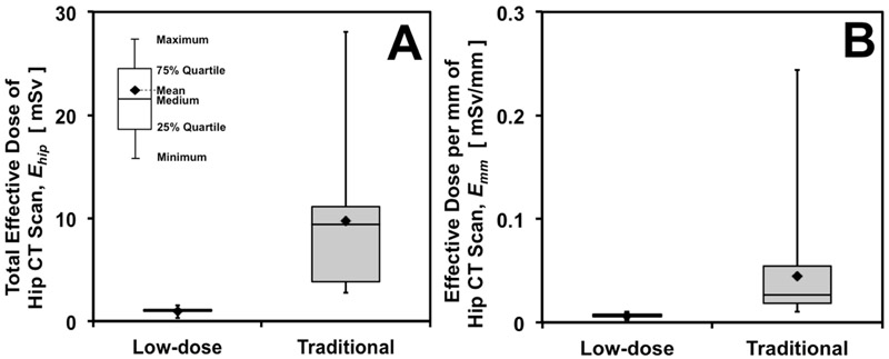
The low-dose CT group received a lower mean effective dose (Ehip) of 0.97 ± 0.28 (range 0.29-1.56) mSv compared to that of 9.68 ± 6.67 (range 2.75-28.04) mSv of the traditional CT group (p<0.001). This difference represented a 90.0% reduction in radiation exposure in the low-dose CT group. The minimal Ehip of the traditional CT group (2.75 mSv) was higher than the maximal Ehip of the low-dose CT group (1.56 mSv). The maximal Ehip in the traditional CT group was 97 times the minimum Ehip in the low-dose CT group. The low-dose CT group received lower mean effective dose per the body length (mm) scanned (Emm) of 0.006 mSv, compared to that of 0.045 mSv of the traditional CT group (p<0.001). (Fig. 1) This represented an 86.7% reduction in radiation exposure per length scanned.
Figure 1.
Radiation exposure resulting from the hip CT scan, comparing the low-dose CT protocol versus the traditional CT protocol: (A) the effective dose of the overall hip CT scan, and (B) the effective dose per unit millimeter scanned.
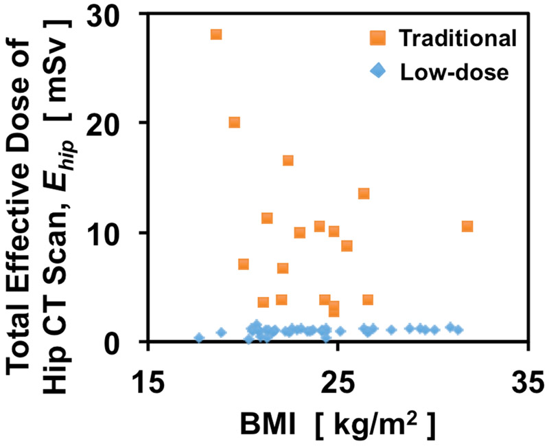
Ehip had poor but significant correlation to the patient’s BMI in the low-dose CT group (R2=0.14, slope=0.03, p=0.02), and did not correlate to the patient’s BMI in the traditional CT group (R2=0.13, p=0.14). (Fig. 2)
Figure 2.
Poor and Non-correlation between the hip CT scan effective dose and the patient’s BMI, respectively in the low-dose and the traditional CT protocol groups.
All patients in the low-dose CT group had documented radiation dose for the scout topogram (DLPscout), and the resulting mean effective dose, Escout, was 0.07 mSv (range, 0.05-0.24). This resulted in a total effective dose of 1.04 mSv for the low-dose CT group. Only six of the 18 patients in the traditional CT group had documented DLPscout, and the resulting median value of Escout was 0.10 mSv (range, 0.04-0.15).
Discussion
A low-dose hip CT scan protocol consistently achieved a lower radiation dose compared to the traditional CT scans. Using the low-dose CT protocol resulted in a nearly 90% reduction in radiation exposure, as well as decreased variability, while maintaining adequate image quality for clinical assessment and preoperative surgical planning. The majority of radiation reduction occurred as a result of reduction in effective dose per mm of CT scan length (86.7% reduction per length scanned), while limitations in scan field also appeared to play a lesser role. The scanning parameters in such a low-dose protocol, including the kVp and mAs values and scanning field, can be easily applied at any institution through collaboration with the radiologists. Monitoring of radiation exposure remains important during implementation of such a low-dose CT protocol.
A thorough correction of the bony deformity in addition to management of soft tissue pathology is important to the long-term outcome of hip preservation surgery. 1-3 The clinical information provided by plain films may be insufficient to fully characterize the complexity and variability of three-dimensional morphology. 20 Various plain radiographic views are commonly utilized to assess the degree of cam morphology with the 45 degrees Dunn view generally used to profile the maximum cam deformity. The AP pelvis, 45 degrees Dunn, and frog-lateral views provide useful assessments of deformity at 12:00, 1:30, and 3:00 positions. However, plain radiographic assessment of acetabular morphology is limited by alterations of such morphology from non-standard pelvic tilt or rotation. Additionally, assessment of femoral head coverage at various positions on the clock-face is quite challenging with plain radiographs, nor is it possible to accurately assess the femoral torsion.
CT scan can provide further characterization of the extent of cam morphology beyond these regions, in both its area and volume, which may significantly affect the ease and feasibility of arthroscopic access. Preoperative hip CT scan21, 22, with 3D reconstruction when available23-26, supplements comprehensive information for surgical planning and may play a role in optimizing deformity correction, therefore minimizing the risk of residual impingement, recurrent pain, and revision surgery. 4, 5 In the present study, the low-dose hip CT scan protocol accomplished these goals regardless of the patient’s age, for those with a BMI less than 35 kg/m2, with a consistent substantial reduction in radiation exposure. In addition, CT protocols may also utilize selected cuts through the knee to allow precise measurement of femoral version.
The risk of tissue damage from ionizing radiation is well recognized but difficult to quantify. To date, there has been no clear dose threshold to determine how much radiation exposure would result in cancer. However, increased radiation exposure has been related to increased risk of various cancers27-33, indicating the importance to minimize radiation exposure as much as possible. Among CT scans, the abdominal and pelvic scans contributed the most to the incidence of radiation-related cancers34, 35, emphasizing the importance of limiting radiation hazard to the sensitive organs in such an area, particularly in a young population undergoing hip preservation. The mean effective dose of 0.97 mSv in the low-dose CT group is approximately 34% of the average annual background radiation (as well as the average annual radiation exposure due to medical procedures) in the United States (both of which are approximately 3 mSv). 30 It is also not more than the previously reported effective dose of 1.2 to 1.4 mSv resulting from two standard hip or pelvis plain film x-rays36. The radiation from low-dose CT in this study also equals the cumulative cosmic radiation exposure of approximately 12 round-trip international air travels. 37, 38 Recent data on the radiation exposure from modern digital radiographs has indicated a radiation exposure of 0.24 mSv from the AP pelvis radiation, 0.12 mSv for Dunn/frog lateral radiographs, and 0.89 mSv from a cross-table lateral radiograph. 39 This indicates the current low dose CT protocol results in similar radiation to four AP pelvis radiographs, or a similar radiation exposure to the cross-table lateral radiograph alone. (Table 2)
Table 2.
Radiation dose of common medical radiology practice14, 15, 36, 41 and daily life radiation exposures30, 37 and the ratio comparing to the radiation dose from hip CT scans performed with low-dose CT protocol or traditional protocol; ×: times.
| Average effective dose [mSv] |
Ratio to low-dose CT |
Ratio to traditional CT |
|
|---|---|---|---|
| Hip CT scans for preservation surgery | |||
| Low-dose CT scan | 0.97 | (1×) | 11% |
| Traditional CT scan | 9.68 | 10× | (1×) |
| Diagnostic plain films | |||
| Chest, PA+Lat | 0.1 | 10% | 1% |
| Chest, PA | 0.02 | 2% | 0.2% |
| Abdomen, 1 view | 0.7 | 72% | 7% |
| Pelvis, 1 view | 0.6 | 62% | 6% |
| Hip series, 5 views | 3.5 | 3.6× | 36% |
| Low-dose digital hip series, 4 views | 1.49 | 1.5× | 15% |
| Dental panoramic radiography | 0.01 | 1% | 0.1% |
| Intraoperative fluoroscopy/CT scans | |||
| C-arm for arthroscopic hip procedures | 0.01 | 1% | 0.1% |
| C-arm for pedicle screw insertion | 0.27 | 28% | 3% |
| Low-dose O-arm for pedicle screws insertion | 1.17 | 1.2× | 12% |
| Standard O-arm for pedicle screws insertion | 12.79 | 13× | 1.3× |
| CT procedures | |||
| Chest | 7 | 7.2× | 72% |
| Abdomen | 8 | 8.3× | 83% |
| Hip/Pelvis | 6 | 6.2× | 62% |
| Dental | 0.2 | 21% | 2% |
| Daily life radiation exposures | |||
| US annual background radiation | 3.1 | 3.2× | 32% |
| US annual radiation exposure from medical procedures | 3 | 3.1× | 31% |
| International travel | 0.04 | 4% | 0.4% |
| ICRP recommended annual occupational radiation exposure | 50 | 52× | 5.2× |
The ICRP (International Commission on Radiological Protection) recommended a maximal occupational radiation exposure is 50 mSv annually, or 100 mSv within five years. 19 While the average effective dose of the traditional CT group of 9.68 mSv is still less than this threshold, patients also receive additional radiation from radiographs, fluoroscopy, and other unrelated medical imaging, in addition to the background (3.1 mSv annually in the US). As practicing surgeons, we believe it is imperative to minimize the radiation exposure to our patients, especially given that higher radiation dose may not provide additional information for surgical planning.
Efforts in radiology have been successful in reducing radiation effective dose while maintaining clinically satisfactory image quality in a variety of settings40 such as the EOS12, 13 system frequently used for spine and limb alignment assessment. The low-dose O-arm scanning protocol for intra-operative pedicle screw insertion results in nearly 90% reduction of radiation exposure. 14, 15 Such ALARA (as-low-as-reasonably-achievable) principle has been applied to CT scans of the neck33, chest7, abdomen8, 11, and coronary angiography9, 10, leading to 23-88% effective dose reduction.
For patients undergoing arthroscopic hip procedures, a recent study reported median cumulative effective doses of 2.4 mSv, 3.5 mSv and 0.01 mSv resulting from preoperative CT scan, plain film hip series studies and intraoperative fluoroscopy, respectively. 41 Each of the above radiation dose parameters correlates with the patient’s BMI. In addition, the CT scan for templating total hip replacement surgery can result in an effective dose as low as 2.5-3.1 mSv by applying a low-dose CT protocol. 16-18 In the current study, the low-dose CT protocol resulted in less radiation exposure than reported in these studies (1.0 mSv vs. 2.4-3.1 mSv). We did not find meaningful correlation between the CT scan effective dose and the patient’s BMI, given those whose BMI was less than 35 kg/m2. Based on our experience, utilization of our low-dose CT protocol in those with a BMI greater than 35 kg/m2 begins to suffer from suboptimal image quality, and may require adjustment with associated increases in radiation exposure but still remain much below traditional CT.
The Ehip of 0.97 mSv approximated four AP pelvis radiographs reported in the literature, or one-third of average U.S. annual background radiation exposure. Such a protocol significantly improves the risk-benefit ratio for patients of child-bearing age who undergo hip preservation procedures by maximizing preoperative information while minimizing ionizing radiation exposure. This resonates with the core value of “Image Gently Campaign”, which highlights safe and effective imaging for young patients. 42 Future investigation including more institutions and patients may further validate the generalizability of the low-dose protocol and potentially investigate the feasibility of additional reductions in radiation exposure in certain subgroups.
Limitations
The present study has several limitations. First, the traditional CT scans came from a variety of sources with various scanning parameters, as reflected by the wider distribution of the radiation dose within the group. This may in fact represent the actual variability of CT protocols used across multiple centers when the scan is aimed for hip preservation surgery, even though low-dose CT protocols have been reported for the abdomen and for total hip replacement planning. Secondly, the image quality was assessed qualitatively based on the subjective clinical judgment of the surgeons. Further characterization of the differences in image quality would require analyses beyond the scope of this study and were not felt to be clinically important. For clinical use, a CT scan with adequate image clarity that minimizes radiation is our clinical target.
Conclusions
Low-dose hip CT protocol for the purpose of hip preservation surgical planning resulted in 90% reduction in radiation exposure compared to traditional CT.
Supplementary Material
Footnotes
The present study was approved by the institutional IRB #201406131
Publisher's Disclaimer: This is a PDF file of an unedited manuscript that has been accepted for publication. As a service to our customers we are providing this early version of the manuscript. The manuscript will undergo copyediting, typesetting, and review of the resulting proof before it is published in its final citable form. Please note that during the production process errors may be discovered which could affect the content, and all legal disclaimers that apply to the journal pertain.
References
- 1.Clohisy JC, Nepple JJ, Larson CM, Zaltz I, Millis M, Academic Network of Conservation Hip Outcome Research M. Persistent structural disease is the most common cause of repeat hip preservation surgery. Clin Orthop Relat Res. 2013;471:3788–3794. [DOI] [PMC free article] [PubMed] [Google Scholar]
- 2.Bogunovic L, Gottlieb M, Pashos G, Baca G, Clohisy JC. Why do hip arthroscopy procedures fail? Clin Orthop Relat Res. 2013;471:2523–2529. [DOI] [PMC free article] [PubMed] [Google Scholar]
- 3.Wenger DE, Kendell KR, Miner MR, Trousdale RT. Acetabular labral tears rarely occur in the absence of bony abnormalities. Clin Orthop Relat Res. 2004;426:145–150. [DOI] [PubMed] [Google Scholar]
- 4.Heyworth BE, Shindle MK, Voos JE, Rudzki JR, Kelly BT. Radiologic and intraoperative findings in revision hip arthroscopy. Arthroscopy. 2007;23:1295–1302. [DOI] [PubMed] [Google Scholar]
- 5.Philippon MJ, Schenker ML, Briggs KK, Kuppersmith DA, Maxwell RB, Stubbs AJ. Revision hip arthroscopy. Am J Sports Med. 2007;35:1918–1921. [DOI] [PubMed] [Google Scholar]
- 6.Zaltz I, Kelly BT, Hetsroni I, Bedi A. The crossover sign overestimates acetabular retroversion. Clin Orthop Relat Res. 2013;471:2463–2470. [DOI] [PMC free article] [PubMed] [Google Scholar]
- 7.Zhu X, Yu J, Huang Z. Low-dose chest CT: optimizing radiation protection for patients. AJR Am J Roentgenol. 2004; 183: 809–816. [DOI] [PubMed] [Google Scholar]
- 8.Sagara Y, Hara AK, Pavlicek W, Silva AC, Paden RG, Wu Q. Abdominal CT: comparison of low-dose CT with adaptive statistical iterative reconstruction and routine-dose CT with filtered back projection in 53 patients. AJR Am J Roentgenol. 2010;195:713–719. [DOI] [PubMed] [Google Scholar]
- 9.Stolzmann P, Donati OF, Scheffel H, et al. Low-dose CT coronary angiography for the prediction of myocardial ischaemia. Eur Radiol. 2010;20:56–64. [DOI] [PubMed] [Google Scholar]
- 10.Scheffel H, Alkadhi H, Leschka S, et al. Low-dose CT coronary angiography in the step-and-shoot mode: diagnostic performance. Heart. 2008;94:1132–1137. [DOI] [PubMed] [Google Scholar]
- 11.Dewes P, Frellesen C, Scholtz JE, et al. Low-dose abdominal computed tomography for detection of urinary stone disease - Impact of additional spectral shaping of the X-ray beam on image quality and dose parameters. Eur J Radiol. 2016;85:1058–1062. [DOI] [PubMed] [Google Scholar]
- 12.Hui SC, Pialasse JP, Wong JY, et al. Radiation dose of digital radiography (DR) versus micro-dose x-ray (EOS) on patients with adolescent idiopathic scoliosis: 2016 SOSORT-IRSSD "John Sevastic Award" Winner in Imaging Research. Scoliosis Spinal Disord. 2016;11:46. [DOI] [PMC free article] [PubMed] [Google Scholar]
- 13.Luo TD, Stans AA, Schueler BA, Larson AN. Cumulative Radiation Exposure With EOS Imaging Compared With Standard Spine Radiographs. Spine Deform. 2015;3:144–150. [DOI] [PubMed] [Google Scholar]
- 14.Su AW, McIntosh AL, Schueler BA, et al. How Does Patient Radiation Exposure Compare With Low-dose O-arm Versus Fluoroscopy for Pedicle Screw Placement in Idiopathic Scoliosis? J Pediatr Orthop. 2017;37:171–177. [DOI] [PubMed] [Google Scholar]
- 15.Su AW, Luo TD, McIntosh AL, et al. Switching to a Pediatric Dose O-Arm Protocol in Spine Surgery Significantly Reduced Patient Radiation Exposure. J Pediatr Orthop. 2016;36:621–626. [DOI] [PubMed] [Google Scholar]
- 16.Huppertz A, Radmer S, Asbach P, et al. Computed tomography for preoperative planning in minimal-invasive total hip arthroplasty: radiation exposure and cost analysis. Eur J Radiol. 2011;78:406–413. [DOI] [PubMed] [Google Scholar]
- 17.Huppertz A, Lembcke A, Sariali el H, et al. Low Dose Computed Tomography for 3D Planning of Total Hip Arthroplasty: Evaluation of Radiation Exposure and Image Quality. J Comput Assist Tomogr. 2015;39:649–656. [DOI] [PubMed] [Google Scholar]
- 18.Geijer M, Rundgren G, Weber L, Flivik G. Effective dose in low-dose CT compared with radiography for templating of total hip arthroplasty. Acta Radiol. 2017;58:1276–1282. [DOI] [PubMed] [Google Scholar]
- 19.The 2007 Recommendations of the International Commission on Radiological Protection. ICRP publication 103. Ann ICRP. 2007;37:1–332. [DOI] [PubMed] [Google Scholar]
- 20.Clohisy JC, Carlisle JC, Trousdale R, et al. Radiographic evaluation of the hip has limited reliability. Clin Orthop Relat Res. 2009;467:666–675. [DOI] [PMC free article] [PubMed] [Google Scholar]
- 21.Lynch TS, Terry MA, Bedi A, Kelly BT. Patient Evaluation, Current Indications, and Outcomes. Hip Arthroscopic Surgery. 2013;41:1174–1189. [DOI] [PubMed] [Google Scholar]
- 22.Dolan MM, Heyworth BE, Bedi A, Duke G, Kelly BT. CT reveals a high incidence of osseous abnormalities in hips with labral tears. Clin Orthop Relat Res. 2011;469:831–838. [DOI] [PMC free article] [PubMed] [Google Scholar]
- 23.Khan O, Witt J. Evaluation of the magnitude and location of Cam deformity using three dimensional CT analysis. Bone Joint J. 2014;96-B:1167–1171. [DOI] [PubMed] [Google Scholar]
- 24.Nakahara I, Takao M, Sakai T, Miki H, Nishii T, Sugano N. Three-dimensional morphology and bony range of movement in hip joints in patients with hip dysplasia. Bone Joint J. 2014;96-B:580–589. [DOI] [PubMed] [Google Scholar]
- 25.Coobs BR, Xiong A, Clohisy JC. Contemporary Concepts in the Young Adult Hip Patient: Periacetabular Osteotomy for Hip Dysplasia. J Arthroplasty. 2015;30:1105–1108. [DOI] [PubMed] [Google Scholar]
- 26.Garcia-Cimbrelo E. CORR Insights(R): Femoral Morphology in the Dysplastic Hip: Three-dimensional Characterizations With CT. Clin Orthop Relat Res. 2017;475:1055–1057. [DOI] [PMC free article] [PubMed] [Google Scholar]
- 27.Brenner DJ, Hall EJ. Computed tomography--an increasing source of radiation exposure. N Engl J Med. 2007;357:2277–2284. [DOI] [PubMed] [Google Scholar]
- 28.Mathews JD, Forsythe AV, Brady Z, et al. Cancer risk in 680,000 people exposed to computed tomography scans in childhood or adolescence: data linkage study of 11 million Australians. Bmj. 2013;346:f2360. [DOI] [PMC free article] [PubMed] [Google Scholar]
- 29.BEIR VII Health Risks from Exposure to Low Levels of Ionizing Radiation, Phase 2. Available at: http://www.nap.edu/openbook.php?isbn=030909156X. Accessed July 21st, 2018. [PubMed]
- 30.Measurements NCoRP. Available at: http://www.ncrppublications.org/Reports/160. Accessed July 21st, 2018.
- 31.Ronckers CM, Land CE, Miller JS, Stovall M, Lonstein JE, Doody MM. Cancer mortality among women frequently exposed to radiographic examinations for spinal disorders. Radiat Res. 2010;174:83–90. [DOI] [PMC free article] [PubMed] [Google Scholar]
- 32.Ronckers CM, Doody MM, Lonstein JE, Stovall M, Land CE. Multiple diagnostic X-rays for spine deformities and risk of breast cancer. Cancer Epidemiol Biomarkers Prev. 2008;17:605–613. [DOI] [PubMed] [Google Scholar]
- 33.Doody MM, Lonstein JE, Stovall M, Hacker DG, Luckyanov N, Land CE. Breast cancer mortality after diagnostic radiography: findings from the U.S. Scoliosis Cohort Study. Spine (Phila Pa 1976). 2000;25:2052–2063. [DOI] [PubMed] [Google Scholar]
- 34.Berrington de Gonzalez A, Mahesh M, Kim KP, et al. Projected cancer risks from computed tomographic scans performed in the United States in 2007. Arch Intern Med. 2009;169:2071–2077. [DOI] [PMC free article] [PubMed] [Google Scholar]
- 35.Smith-Bindman R, Lipson J, Marcus R, et al. Radiation dose associated with common computed tomography examinations and the associated lifetime attributable risk of cancer. Arch Intern Med. 2009;169:2078–2086. [DOI] [PMC free article] [PubMed] [Google Scholar]
- 36.Mettler FA Jr., Huda W, Yoshizumi TT, Mahesh M. Effective doses in radiology and diagnostic nuclear medicine: a catalog. Radiology. 2008;248:254–263. [DOI] [PubMed] [Google Scholar]
- 37.Friedberg W, Copeland K, Duke FE, O'Brien K 3rd, Darden EB Jr. Radiation exposure during air travel: guidance provided by the Federal Aviation Administration for air carrier crews. Health Phys. 2000;79:591–595. [DOI] [PubMed] [Google Scholar]
- 38.Bottollier-Depois JF, Chau Q, Bouisset P, Kerlau G, Plawinski L, Lebaron-Jacobs L. Assessing exposure to cosmic radiation during long-haul flights. Radiat Res. 2000;153:526–532. [DOI] [PubMed] [Google Scholar]
- 39.Nepple JJ, Martel JM, Kim YJ, Zaltz I, Clohisy JC, Group AS. Do plain radiographs correlate with CT for imaging of cam-type femoroacetabular impingement? Clin Orthop Relat Res. 2012;470:3313–3320. [DOI] [PMC free article] [PubMed] [Google Scholar]
- 40.Marcus RP, Koerner E, Aydin RC, et al. The evolution of radiation dose over time: Measurement of a patient cohort undergoing whole-body examinations on three computer tomography generations. Eur J Radiol. 2017;86:63–69. [DOI] [PubMed] [Google Scholar]
- 41.Canham CD, Williams RB, Schiffman S, Weinberg EP, Giordano BD. Cumulative Radiation Exposure to Patients Undergoing Arthroscopic Hip Preservation Surgery and Occupational Radiation Exposure to the Surgical Team. Arthroscopy. 2015;31:1261–1268. [DOI] [PubMed] [Google Scholar]
- 42.The Image Gently Alliance. Available at: http://www.imagegently.org/. Accessed July 21st, 2018.
Associated Data
This section collects any data citations, data availability statements, or supplementary materials included in this article.


