Abstract
Differential diagnosis of supraventricular tachycardia (SVT) and ventricular tachycardia (VT) is of paramount importance for appropriate patient management. Several diagnostic algorithms for discrimination of VT and SVT based on surface electrocardiogram (ECG) analysis have been proposed. Following established diagnosis of VT, a specific origination tachycardia site can be supposed according to QRS complex characteristics. This review aims to cover comprehensive and comparative description of the main VT diagnostic algorithms and to present ECG characteristics which permit to suggest the most common VT origination sites.
Keywords: Arrhythmias, Electrocardiogram, Supraventricular, Tachycardia, Ventricular
1. Introduction
Electrocardiographic diagnosis and localization of cardiac tachyarrhythmia origin are sometimes challenging. During the last decades, several algorithms for differentiation of supraventricular tachycardia (SVT) and ventricular tachycardia (VT) have been proposed. Electrocardiogram (ECG) characteristics of ectopic QRS complexes have also been characterized in order to define their origin in the heart ventricles. Based on surface ECG analysis it is possible to perform relatively accurate differential diagnosis between SVT and VT. Our review aims to provide contemporary algorithms of VT discrimination based on 12-lead ECG analysis, and characteristics which help to understand an origination/exit site of VT or premature ventricular contractions (PVC).
2. Algorithms for diagnosis of ventricular tachycardia
The term “tachycardia” indicates heart rate > 100 beats per minute.[1] A tachycardia can be classified as wide-complex (QRS > 120 ms) and narrow-complex (QRS < 120 ms). SVT has its origin in the atria or in the atrioventricular (AV) node while ventricular tachycardia is originated in the ventricular myocardium or in the conduction system below the AV node. Wide complex tachycardia (WCT) can be VT (80%), SVT (15%) with right or left bundle branch aberration (either atrial flutter, focal atrial tachycardia, AV nodal reciprocating tachycardia, reciprocating tachycardia utilizing an accessory pathway).[2]
When a narrow QRS tachycardia is seen on ECG, it is usually SVT. In rare cases VT originating from the conduction system (fascicular tachycardia, interfascicular tachycardia) can be characterized by QRS ≤ 120 ms.[3] The main challenge is encountered when a tachycardia is regular, with wide QRS, and no AV dissociation can be identified. Discrimination of WCT has an important role due to its clinical significance.[4]
2.1. Brugada algorithm
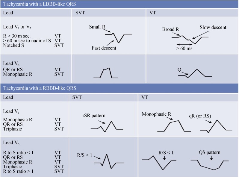
Brugada and co-authors presented systemic analysis of numerous ECGs and proposed an algorithm based on 4 steps that could relatively accurate diagnose a VT (Figure 1): (1) absence of an RS complex in all precordial leads; (2) an RS complex in any precordial lead with an RS interval >100 ms; (3) AV dissociation; and (4) morphological criteria for VT present in both leads V1 and V6 (Figure 2).
Figure 1. Brugada algorithm for the differential diagnosis of WCTs.
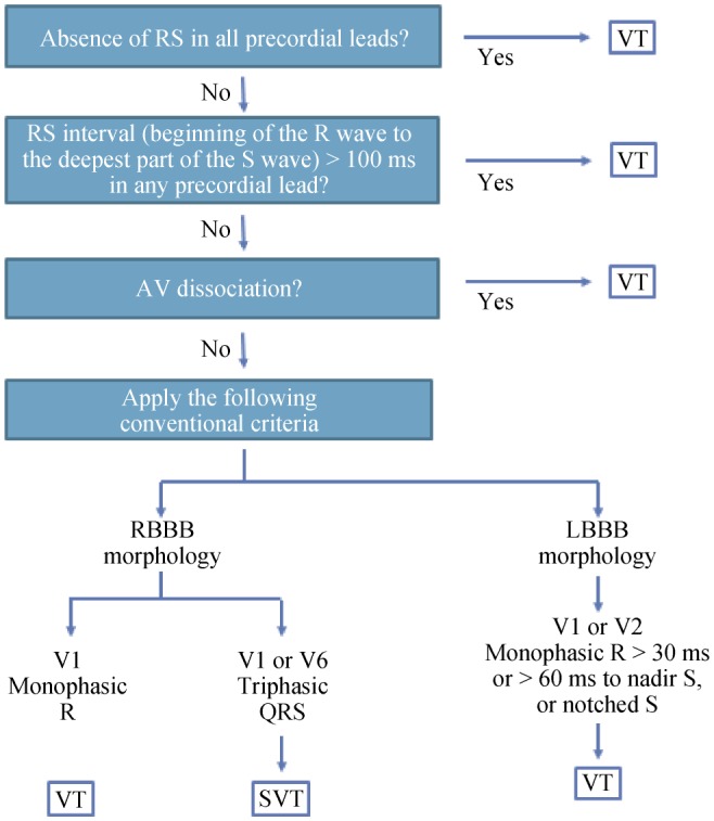
With modifications from Brugada, et al.[5] AV: atrioventricular; LBBB: left bundle branch block; RBBB: right bundle branch block; SVT: supraventricular tachycardia; VT: ventricular tachycardia; WCTs: wide complex tachycardias.
Figure 2. Morphological criteria for diagnosis of ventricular tachycardias used by Brugada, et al.[5].
With modifications from Eckardt L, et al.[7] LBBB: left bundle branch block; RBBB: right bundle branch block; SVT: supraventricular tachycardia; VT: ventricular tachycardia.
In this study, a total of 554 ECGs with WCTs were analyzed by two independent observers that reported 384 VT and 170 SVT, VT was diagnosed when any criterion was present. In the case that the first step was absent, the rest of the criteria were evaluated, and if any of them was not present, SVT with aberrant conduction was diagnosed by exclusion. The sensitivity of these four steps criteria for the diagnosis of VT was 98.7%, and the specificity was 96.5%.[5]
2.2. Vereckei algorithm
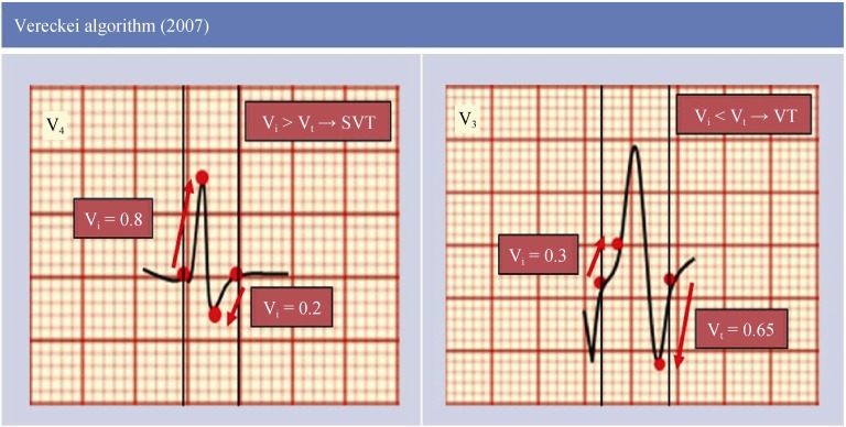
Vereckei, et al.[6] proposed a new simplified algorithm. The algorithm comprised the following four steps (Figure 3): (1) AV dissociation; (2) initial R wave in lead aVR; (3) morphology of a wide QRS tachycardia does not correspond to BBB (Figure 2)[7] or fascicular block (Table 1); and (4) Vi/Vt ratio, obtained by measuring the voltage of the initial 40 ms (Vi) and the terminal 40 ms of a QRS (in millivolts) in any ECG lead; the Vi/Vt ratio represents ventricular activation velocity. When the ratio is < 1, the diagnosis of VT is made. If the Vi/Vt is > 1, the diagnosis is SVT (Figure 4).
Figure 3. Vereckei algorithm for the differential diagnosis of WCT.
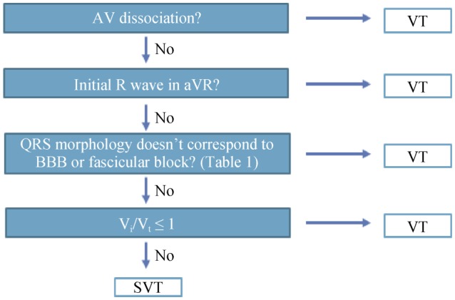
With modifications from Vereckei, et al.[6] AV: atrioventricular; BBB: bundle branch block; SVT: supraventricular tachycardia; VT: ventricular tachycardia; WCT: wide complex tachycardia.
Table 1. Criteria for the diagnosis of fascicular blocks (morphological criteria for BBB, Figure 2).
| Fascicular blocks | |
| LAFB Qualifying statements S1) QRS duration < 0.12 s (adults) S2) QRS axis ≤ –30° S3) rS pattern in II and III and aVF S5) R peak time ≥ 0.045 s in aVL S6) Slurred R downstroke in aVL S7) Slurred S in V5 or V6 Criteria for LAFB a) S1 and S2 and S3 and S4 and S5 or b) S1 and S2 and S3 and S4 and S6 or c) S1 and S2 and S3 and S4 and S7 Qualifying statement S3 is usually present with criteria a, b, and c above. If there is a QS in lead II, LAFB cannot be differentiated from inferior MI. |
LPFB Qualifying statements S1) QRS duration < 0.12 s S2) 180° > QRS axis > 90° S3) qR pattern in III and aVF with Q duration ≤ 0.04 sec S4) Absence of other causes of right axis deviation Criteria for LBBB Criteria for LPFB a) S1 and S2 and S3 a) S1 and S2 and S3 and S4 |
With modifications from Vereckei, et al.[6] BBB: bundle branch block; LAFB: left anterior fascicular block; LBBB: left bundle branch block; LPFB: left posterior fascicular block; MI: myocardial infarction.
Figure 4. Schematic electrocardiogram representations.
In lead V4 there are vertical lines denoting the onset of QRS complex, the initial and terminal 40 ms of the QRS complex is marked by small red points. During the initial 40 ms of the QRS, the impulse advanced vertically 0.8 mV, then the Vi = 0.8 and during the terminal 40 ms of the QRS, the impulse advanced vertically 0.2 mV, consequently the Vt = 0.2, and thus the Vi/Vt > 1 suggesting the diagnosis of SVT. In lead V3; Vi = 0.3 and Vt = 0.65 in this example, and therefore the Vi/Vt < 1 suggesting the diagnosis of VT. SVT: supraventricular tachycardia; VT: ventricular tachycardia.
If any criterion is present the analysis is terminated and VT is diagnosed, if no criteria are found SVT is diagnosed. Vereckei, et al.[6] reported sensitivity 95.7% for VT diagnosis and specificity 72.4%, even though limitations were present in this algorithm unable to recognize certain WCT (bundle branch re-entry VT, fascicular VT, and SVT involving an atriofascicular accessory pathway are associated with typical BBB pattern indistinguishable from that associated with SVT with functional aberrancy or pre-existent BBB, unless A-V dissociation is present).
2.3. aVR lead algorithm
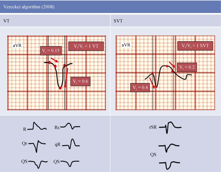
In order to further simplify VT diagnosis, Vereckei, et al.[8] proposed another algorithm based on the direction and velocity of initial and terminal ventricular activation in the aVR lead. The QRS morphological criteria were completely eliminated and only the aVR lead was evaluated, this diagnostic method was also based on four steps (Figure 5): (1) initial R-wave in aVR; (2) initial r- or q-wave with width > 40 ms; (3) notching on the descending limb of a negative onset, predominantly negative QRS complex; and (4) Vi/Vt ratio, obtained by measuring the voltage of the initial 40 ms (Vi) and the terminal 40 ms of a QRS (in millivolts) in the aVR lead. When the ratio is < 1, the diagnosis of VT is made. If the Vi/Vt is > 1, the diagnosis is SVT (Figure 6).
Figure 5. Vereckei algorithm for the differential diagnosis of wide QRS tachycardia based on the aVR lead.
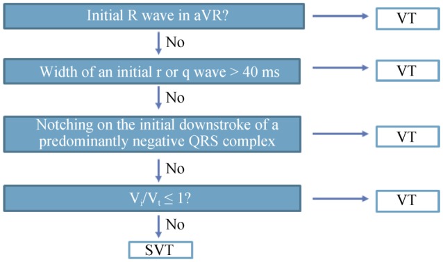
With modifications from Vereckei, et al.[8] SVT: supraventricular tachycardia; VT: ventricular tachycardia.
Figure 6. Schematic electrocardiogram representations.
Vi/Vt ratio criterion in the fourth step of the aVR algorithm. In the inferior part, most common lead aVR electrocardiogram patterns in SVT and VT.[8] SVT: supraventricular tachycardia; VT: ventricular tachycardia.
If any criterion is present the analysis is terminated and VT is diagnosed if no criteria are found SVT is diagnosed. Vereckei, et al.[8] reported sensitivity 96.5% and specificity 75% for VT diagnosis, and suggested that this single lead algorithm had the advantage of being a suitable tool for emergency situations in which a quick diagnosis is required, even though the required time for its evaluation was not compared with the previous algorithms. Factors that may influence the Vi/Vt ratio like anteroseptal myocardial infarction, scar at a late activated ventricular site, fascicular VT, and VT exit site close to the His-Purkinje system were described as limitations. Another limitation was the limited number of VTs occurring in the absence of structural heart disease that can be confused with SVT, these VTs present a narrower QRS complex compared with QRS complexes in patients with ischemic heart disease and can be confused with SVT when other algorithms are applied.
2.4. RWPT ≥ 50 ms in lead II, as a single criterion for VT-Pava's criterion
Pava, et al.[9] proposed a new VT criterion based on the analysis of the lead II (Figure 7), considering that this lead is usually present on ECGs registered during acute clinical situations. The criterion is applied by measuring the interval from the QRS onset to the peak of the R-wave. In this study, R-wave peak time (RWPT) of ≥ 50 ms in lead II had a sensitivity of 93%, specificity of 99% and positive predictive value of 98% in identifying VT.
Figure 7. Measurement of the RWPT in lead II.
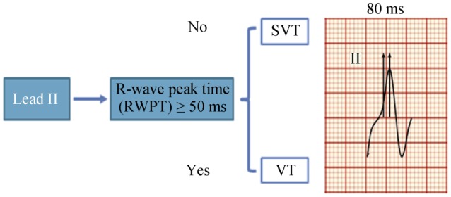
RWPT measured from the “isoelectric” line to the point of first change in polarity, was >50 ms (80 ms), with modifications from Pava LF, et al.[9] RWPT: R-wave peak time; SVT: supraventricular tachycardia; VT: ventricular tachycardia.
This criterion has not been compared with the previously described algorithms, the electrophysiological study was used as a gold standard to determine if VT or SVT was present. Difficult determination of QRS onset during fast VT is a limitation of this method, this method still waits for its validation by other researchers.[9]
2.5. VT score algorithm
In 2015, a VT score algorithm for the differentiation between VT and SVT was published. This novel score was based on seven characteristics and showed high specificity for VT diagnosis (Table 2).[10] Points attributable to each characteristic are summarized, and the resultant number represents a total score. A VT score ≥ 1 is considered diagnostic for VT. A VT score = 0 indicates SVT diagnosis. A VT score ≥ 3 indicates VT with the highest specificity, but low sensitivity (Table 3).[10]
Table 2. VT score-criteria for the diagnosis of VT.
| Criteria | ECG examples | Point |
| 1. Initial R wave in V1 |
 Criterion proposed by Sandler and Marriott.[11] |
1 |
| 2. Initial r > 40 ms in V1 or V2 |
 Criterion proposed by Swanick, et al.[12], and later validated by Kindwall, et al.[13] |
1 |
| 3. Notched S in V1 |
 Criterion proposed by Kindwall, et al.[13] |
1 |
| 4. Initial R wave in aVR |
 Criterion proposed by Vereckei, et al.[8] |
1 |
| 5. Lead II RWPT ≥ 50 ms |
 Criterion proposed by Pava, et al.[9] |
1 |
| 6. Lack of RS complex in leads V1-V6 | If the QRS complexes from V1-V6 have the following configurations --- > QS, R, qR, Qr, rSR′, Rsr′, this criterion is fulfilled. Criterion proposed by Brugada, et al.[5] | 1 |
| 7. AV dissociation |  |
2 |
With modifications from Jastrzebski, et al.[10] ECG figures reproduced with permission. RWPT: wave peak time; VT: ventricular tachycardia.
Table 3. Diagnostic performance of the VT score, as assessed in the validation cohort of 684 WCTs.
| Parameter | Specificity | Sensitivity |
| VT score ≥ 1 | 63.2% | 93.3% |
| VT score ≥ 2 | 88.3% | 76.4% |
| VT score ≥ 3 | 99.6% | 56.9% |
| VT score ≥ 4 | 100% | 32.6% |
With modifications from Jastrzebski, et al.[10] VT: ventricular tachycardia; WCTs: wide complex tachycardias.
2.6. Comparison of VT algorithms diagnostic value
Comparison of the VT score with other ECG-based methods was performed (Table 4). Chai and collaborators performed a comparison of Brugada, Vereckei and other algorithms. Thirteen studies were included in this meta-analysis, and 1918 electrocardiograms were analyzed. The Brugada algorithm showed the highest pooled sensitivity 0.92 and moderate pooled specificity 0.71 for discrimination SVT from VT,[14] as compared with other algorithms. The VT score and lead II RWPT criterion were not included in this meta-analysis. Table 5 summarizes the sensitivity and specificity of the algorithms reported by each author.
Table 4. Comparison of the VT score with other ECG-based methods.
| Parameter | VT score ≥ 1 | Brugada algorithm | aVR algorithm | Lead II RWPT |
| Accuracy | 83.1% | 80.4% | 73.4% | 70.1% |
| Sensitivity | 96.6% | 91.0% | 79.9% | 62.0% |
| Specificity | 63.4% | 60.4% | 61.2% | 85.3% |
With modifications from Jastrzebski, et al.[10] RWPT: wave peak time; VT: ventricular tachycardia.
Table 5. Comparison of wide QRS tachycardia discrimination algorithms.
In the European Heart Rhythm Association (EHRA) consensus document on the management of supraventricular arrhythmias and 2015 ACC/AHA/HRS Guidelines for the management of adult patients with supraventricular tachycardia the following algorithms and criteria were advised for VT discrimination: the Brugada, Vereckei (aVR algorithm) and lead II (Pava's) criterion.[2]–[15]
3. Specific VT/PVC origin
Discrimination of VT from SVT is the first step in the direction of tachyarrhythmia treatment. The site of origin (SoO) of VT needs to be defined in certain cases. This is of paramount importance for interventional procedures, such as catheter ablation. The other important clinical aspect is the definition whether VT origination site is consistent with ventricular lesion site (for instance, post-myocardial infarction scar, or abnormal findings detected by visualization methods-magnetic resonance imaging, echocardiography, etc.). In this section, we review characteristics of QRS complexes which help to suggest commonly encountered VT/PVC origination sites.
3.1. Outflow tract ventricular tachycardia
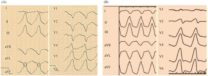
Most commonly, ventricular arrhythmias (VA) in patients without structural heart disease are originated in the right ventricular outflow tracts (RVOT) and left ventricular outflow tracts (LVOT).[16] Outflow tract arrhythmias are characterized by inferior axis, positive QRS complexes in II, III and aVF (Figure 8).[17] Not infrequently, distinguishing RVOT from LVOT based on surface ECG analysis is challenging. Bellow we describe some features that can help in differential diagnosis (Table 6).
Figure 8. Common characteristics found in outflow tract ventricular tachycardia (inferior axis deviation, positive QRS complexes in II, III and aVF).
(A): RVOT tachycardia; (B): LVOT tachycardia. LVOT: left ventricular outflow tract; RVOT: right ventricular outflow tracts.
Table 6. ECG features VA arising from RVOT and LVOT.
| Location of VA | BBB | Axis | Precordial transition | I | V1 | V6 | Other features |
| RVOT | |||||||
| Anteroseptal | LBBB | Inferior | ≤ V3 | rS | rS | R | Negative, isoelectric, or multiphasic I |
| Posterior, free Wall | LBBB | Inferior | ≥ V3 | R | rS | R | Positive I, broad late notched inferior leads |
| LVOT | |||||||
| Supravalvular | |||||||
| LCC | LBBB | Inferior | ≤ V2 | rS | rS, RS | R | QS or RS in lead I; notched M or W in V1 |
| RCC | LBBB | Inferior | ≤ V3 | R | rS, RS | R | Broad R in V2 |
| LCC/RCC junction | LBBB | Inferior | V3 | R/Rsr' | qrS | R | Notched on the downward deflection or W pattern in V1 |
| Infravalvular | |||||||
| AMC | RBBB | Inferior | Positive concordance | R/Rs | qR | R | No S in V6 |
| Septal-para-Hisian | LBBB | Left inferior | Early | Rs | QS, Qr | Rs | QS amplitude lead II/III ratio > 1 |
| Anterior interventricular great cardiac vein junction | LBBB | Inferior | Early | rS | rS, QS | R | Precordial pattern break, MDI > 0.55 |
With modifications from Della Rocca, et al.[18] AMC: aortomitral cotinuity; BBB: bundle branch block; ECG: electrocardiogram; LBBB: left bundle branch block; LCC: left coranary cusp; LVOT: left ventricular outflow tract; MDI: maximum deflection index; RBBB: right bundle branch block; RCC: right coronary cusp; RVOT: right ventricular outflow tract; VA: ventricular arrhythmia.
There is a substantial body of research published on discrimination between RVOT and LVOT VT origin. Due to close anatomical relationship of posteroseptal RVOT with LVOT, distal coronary sinus, coronary arteries, these algorithms applied to standard ECG leads' positions are sometimes non-specific.
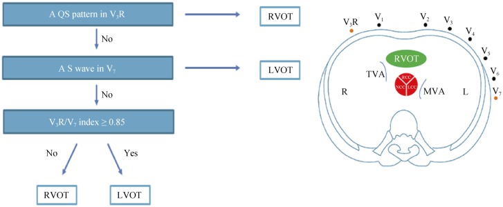
Recently, a study by Cheng and colleagues has been published, the authors evaluated the right precordial and posterior leads with calculation of the V3R/V7 index (defined as R-wave amplitude in lead V3R divided by that in lead V7 when measured in ectopic QRS). ECG leads V5 and V6 are placed into positions V3R and V7 (Figure 9).[19]
Figure 9. Criteria for differentiating LVOT from RVOT.
With modifications from Cheng D, et al.[19] LVOT: left ventricular outflow tract; RVOT: right ventricular outflow tract.
If QS pattern is present in lead V3R, the origin of VAs is RVOT, then if an S-wave is observed in lead V7, the origin of VA is LVOT. If the QRS morphology does not show these characteristics, then the V3V/V7 index is measured. Index < 0.85 indicates RVOT origin, whereas index ≥ 0.85 predicts LVOT origin. The V3V/V7 index ≥ 0.85 predicted an LVOT origin with 87% sensitivity and 96% specificity. The major limitations of this study were the small sample size, lack of validation in larger populations, and even if the origin of VA was defined by the successful ablation sites, it may not represent the true origin, but a site which is anatomically close enough to affect the focus.[19]
3.2. Ventricular arrhythmias originating from the aortic root
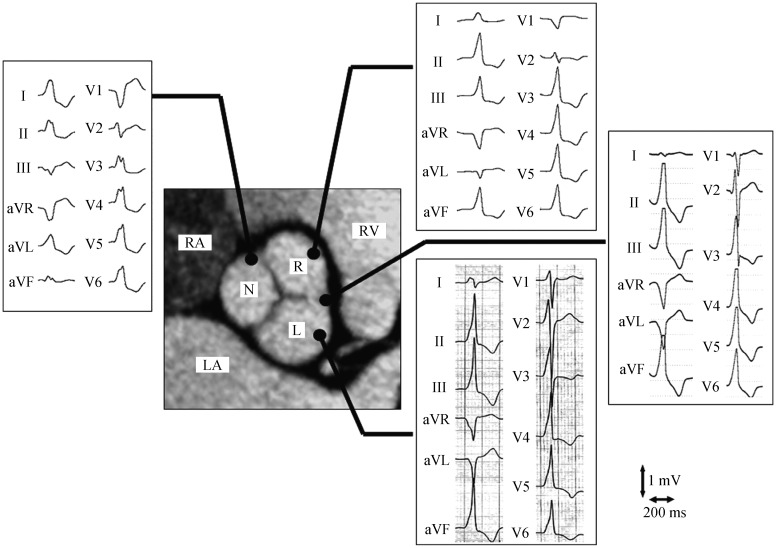
Regarding LVOT, Yamada, et al.[20] reported that VT arising from the aortic root are more commonly originated from the left coronary cusp (LCC), as compared with the right coronary cusp (RCC), non-coronary cusp (NCC) and at the junction between the LCC and RCC (L-RCC). Surface ECG is a useful tool that allows differentiating the site of origin (Figure 10), such us, an R-wave in lead aVL rules out origin in the LCC, RCC, or L-RCC. Right bundle branch block (RBBB) and inferior axis indicate a VT arising from the LCC. To distinguish between the most common origins (LCC and RCC), the R-wave amplitude ratio in leads II and III might be a practical differential element. A III/II ratio > 0.9 determines LCC origin.[20]
Figure 10. Typical electrocardiograms for VT originating from the aortic root.
With permission from Yamada T, et al.[20] L: left coronary cusp; LA: left atrium; N: non-coronary cusp; R: right coronary cusp; RA: right atrium; RV: right ventricle; VT: ventricular tachycardia.
3.3. Fascicular VT/PVC
Described by Belhassen B, et al.[21], fascicular VT is an idiopathic reentry arrhythmia, which is characterized by a RBBB pattern and which involves altered Purkinje fibers on the LV septal wall.[18] Left posterior fascicular VT is present with left superior axis and RS complex in V5 and V6 and RBBB. Left anterior fascicular VT shows RBBB morphology with right axis deviation. The upper septal fascicular VT is rare and exhibits a narrow QRS complex with normal or right axis deviation. Left posterior fascicular VT is the most common compared to other fascicular VTs,[22]–[24] the ECG features of fascicular VT are summarized in Table 7.
Table 7. Different types of fascicular tachycardias.
| Type of tachycardia | QRS width, ms | QRS pattern | QRS axis |
| Posterior fascicle (most frequent) | < 120–140 | RBBB | Left |
| Anterior fascicle | < 120–140 | RBBB | Right |
| Upper septal fascicle | 120 | RBBB or normal | Normal |
With modifications from Kapa, et al.[24] RBBB: right bundle branch block.
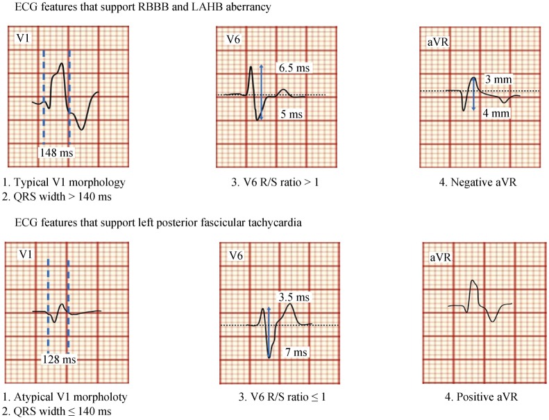
Michowitz, et al.[25] demonstrated that left posterior fascicular VT (LPFVT) is frequently misdiagnosed as SVT with aberrant RBBB and left anterior hemiblock (LAHB), and proposed a novel diagnostic method based on four variables for distinguishing LPFVTs (Figure 11). The authors reported sensitivity and specificity of 82.1% and 78.3%. It was stated that patients with 3 of 4 positive criteria had a high probability of LPFVT, in contrast to patients with ≤ 1 positive criterion, in which RBBB and LAHB was always present.
Figure 11. Schematic electrocardiogram representations.
Differentiating the QRS morphology of LPFVT from RBBB and LAHB aberrancy, when 3 or 4 criteria are positive the diagnosis of left posterior fascicular VT is likely. LAHB: left anterior hemiblock; LPFVT: left posterior fascicular ventricular tachycardia; RBBB: right bundle branch block; VT: ventricular tachycardia.
3.4. Papillary muscle (PM) VT/PVC
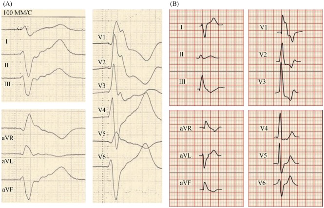
VA originated in the left anterolateral PM exhibit an RBBB pattern with right inferior axis,[26] compared with VA arising from the left inferoseptal PM have RBBB pattern with right or left superior axis (Figure 12).[27] VA arising from the right ventricular papillary muscles (RVPM) where analyzed by Crawford and coworkers, these arrhythmias can be originated from the posterior or anterior RVPM showing a superior axis with late R-wave transition (> V4), though septal right ventricular PM arrhythmias usually present an inferior axis with an earlier R-wave transition in the precordial leads (< V4).[28]
Figure 12. VT from the inferoseptal papillary muscle (A) and VT from the anteroseptal papillary muscle (B: schematic ECG representation).
VT: ventricular tachycardia.
3.5. Differentiation of PM from fascicular and mitral annular ventricular arrhythmias
Ventricular arrhythmias can originate from PMs in structurally normal and diseased hearts. The PMs are the parts of the mitral valve (MV) and tricuspid valve apparatus.[29] VA originating from the PMs, fascicles, and mitral annulus have RBBB pattern.
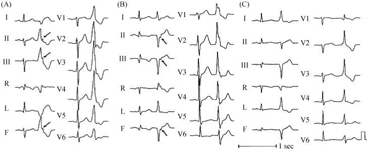
Tada, et al.[30] reported that mitral annulus (MA) VA most commonly originate from the anterolateral (58%), posteroseptal (31%) and posterior MA (11%). In this study, all patients with VA had right bundle branch block morphology, R or Rs pattern in leads V2-V6 and precordial transition in V1 or V2. The anterolateral MA SoO was associated with an inferior axis and negative polarity in leads I and aVL. A posteroseptal or posterior SoO was associated with a superior axis and positive polarity in leads I and aVL (Figure 13).
Figure 13. Representative 12-lead electrocardiograms of premature ventricular contractions originating from the anterolateral (A), posterior (B), and posteroseptal (C) portions of the mitral annulus.
Arrows indicate “notching” of the late phase of the QRS complex in the inferior leads, with permission from Tada H, et al.[30]
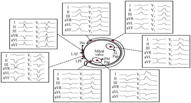
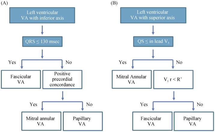
Differentiation of VT/PVC originating from these structures is important for management, since fascicular VT can be treated by calcium antagonists, ablation of VT from PM may require additional ultrasound visualization. Although ECG-based differentiation can be challenging, there are several useful characteristics that can be taken into account (Figure 14).[31] An algorithm for differentiation of fascicular, PM, and mitral annulus VT was proposed by Al'Aref, et al.[31] and included the following features: QRS duration, precordial lead transition, and V1 QRS morphology (Figure 15). This novel algorithm reported an acceptable accuracy rate for the diagnosis of papillary muscle VAs (83%), fascicular VAs (87%), and mitral annular VAs (89%).
Figure 14. Representative 12-lead electrocardiograms of papillary muscle, fascicular, and mitral annular ventricular arrhythmias with corresponding locations on schematic diagram.
With permission from Al'Aref SJ, et al.[31] AL: anterolateral; LAF: left anterior fascicle; LPF: left posterior fascicular; Pap: papillary muscle; PM: posteromedial.
Figure 15. Algorithm for differentiation of focal left VA.
(A): Flow chart shows algorithm for differentiation of inferior axis VA into papillary, fascicular, or mitral annular arrhythmia based on QRS duration and positive precordial concordance; (B): Flow chart shows algorithm for differentiation of superior axis VA into papillary, fascicular, or mitral annular arrhythmia based on QRS morphology in leads V1 and V5. VA: ventricular arrhythmia.
3.6. Ventricular arrhythmias originating from the tricuspid annulus
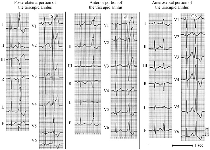
Although VAs originating from the tricuspid annulus are not infrequent, their prevalence is lower compared to other sites. Tada, et al.[30] in his study described that 8% (34 patients) of the total evaluated 454 patients exhibited a VT arising from the tricuspid annulus in which the preferential SoO was the septal portion, especially the anteroseptal portion, as well as it was described VT arising from the free wall. Many ECG features were found, in all patients QRS complexes during the VT had LBBB morphology, lead I had a R or r pattern, negative component in aVR (QS, qs, Qr, or qr pattern), positive component (r or R) in aVL. VT arising from the free wall portion of the annulus had a wider QRS (167 ± 21 ms) in comparison with VT arising from septal portion (143 ± 16 ms). The mid or late component of the notched QRS was more frequent in VT originated in the free wall than from the septal portion. In VT arising from the free wall the R-wave transition occurred after lead V3 and in VT arising from the septal portion of the annulus occurred in lead V3. V1-V3 Q-wave amplitude was higher in VT originated in the free wall portion than in the VT arising from the septal portion (Figure 16).[32]
Figure 16. Representative 12-lead electrocardiograms of premature ventricular contractions originating from the posterolateral, anterior, and anteroseptal portions of the tricuspid annulus.
Arrows indicate the second peak of the “notched” QRS complex in the limb leads, with permission from Tada H, et al.[32]
3.7. Epicardial origin
As reported, about 15% of ventricular arrhythmias have an epicardial site of origin,[33] not amenable to conventional endocardial catheter ablation. This is why electrophysiologists pay special attention to the pre-procedure screening of possible epicardial VT exit sites. Several attempts have been made to develop approaches for identification of ECG characteristics specific for epicardial VT origin. These criteria are summarized in Table 8 and Figure 17.[36]
Table 8. Electrocardiographic criteria proposed for the identification of epicardial VTs.
| Author | Underlying heart disease | Limitations | ECG criteria |
| Berruezo, et al.[36] | CAD: 72% DCM: 28% |
RBBB VT | Pseudodelta wave ≥ 34 ms Intrinsicoid deflection V2 ≥ 85 ms Shortest RS complex ≥ 121 ms |
| Daniels, et al.[35] | No SHD | Described for LVOT VT | Precordial maximum deflection index ≥ 0.55 |
| Bazan, et al.[37]
Vallès, et al.[38] |
NICM | Absence of Q-wave in sinus rhythm | Q-wave in lead I for anterolateral epi VT Q-wave in inferior lead for inferior epi VT |
| Bazan, et al.[39] | CAD: 2, DCM: 4, ARVC: 2, No SHD: 5 | No tested in ARVC VTs. Absence of Q-wave in sinus rhythm | Q-wave in lead I / QS in lead V2 for anterior epi RV VT Q-wave in leads II, III, and aVF is inferior epi RV VT |
ARVC: arrhythmogenic right ventricular cardiomyopathy; CAD: coronary artery disease; DCM: dilated cardiomyopathy; ECG: electrocardiogram; LVOT: left ventricular outflow tract; NICM: non-ischemic cardiomyopathy; RBBB: right bundle branch block; RV: right ventricle; SHD: structural heart disease; VT: ventricular tachycardia.
Figure 17. VT with epicardial origin.
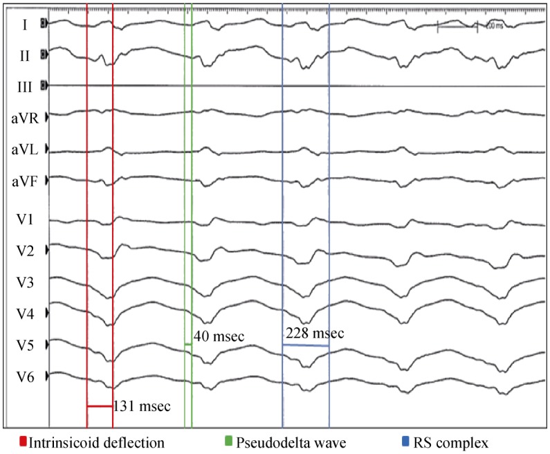
A pseudo delta wave ≥ 34 ms (measured from the earliest ventricular activation to the earliest fast deflection in any precordial lead), intrinsicoid deflection V2 ≥ 85 ms (defined as the interval measured from the earliest ventricular activation to the peak of QRS in V2), shortest RS complex ≥ 121 ms (defined as the interval measured from the earliest ventricular activation to the nadir of the first S wave in any precordial lead). VT: ventricular tachycardia.
It should be acknowledged that these criteria are more specific for non-ischemic VTs, and in patients with post-myocardial infarction VT/PVC they are of less specificity.[36] Once an epicardial SoO is suspected, an operator may consider that this site can be approached via cardiac venous system (through coronary sinus---for some left ventricular basal localizations) or via pericardial access. Pericardial access is performed surgically or via sybxyphoid or modified puncture.[40],[41] Although transcutaneous pericardial puncture is less invasive, it can be associated with major life-threatening complications.[42]
4. VT in structural heart disease
SoO of VTs in patients without structural heart disease (SHD) can be defined by ECG features with high accuracy. Nevertheless, in patients with SHD, such us, myocardial infarction (MI), idiopathic cardiomyopathy (ICM), Chagas disease, sarcoid, etc., determine a certain SoO can represent some difficulties due to the substitution of part of the myocardium with scar what causes changes in the QRS morphology. ECG features of patients with SHD and patients without SHD are differentiated by the presence of QRS complexes with low amplitude, long duration, notches and a reentrant mechanism. In most cases of SHD, VT with RBBB or LBBB pattern arise from the LV.[43]
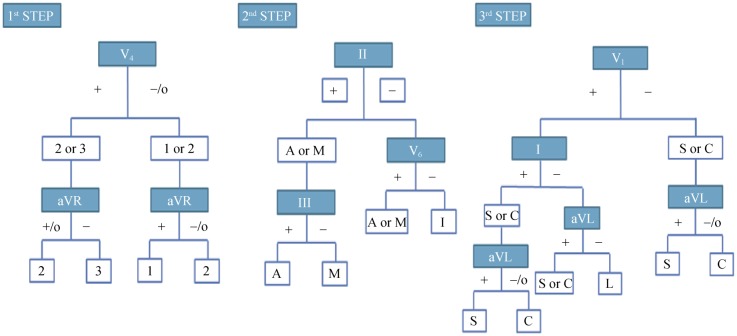
In his study, Kuchar and collaborators analyzed ECG features in a group of 22 patients with prior MI in order to predict the origin of VA.[44] Afterwards a three steps algorithm (Figure 19) was elaborated and tested in 44 patients with sustained ventricular tachycardia. The LV was divided into 3 different areas using two different fluoroscopy views, the right anterior oblique (RAO) at 30° and the left anterior oblique (LAO) at 60° (Figure 18).
Figure 19. Algorithm proposed by Kuchar and collaborators for the identification of exit sites of VT.
A: anterior; C: central; I: inferior; L: lateral; M: middle; S: septal; VT: ventricular tachycardia; o: isoelectric; (+): positive; (–): negative.
Figure 18. Schematic representation of anatomical areas in right anterior oblique (RAO) and left anterior oblique (LAO) views.
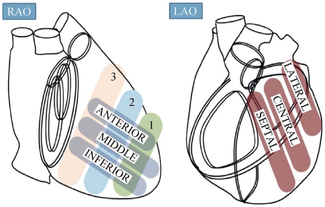
LAO: left anterior oblique; RAO: right anterior oblique.
This algorithm seems to be suitable for diagnosis of VT arising from anterior, inferior, septal and lateral sites offering a success rate of 82% to 90% approximately, the rest of the anatomical points were poorly identify. The impossibility for identifying VT arising from right ventricular exits sites was described as a limitation.[44]
5. Conclusions
Discrimination of VT from SVT on surface ECG is possible in the majority of patients, and localization of VT SOO is very important in everyday practice for invasive electrophysiologists. There are some limitations of surface ECG analysis, and differential diagnosis of WCT is sometimes very difficult. With a certain sensitivity and specificity VT can be diagnosed using a set of algorithms, and VT localization can be suggested. Nowadays, surface ECG stays the most innocuous and practical diagnostic method to identify and classify tachyarrhythmias.
Acknowledgments
This work was supported by RFBR grant (#18-29-02036). The authors had no conflicts of interest to disclose.
References
- 1.Blomström-Lundqvist C, Scheinman MM, Aliot EM, et al. Writing Committee to Develop Guidelines for the Management of Patients With Supraventricular A. ACC/AHA/ESC guidelines for the management of patients with supraventricular arrhythmias-executive summary: a report of the American College of Cardiology/American Heart Association Task Force on Practice Guidelines and the European Society of Cardiology Committee for Practice Guidelines (Writing Committee to Develop Guidelines for the Management of Patients With Supraventricular Arrhythmias) Circulation. 2003;108:1871–1909. doi: 10.1161/01.CIR.0000091380.04100.84. [DOI] [PubMed] [Google Scholar]
- 2.Katritsis DG, Boriani G, Cosio FG, et al. European Heart Rhythm Association (EHRA) consensus document on the management of supraventricular arrhythmias, endorsed by Heart Rhythm Society (HRS), Asia-Pacific Heart Rhythm Society (APHRS), and Sociedad Latinoamericana de Estimulacion Cardiaca y Electrofisiologia (SOLAECE) Europace. 2017;19:465–511. doi: 10.1093/europace/euw301. [DOI] [PubMed] [Google Scholar]
- 3.Cohen HC, Gozo EG, Jr, Pick A. Ventricular Tachycardia with narrow QRS complexes (left posterior fascicular tachycardia) Circulation. 1972;45:1035–1043. doi: 10.1161/01.cir.45.5.1035. [DOI] [PubMed] [Google Scholar]
- 4.Lebedev DS. Wide QRS tachycardia: differential diagnosis and treatment. J Arrhythmol. 1998;7:65–73. [Google Scholar]
- 5.Brugada P, Brugada J, Mont L, et al. A new approach to the differential diagnosis of a regular tachycardia with a wide QRS complex. Circulation. 1991;83:1649–1659. doi: 10.1161/01.cir.83.5.1649. [DOI] [PubMed] [Google Scholar]
- 6.Vereckei A, Duray G, Szénási G, et al. Application of a new algorithm in the differential diagnosis of wide QRS complex tachycardia. Eur Heart J. 2007;28:589–600. doi: 10.1093/eurheartj/ehl473. [DOI] [PubMed] [Google Scholar]
- 7.Eckardt L, Breithardt G, Kirchhof P. Approach to wide complex tachycardias in patients without structural heart disease. Heart. 2006;92:704–711. doi: 10.1136/hrt.2005.063792. [DOI] [PMC free article] [PubMed] [Google Scholar]
- 8.Vereckei A, Duray G, Szénási G, et al. New algorithm using only lead aVR for differential diagnosis of wide QRS complex tachycardia. Heart Rhythm. 2008;5:89–98. doi: 10.1016/j.hrthm.2007.09.020. [DOI] [PubMed] [Google Scholar]
- 9.Pava LF, Perafán P, Badiel M, et al. R-wave peak time at DII: a new criterion for differentiating between wide complex QRS tachycardias. Heart Rhythm. 2010;7:922–926. doi: 10.1016/j.hrthm.2010.03.001. [DOI] [PubMed] [Google Scholar]
- 10.Jastrzebski M, Sasaki K, Kukla P, et al. The ventricular tachycardia score: a novel approach to electrocardiographic diagnosis of ventricular tachycardia. Europace. 2016;18:578–584. doi: 10.1093/europace/euv118. [DOI] [PubMed] [Google Scholar]
- 11.Sandler IA, Marriott HJ. The differential morphology of anomalous ventricular complexes of RBBB-type in lead V1; ventricular ectopy versus aberration. Circulation. 1965;31:551–556. doi: 10.1161/01.cir.31.4.551. [DOI] [PubMed] [Google Scholar]
- 12.Swanick EJ, LaCamera F, Jr, Marriott HJ. Morphologic features of right ventricular ectopic beats. Am J Cardiol. 1972;30:888–891. doi: 10.1016/0002-9149(72)90015-x. [DOI] [PubMed] [Google Scholar]
- 13.Kindwall KE, Brown J, Josephson ME. Electrocardiographic criteria for ventricular tachycardia in wide complex left bundle branch block morphology tachycardias. Am J Cardiol. 1988;61:1279–1283. doi: 10.1016/0002-9149(88)91169-1. [DOI] [PubMed] [Google Scholar]
- 14.Chai Q, Liu G, Zhang H, et al. The value of brugada algorithm in the differential diagnosis of broad QRS complex tachycardia: a meta-analysis. J Cardiovasc Dis Diagn. 2018;6:314–314. [Google Scholar]
- 15.Page RL, Joglar JA, Caldwell MA, et al. 2015 ACC/AHA/HRS Guideline for the management of adult patients with supraventricular tachycardia. J Am Coll Cardio. 2016;67:e27–e115. doi: 10.1016/j.jacc.2015.08.856. [DOI] [PubMed] [Google Scholar]
- 16.Hutchinson MD, Garcia FC. An organized approach to the localization, mapping, and ablation of outflow tract ventricular arrhythmias. J Cardiovasc Electrophysiol. 2013;24:1189–1197. doi: 10.1111/jce.12237. [DOI] [PubMed] [Google Scholar]
- 17.Scanavacca M, Lara S, Hardy C, et al. How to identify & treat epicardial origin of outflow tract tachycardias. J Atr Fibrillation. 2015;7:1195–1195. doi: 10.4022/jafib.1195. [DOI] [PMC free article] [PubMed] [Google Scholar]
- 18.Della Rocca DG, Gianni C, Mohanty S, et al. Localization of ventricular arrhythmias for catheter ablation. Card Electrophysiol Clin. 2018;10:333–354. doi: 10.1016/j.ccep.2018.02.006. [DOI] [PubMed] [Google Scholar]
- 19.Cheng D, Ju W, Zhu L, et al. A novel electrocardiographic criterion for differentiating left from right ventricular outflow tract arrhythmias origins. Circ Arrhythm Electrophysiol. 2018;11:e006243–e006243. doi: 10.1161/CIRCEP.118.006243. [DOI] [PubMed] [Google Scholar]
- 20.Yamada T, McElderry HT, Doppalapudi H, et al. Idiopathic ventricular arrhythmias originating from the aortic root prevalence, electrocardiographic and electrophysiologic characteristics, and results of radiofrequency catheter ablation. J Am Coll Cardiol. 2008;52:139–147. doi: 10.1016/j.jacc.2008.03.040. [DOI] [PubMed] [Google Scholar]
- 21.Belhassen B, Rotmensch HH, Laniado S. Response of recurrent sustained ventricular tachycardia to verapamil. Br Heart J. 1981;46:679–682. doi: 10.1136/hrt.46.6.679. [DOI] [PMC free article] [PubMed] [Google Scholar]
- 22.Prystowsky EN, Padanilam BJ, Joshi S, et al. Ventricular arrhythmias in the absence of structural heart disease. J Am Coll Cardiol. 2012;59:1733–1744. doi: 10.1016/j.jacc.2012.01.036. [DOI] [PubMed] [Google Scholar]
- 23.Lerman BB, Stein KM, Markowitz SM. Mechanisms of idiopathic left ventricular tachycardia. J Cardiovasc Electrophysiol. 1997;8:571–583. doi: 10.1111/j.1540-8167.1997.tb00826.x. [DOI] [PubMed] [Google Scholar]
- 24.Kapa S, Gaba P, DeSimone CV, et al. Fascicular ventricular arrhythmias: pathophysiologic mechanisms, anatomical constructs, and advances in approaches to management. Circ Arrhythm Electrophysiol. 2017;10:e002476–e002476. doi: 10.1161/CIRCEP.116.002476. [DOI] [PubMed] [Google Scholar]
- 25.Michowitz Y, Tovia-Brodie O, Heusler I, et al. Differentiating the QRS morphology of posterior fascicular ventricular tachycardia from right bundle branch block and left anterior hemiblock aberrancy. Circ Arrhythm Electrophysiol. 2017;10:e005074–e005074. doi: 10.1161/CIRCEP.117.005074. [DOI] [PubMed] [Google Scholar]
- 26.Yamada T, Mcelderry HT, Okada T, et al. Idiopathic focal ventricular arrhythmias originating from the anterior papillary muscle in the left ventricle. J Cardiovasc Electrophysiol. 2009;20:866–872. doi: 10.1111/j.1540-8167.2009.01448.x. [DOI] [PubMed] [Google Scholar]
- 27.Doppalapudi H, Yamada T, McElderry HT, et al. Ventricular tachycardia originating from the posterior papillary muscle in the left ventricle. Circ Arrhythm Electrophysiol. 2008;1:23–29. doi: 10.1161/CIRCEP.107.742940. [DOI] [PubMed] [Google Scholar]
- 28.Crawford T, Mueller G, Good E, et al. Ventricular arrhythmias originating from papillary muscles in the right ventricle. Heart Rhythm. 2010;7:725–730. doi: 10.1016/j.hrthm.2010.01.040. [DOI] [PubMed] [Google Scholar]
- 29.Enriquez A, Supple GE, Marchlinski FE, et al. How to map and ablate papillary muscle ventricular arrhythmias. Heart Rhythm. 2017;14:1721–1728. doi: 10.1016/j.hrthm.2017.06.036. [DOI] [PubMed] [Google Scholar]
- 30.Tada H, Ito S, Naito S, et al. Idiopathic ventricular arrhythmia arising from the mitral annulus: a distinct subgroup of idiopathic ventricular arrhythmias. J Am Coll Cardiol. 2005;45:877–886. doi: 10.1016/j.jacc.2004.12.025. [DOI] [PubMed] [Google Scholar]
- 31.Al'Aref SJ, Ip JE, Markowitz SM, et al. Differentiation of papillary muscle from fascicular and mitral annular ventricular arrhythmias in patients with and without structural heart disease. Circ Arrhythm Electrophysiol. 2015;8:616–624. doi: 10.1161/CIRCEP.114.002619. [DOI] [PubMed] [Google Scholar]
- 32.Tada H, Tadokoro K, Ito S, et al. Idiopathic ventricular arrhythmias originating from the tricuspid annulus: prevalence, electrocardiographic characteristics, and results of radiofrequency catheter ablation. Heart Rhythm. 2007;4:7–16. doi: 10.1016/j.hrthm.2006.09.025. [DOI] [PubMed] [Google Scholar]
- 33.Baman TS, Ilg KJ, Gupta SK, et al. Mapping and ablation of epicardial idiopathic ventricular arrhythmias from within the coronary venous system. Circ Arrhythm Electrophysiol. 2010;3:274–279. doi: 10.1161/CIRCEP.109.910802. [DOI] [PubMed] [Google Scholar]
- 34.Fernandez-Armenta J, Berruezo A. How to recognize epicardial origin of ventricular tachycardias? Curr Cardiol Rev. 2014;10:246–256. doi: 10.2174/1573403X10666140514103047. [DOI] [PMC free article] [PubMed] [Google Scholar]
- 35.Daniels DV, Lu YY, Morton JB, et al. Idiopathic epicardial left ventricular tachycardia originating remote from the sinus of valsalva: electrophysiological characteristics, catheter ablation and identification from the 12-lead electrocardiogram. Circulation. 2006;113:1659–1666. doi: 10.1161/CIRCULATIONAHA.105.611640. [DOI] [PubMed] [Google Scholar]
- 36.Berruezo A, Mont L, Nava S, et al. Electrocardiographic recognition of the epicardial origin of ventricular tachycardias. Circulation. 2004;109:1842–1847. doi: 10.1161/01.CIR.0000125525.04081.4B. [DOI] [PubMed] [Google Scholar]
- 37.Bazan V, Gerstenfeld EP, Garcia FC, et al. Site-specific twelve-lead ECG features to identify an epicardial origin for left ventricular tachycardia in the absence of myocardial infarction. Heart Rhythm. 2007;4:1403–1410. doi: 10.1016/j.hrthm.2007.07.004. [DOI] [PubMed] [Google Scholar]
- 38.Vallès E, Bazan V, Marchlinski FE. ECG criteria to identify epicardial ventricular tachycardia in nonischemic cardiomyopathy. Circ Arrhythm Electrophysiol. 2010;3:63–71. doi: 10.1161/CIRCEP.109.859942. [DOI] [PubMed] [Google Scholar]
- 39.Bazan V, Bala R, Garcia FC, et al. Twelve-lead ECG features to identify ventricular tachycardia arising from the epicardial right ventricle. Heart Rhythm. 2006;3:1132–1139. doi: 10.1016/j.hrthm.2006.06.024. [DOI] [PubMed] [Google Scholar]
- 40.Sosa E, Scanavacca M, D'avila A, et al. A new technique to perform epicardial mapping in the electrophysiology laboratory. J Cardiovasc Electrophysiol. 1996;7:531–536. doi: 10.1111/j.1540-8167.1996.tb00559.x. [DOI] [PubMed] [Google Scholar]
- 41.Simonova KA, Mikhailov EN, Tatarsky RB, et al. Epicardial arrhythmogenic substrate in patients with postinfarction ventricular tachycardias: a pilot study. J Arrhythmol. 2019;26:38–46. [Google Scholar]
- 42.Koruth JS, Aryana A, Dukkipati SR, et al. Unusual complications of percutaneous epicardial access and epicardial mapping and ablation of cardiac arrhythmias. Circ Arrhythm Electrophysiol. 2011;4:882–888. doi: 10.1161/CIRCEP.111.965731. [DOI] [PubMed] [Google Scholar]
- 43.Miller JM, Jain R, Dandamudi G, et al. Electrocardiographic localization of ventricular tachycardia in patients with structural heart disease. Card Electrophysiol Clin. 2017;9:1–10. doi: 10.1016/j.ccep.2016.10.001. [DOI] [PubMed] [Google Scholar]
- 44.Kuchar DL, Ruskin JN, Garan H. Electrocardiographic localization of the site of origin of ventricular tachycardia in patients with prior myocardial infarction. J Am Coll Cardiol. 1989;13:893–903. doi: 10.1016/0735-1097(89)90232-5. [DOI] [PubMed] [Google Scholar]


