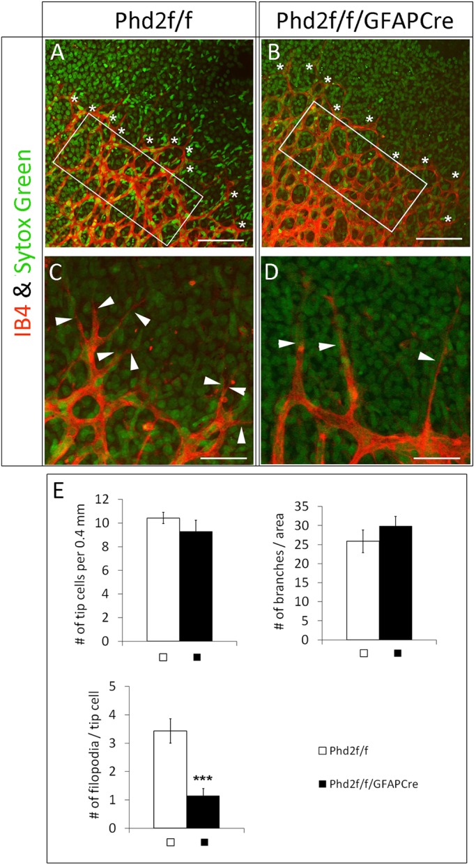
Fig. 6.

Astrocytic PHD2 deficiency hinders tip cell filopodia development. (A,B) Vascular front from P5 retinas double stained with IB4-Alexa 594 and SYTOX Green, with tip cells indicated by white asterisks. (C,D) High-magnification images showing filopodia (white arrowheads). (E) Number of vascular branches per 0.02 mm2 in white rectangles in A and B, number of tip cells per 0.4 mm length of vascular front, and filopodia per tip cell. n=7 mice/group. ***P<0.001. Scale bars: 100 µm (A,B); 40 µm (C,D).
