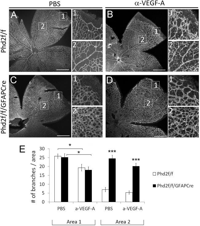
Fig. 9.
Anti-VEGF does not rescue vascular pruning in Phd2f/f/GFAPCre mice. (A-D) IB4-stained P8 retinas from Phd2f/f and Phd2f/f/GFAPCre mice pre-treated with intravitreal injection of anti-VEGF or PBS at P4. Images numbered 1 or 2 are expanded from corresponding boxed areas in the main panels. (E) Quantification of vascular branches per 0.02 mm2 area. Vascular density in area 1 mostly reflects sprouting angiogenesis, because vascular pruning is negligible near the vascular front. At 1.8 µg per eye, anti-VEGF (a-VEGF-A) moderately suppressed angiogenesis. Values in area 2 reflect the sum of the initial angiogenesis and subsequent pruning. n=7 mice/group. *P<0.05; ***P<0.001. Scale bars: 500 µm.

