Abstract
Stress during early life has potential to program and alter the response to stressful events and metabolism in later life. Repeated short exposure of Atlantic salmon to cold water and air during embryonic (E), post-hatch (PH) or both phases of development (EPH) has been shown to alter the methylome and transcriptome and to affect growth performance during later life compared to untreated controls (CO). The aim of this study was to investigate how the transcriptome of these fish responds to subsequent acute stress at the start feeding stage, and to describe methylation differences that might steer these changes. EPH treated fish showed the strongest down-regulation of corticotropin releasing factor 1, up-regulation of glucocorticoid receptor and 3-oxo-5-alpha-steroid 4-dehydrogenase 2 gene expression and a suppressed cortisol response 3 hr after the acute stress, differences that could influence hormesis and be affecting how EPH fish cope and recover from the stress event. Growth hormone 2 and insulin-like growth factor 1 were more strongly down-regulated following acute stress in EPH treated fish relative to E, PH and CO fish. This indicates switching away from growth toward coping with stress following stressful events in EPH fish. Genes implicated in immune function such as major histocompatibility class 1A, T-cell receptor and toll-like receptor also responded to acute stress differently in EPH treated fish, indicating that repeated stresses during early life may affect robustness. Differential DNA methylation was detected in regions mapping <500 bases from genes differentially responding to acute stress suggesting the involvement of epigenetic mechanisms. Stress treatments applied during early development therefore have potential as a husbandry tool for boosting the productivity of aquaculture by affecting how fish respond to stresses at critical stages of production.
Keywords: Development, acute stress response, gene expression, DNA methylation, Atlantic salmon
Exposure to stressful events during early life stages may result in the development of a variety of metabolic, immune, endocrine and neuro-psychiatric disorders (Kapoor et al. 2006; Weinstock 2008; Korosi and Baram 2010; Meaney et al. 2007; Love and Williams 2008; Hu et al. 2008; Auperin and Geslin 2008; Reul et al. 2015). The detailed stress response depends on the nature and effect of the stressor/threatening stimuli, but the initial epinephrine surge, and “fight or flight” response, is generally followed by the activation of the hypothalamic-pituitary-adrenal (HPA) axis, or the functional analog, hypothalamic-pituitary-interrenal (HPI) axis in amphibians and fish (Mateus et al. 2017). The release of corticotropin releasing factor (CRF) from the hypothalamic preoptic area induces the synthesis of the pituitary pro-opiomelanocortin (POMC) to be processed into adrenocorticotropic hormone (ACTH). The binding of ACTH to the melanocortin 2 receptor (MC2R) activates the synthesis and release of glucocorticoid (GC) and mineralocorticoid (MC) hormones from the adrenal gland, or the interrenal cells in lower vertebrates (Vander et al. 1970). The glucocorticoids and their receptor (GR) play pivotal roles in the response to stressful challenges by regulating a diversity of metabolic, endocrine and immune processes (Korosi and Baram 2010; Seckl 2008; Szyf et al. 2007). Stress resilience depends on the proper regulation of basal and stress-induced glucocorticoid levels involving rapid negative feedback control of the HPA/HPI axis (Paragliola et al. 2017). Epigenetic mechanisms affecting this feedback in mammals, amphibians and fish seem to involve the regulation of gr, crf1 and pomc (Boschen et al. 2018; Yehuda et al. 2015; Wu et al. 2014; Li and Leatherland 2013; Palma-Gudiel et al. 2015; Metzger and Schulte 2016; Reul et al. 2015).
Cortisol is the main glucocorticoid hormone in fish and exerts its actions through GR and/or the mineralcorticoid receptor (MR) (Baker et al. 2013a; Baker et al. 2013b; McCormick et al. 2008). In addition to its role in the stress response, cortisol is thought to reduce the ability to resist infection or respond to injuries by reducing inflammatory responses and has an important role in energy maintenance through GR signaling (Rose and Herzig 2013; Chatzopoulou et al. 2016). Maternally derived cortisol and gr transcripts play a key role in developmental programming as demonstrated in zebrafish (Pikulkaew et al. 2011; Nesan et al. 2012; Nesan and Vijayan 2012; Best et al. 2017). The onset of the embryonic synthesis of cortisol varies between fish species, but a cortisol stress-response first develops after hatching in most teleosts (Stouthart et al. 1998; Wilson et al. 2013).
Despite the lack of an early stress-induced cortisol response, frequent stress treatment during the early development of teleosts has been shown to influence the sensitivity to acute stress at later stages. For example, the plasma cortisol response to acute stress is dampened in 5-month old rainbow trout fingerlings exposed to brief stress at the eye pigmentation, hatching, or yolk resorption stages (Auperin and Geslin 2008). Similarly, unpredictable chronic low intensity stresses during early life stages in sea bass modified the cortisol response to acute stress at the juvenile stage (Tsalafouta et al. 2014).
Possession of a functional HPI axis in hatching wild salmon is advantageous in the animals natural habitat at a time when it rises from the gravel and faces many different predators (Hagen et al. 2002). In the aquaculture environment there are also many stresses encountered around hatching time (crowding, handling and cleaning) and the response of the fish at this stage could affect survival and performance in aquaculture. Together with the development of the HPI axis, the growth hormone (GH)- insulin-like growth factor 1 (IGF-1) axis seems to be playing a functional role in fish development and differentiation at very early embryonic stages (Besseau et al. 2013). Recently we showed that frequent exposure to “mild” stress (cold water and air) during embryonic and/or post-hatch stages affected the methylome and transcriptome of Atlantic salmon and growth performance in later life, providing evidence of hormesis, or an adaptive response to low-level repeated stress (Moghadam et al. 2017). Here we extend this study by investigating how Atlantic salmon stressed at early life stages are programmed to respond to acute stresses later in life and whether epigenetic programming during early development is associated with the stress response. The aim of the study was to test how such changes might ultimately affect performance in the aquaculture environment.
Methods
Experimental treatments
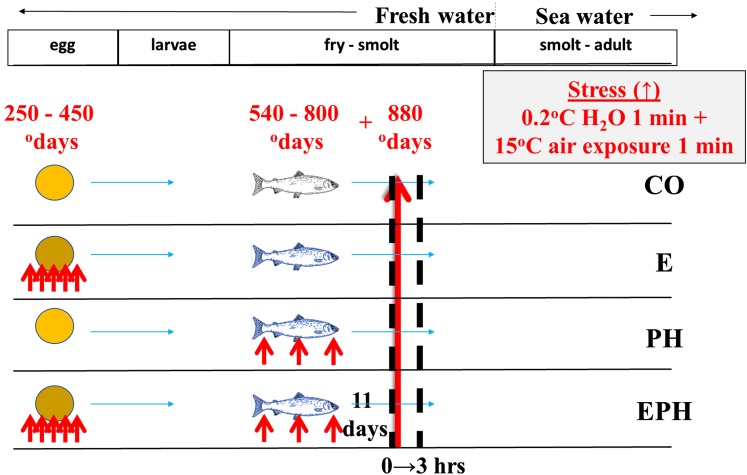
Developing Atlantic salmon were treated as described in Moghadam et al. (2017). In brief, eggs and milt from a full-sib family were fertilized using standard procedures at the Aquaculture Research Station in Tromsø, Norway. Embryos were divided into four triplicate groups of fish which were i: unstressed until 880 odays (CO), ii: stressed during embryogenesis (E), iii: stressed during post-hatch (PH), and iv: stressed during both embryonic and post-hatch stages (EPH). The stress treatment consisted of exposure to cold water at 0.2° for 1 min followed by exposure to 15° air for 1 min, 5 times during embryogenesis from 250 – 450 odays and/or 3 times during post-hatch development from 540 – 800 odays (Figure 1). In between the bouts of stress animals were held at 7°. The results from this stress treatment and the resulting body growth after sea transfer are published in (Moghadam et al. 2017). The weight of ten randomly picked individuals per replicate was checked prior to start feeding (this was 1 year before the measurement after sea transfer by Moghadam et al. 2017).
Figure 1.
Time course of experimental treatments and sampling for control (CO) embryonic (E) post hatch (PH) and embryonic post hatch (EPH) experimental treatments. Small red arrows indicate application of stresses for the three treatment groups (E, PH and EPH) and the large red arrow indicates the timing and application of the acute stress event to all experimental groups. Dashed black lines show when samples were taken for RRBS and RNA-seq.
To test how these different experimental groups responded to an acute stress at a later stage we examined the response of the transcriptome in these fish following a “stress test” at start feeding. To identify associated epigenetic marking that might influence the response of the transcriptome, we looked for differences between the experimental treatments existing in the DNA methylome before the acute stress event. Sixteen thousand fertilized eggs in total were evenly aliquoted to the treatment and control trays. Six fish from each of the CO, E, PH and EPH treatment groups were randomly sampled (two fish from each of the three replicate trays per group) to measure baseline gene expression and DNA methylation at 880 odays (designated as 0 hr). This 0 hr baseline gene expression and DNA methylation time point was 11 days (80 odays) after the repeated stress treatments for the PH and EPH groups and 62 days (430 odays) after the repeated stress treatment for the E group. The 0 hr samples were the same as those used to measure gene expression responses and DNA methylation patterns by Moghadam et al. (2017). Animals from each group were distributed into special containers housed within a common tank and subjected to acute stress consisting of a 1-minute cold shock and 1-minute exposure to air and sampled at 3 hr post-stress. The post-stress samples were chosen for RNA sequencing to assess the gene expression response to the acute stress (with reference to baseline gene expression in animals sampled before the acute stress).
mRNA-seq
Samples (whole fish) were stored in RNA later (per manufacturer’s specifications, Ambion). RNA and DNA from 24 individuals from each time point (6 per experimental group) was isolated as described in Moghadam et al. (2017). Samples were shipped on dry ice, quality checked using a Bioanalyzer (Agilent) and prepared for sequencing using standard procedures by Zymo Research (San Diego, CA, USA). Total RNA was treated with the Ribo-Zero Magnetic Gold Kit (Human/Mouse/Rat) from Illumina (cat. #MRZG126), and stranded libraries were prepared from rRNA depleted samples using ScriptSeq v2 RNA-Seq Library Preparation Kit from Illumina (Cat. #SSV21106). Sequencing of the 24 libraries of single-end 50 bp reads was performed on an Illumina HiSeq 2500 genome analyzer. mRNA-seq produced on average 64.4 million reads per sample, of which around 60.3 million were mapped to the Atlantic salmon genome (consisting of 19.3 million multiple hits and 41.0 million unique hits on average per sample).
Reduced representation bisulphite sequencing
Preparation of samples for RRBS sequencing (0 hr) is described in (Moghadam et al. 2017). In brief, libraries were prepared from TaqαI and MspI digested gDNA and ligated to adapters containing 5′-methyl-cytosine instead of cytosine according to manufacturer’s instructions (Illumina Inc., San Diego, CA, USA). The ligated fragments were bi-sulfite treated, amplified with PCR and 50 bp of the paired-ends were sequenced on a Illumina HiSeq 2500 genome analyzer.
Analysis of differential gene expression and DNA methylation
The sequencing reads were trimmed to remove adapters and low-quality base end regions with Trimmomatic (Bolger et al. 2014). Trimmomatic was set to: a) remove N’s or low quality sequence (below quality score 15) in leading and trailing bases; b) scan using a 4 base sliding window and trim or cut the sequence when the average quality per base drops below 15 and 3, respectively) exclude any of the trimmed and filtered reads with lengths less than 30 bases. Genomic and transcriptomic short reads were mapped to Atlantic salmon genome assembly GCA000233375.4 ICSASG_v2 (Lien et al. 2016) using TopHat version 2.0.8b (Trapnell et al. 2009) and Bismark (Krueger and Andrews 2011) with Bowtie1 (Langmead et al. 2009) (the latter used to extract methylation status). Default parameters for TopHat were used except for library-type stranded and the b2 option sensitive (which calls a package of pre-set parameters that is passed to Bowtie, Langmead et al. 2009). The scripts.count python program in HTSeq (version 0.8.0, Anders et al. 2015) was used to count the reads in the bam file mapping to each feature, ordering the data according to name, skipping all reads with alignment quality less than 10, using exon as the feature type, using gene_id as feature ID and union mode to handle reads overlapping more than one feature. The annotation used to derive the gene counts was created in-house based on RNAseq data from 494 individuals (various life stages and tissues sampled as described in Moghadam et al. 2017 and supplied as supplementary data). Differential gene expression was tested using a model for analysis of read coverage data based on the negative binomial distribution (DESeq2 package in R) (Love et al. 2014). A principle component analysis of the DESeq2 normalized and regularised-logarithmic transformed read abundance data were conducted to visualize any influence of treatment or time post-acute stress on overall patterns of gene expression.
RRBS methylation data (read coverage and % methylation per base position) was analyzed using logistic regression (methylKit package in R) (Akalin et al. 2012). Methylation differences were considered significant where the difference in methylation was >20% and q-value <0.1. Areas containing significant DNA methylation differences between treatments were flagged when they occurred <500kb up- or down-stream of differentially expressed genes.
Cortisol assay and analysis
Cortisol extraction was performed according to de Jesus et al. (1991) on 15 samples per treatment (5 per replicate) at 0, 1, 3 and 24 hr post-acute stress. In brief, 0.2 to 0.6 g of embryonic and larval samples were homogenized in 5x (w/v) ice-cold phosphate buffer saline (pH 7.4) with a rotor homogenizer. Cortisol was extracted from 2 × 250 μl of homogenate with 3 ml of diethyl ether. The water phase of the extract was allowed to freeze by placing tubes in -80° and the combined diethyl ether layer was transferred into a new tube. The ether was evaporated by placement of tubes in a 45° water bath for 1 h. Samples were then reconstituted in 250 μl of enzyme immunoassay buffer (EIA buffer Cayman Chemical, MI, USA). Cortisol was quantified using a commercial enzyme immunoassay kit (Cayman Chemical, MI, USA), which was previously used to evaluate zebrafish whole-trunk cortisol measurements (Pavlidis et al. 2011). Displacement curves (three different homogenates in serial dilutions 1:10 to 1:75) demonstrated linear parallelism (standard curve r2 = 0.997 ± 0.001). The recovery of cortisol added to pool homogenates was 94.5 ± 1.2% (mean ± SEM, n = 2).
Data availability
The GSA Figshare portal was used to upload supplemental files (captions below). All raw sequences were deposited to the NCBI Short Read Archive under the BioProject IDs PRJNA388534 and PRJNA510456. Supplemental material available at Figshare: https://doi.org/10.25387/g3.7586393.
Results
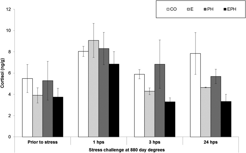
No significant differences in growth rate or survival were detected between the replicates for any of the treatments used in this study. The four treatment groups showed similar basal levels of cortisol prior to the acute stress at 880 0days and the levels peaked 1 hr after the acute stress in all treatment groups (Figure 2). EPH treated fish tended to have the lowest cortisol levels and returned to pre-stress levels at three hours post-stress. Based on the cortisol levels, preliminary qPCR results (not shown) and the literature, we chose to study the response to acute stress at 3 hr under the assumption that most major differences in gene expression would be occurring two hours after the cortisol peak.
Figure 2.
Cortisol concentrations measured in CO, E, PH and EPH treated fish prior to stress and at 1, 3 and 24 hr post-stress (hps).
Genome-wide gene expression changes
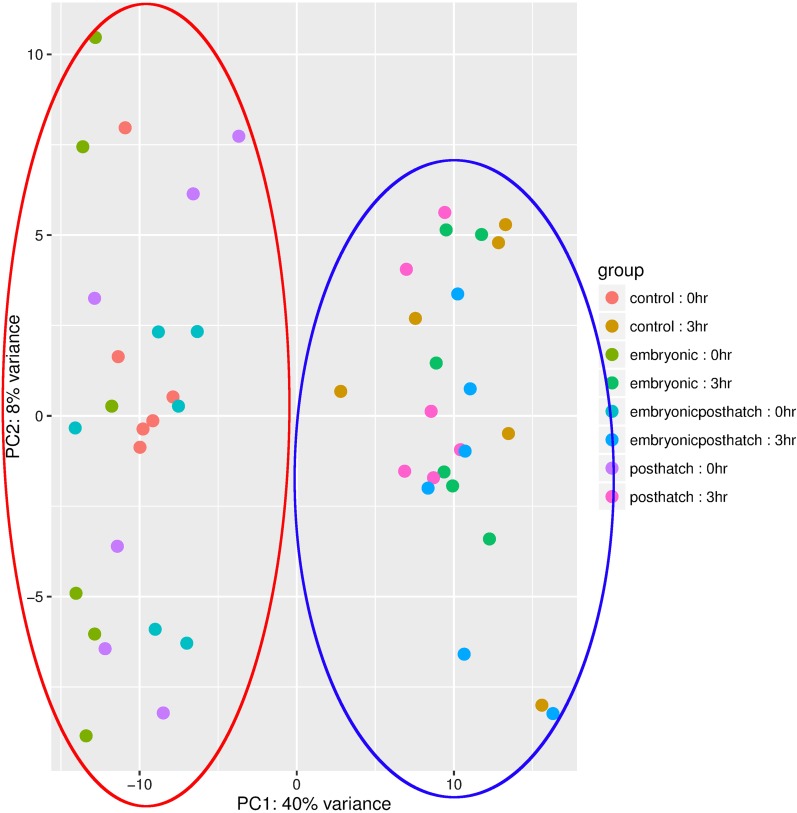
To visualize any influence of treatment or time post-acute stress on overall patterns of gene expression, we performed a principle component analysis and showed that samples cluster according to hours post-acute stress (0 hr vs. 3 hr, Figure 3). The first component explained most (40%) of the variance. No clustering was apparent for the different treatments.
Figure 3.
Principle component analysis of treatment vs. time after acute stress. The red circle highlights samples taken at 0 hr and the purple circle highlights samples taken at 3 hr post-stress.
The expression of large numbers of genes were up- and down-regulated in all experimental groups 3 hr after the acute stress when compared to the pre-stress levels (0 hr) (Supplementary Figure 1). There was a greater trend toward down-regulated gene expression in response to the acute stress in all groups (3746, 2664, 407 and 1517 genes more than onefold down-regulated with Padj < 0.05 in CO, E, PH and EPH groups respectively) than up-regulated (637, 883, 149 and 854 genes up-regulated respectively with Padj < 0.05). The largest number of differentially expressed genes were detected for animals previously stressed during embryonic development (E) (Supplementary Figure 1B) while the least were found for animals stressed after hatching (PH) (Supplementary Figure 1C). Early exposure to stress at both embryo and post-hatch stages (EPH) tended to lead to an intermediate acute stress response based on the intermediate number of differentially expressed genes in this group.
As the expression of many genes were affected by the acute stress, we focused in on particular genes that were differentially expressed exclusively in the EPH treated fish compared to the other treatment groups or control fish. Fish treated with an extended period of stress in this way were shown to adaptively respond, and have superior growth, compared to E and PH stress treatments (Moghadam et al. 2017), and therefore we were interested to see how the response to acute stress differs in these fish, particularly for genes affecting the HPI and somatotropic axes, cortisol inactivation and immune response.
Genes influencing the HPI axis and cortisol inactivation
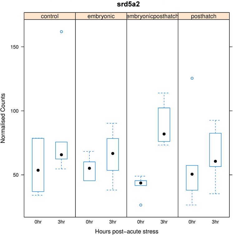
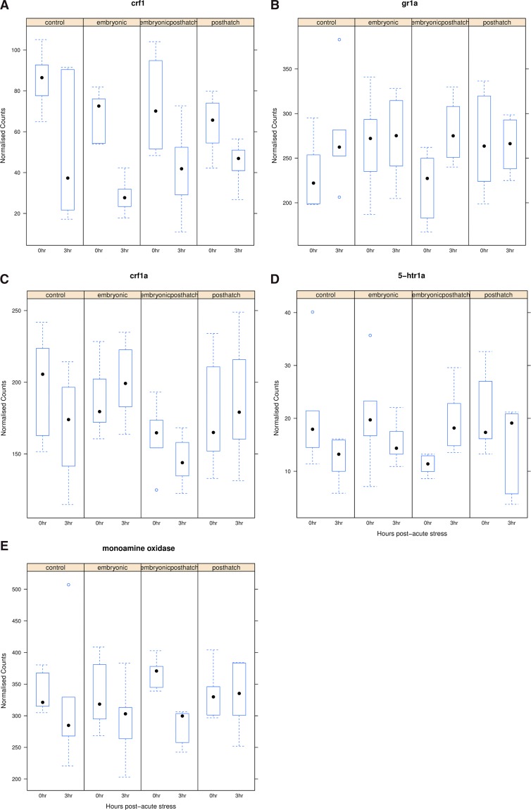
3-oxo-5-alpha-steroid 4-dehydrogenase 2 (srd5a2) was significantly up-regulated in response to acute stress in EPH treated fish, but unresponsive with other treatments (Figure 4). The enzyme encoded by this gene plays a role in metabolism and inactivation of cortisol. The expression of arginine vasopressin-induced 1 (avpi1), crf1 (Figure 5A) and crf binding proteins (crfbp) and pomc A1 and A2 genes was reduced, while the expression of many glucocorticoid and mineralocorticoid receptor genes (gr1a, gr1-like, mr1a and mr1blike, e.g., gr1a, Figure 5B) was increased, or stabile, in response to acute stress across all treatments and control. Although insignificant, CO and EPH treated fish tended to show up-regulated gr1a expression, while relatively little change in response to acute stress was observed for E and PH treated fish (Figure 5B). Responses to acute stress were insignificant for the crf receptors (e.g., crf1a Figure 5C) but, crfr2a tended to be down-regulated in CO and E, unchanged in PH and up-regulated in EPH treated animals. The brain serotonergic system plays a key role in coordinating stress responses, and the serotonin receptors, or 5-hydroxytryptamine receptors (5-HTR), (e.g., 5-htr1a, Figure 5D) tended to be down-regulated in expression under all treatments in response to the acute stress, except for 5-htr1b which was non-significantly up-regulated in the PH and EPH groups (with relatively low levels before the acute stress in EPH treated fish). The enzymatic degradation of serotonin is mainly served by monoamine oxidase, which displayed down-regulated (padj < 0.01) gene expression in the EPH treatment group (Figure 5E), but did not respond to acute stress in the CO, E and PH treatment groups. In summary, a specific response of the EPH fish to acute stress is suggested by the differential expression of various genes along the HPI axis.
Figure 4.
Box plot of changes in the expression of 3-oxo-5-alpha-steroid 4-dehydrogenase 2 (srd5a2) for control (CO), embryonic (E), post-hatch (PH) and embryonic post-hatch (EPH) treated fish at 0 and 3 hr post-acute stress.
Figure 5.
Box plot of changes in the expression of corticotropin releasing factor 1 (crf1, A), glucocorticoid receptor 1a (gr1a, B), corticotropin releasing factor receptor 1 (crf1a, C), 5-hydroxytryptamine receptor 1A (5-htr1a, D) and monoamine oxidase (E) for control (CO), embryonic (E), post-hatch (PH) and embryonic post-hatch (EPH) treated fish at 0 and 3 hr post-acute stress.
Response of the somatotropic axis
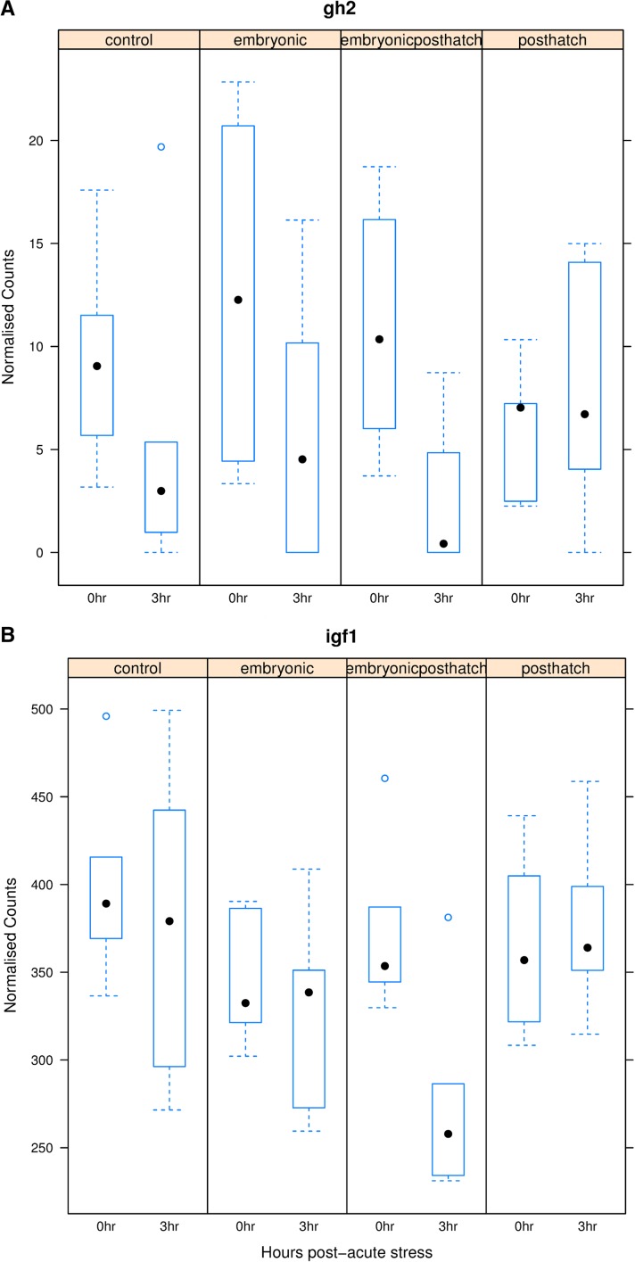
Expression of growth hormone 2 (gh2) (Figure 6A) and insulin-like growth factor 1 (igf1) (Figure 6B) was significantly suppressed for EPH treated fish (padj < 0.05), while expression was relatively unresponsive or not significantly down-regulated for the controls and other treatments. Somatostatin 1A (sst1a) showed little response to acute stress while the expression of the GH releasing peptide ghrelin tended to be suppressed with acute stress for all treatments. The down-regulation of these genes in the somatotropic axis in response to acute stress after EPH treatment contrasts with the response of genes involved in the HPI axis, wherein gr became more strongly up-regulated following acute stress in EPH treated fish.
Figure 6.
Box plot of changes in the expression of growth hormone 2 (gh2, A) and insulin-like growth factor 1 (igf1, B) for control (CO), embryonic (E), post-hatch (PH) and embryonic post-hatch (EPH) treated fish at 0 and 3 hr post-acute stress.
Expression differences After repeated stress treatments and response to acute stress
To determine whether genes that were differentially expressed between the E, PH and EPH treated fish (before the acute stress at 0 hr; Moghadam et al. 2017) were also differentially expressed in response to the acute stress (3 hr), we performed a Pearson correlation analysis of fold-difference in expression (Supplementary Figure 2). The acute stress had the effect of adding a PH stress treatment such that genes that were up-regulated in EPH compared to E treatment (at 0 hr) were generally the same as those that were up-regulated after acute stress was applied to the E treatment group (0 vs. 3 hr, r = 0.8, Supplementary Figure 2K). A weaker positive correlation was observed comparing up-regulation in CO relative to EPH treatment with the response to acute stress over 3 hr in CO treated animals (r = 0.6, Supplementary Figure 2A).
Genes whose expression was up- or down-regulated in the EPH compared to the PH treatment were often affected in the reverse fashion (down- or up-regulated respectively) when EPH treated animals were subjected to an acute stress (r=-0.62, Supplementary Figure 2F). A similar negative correlation was observed for genes that were down- or up-regulated in E treated fish relative to CO compared to the response to acute stress in CO and in E treated animals (r=-0.56 and -0.45, Supplementary Figures 2 I and J respectively). These reversals occurred 3 hr after the acute stress is applied and could be a transient response. A possible explanation is that many of these genes are down-regulated due to broad scale switching in favor of genes influencing the acute stress response of the fish. Further research is needed to see whether the expression bounces back or remains suppressed as the animals recover from the acute stress.
Methylation differences
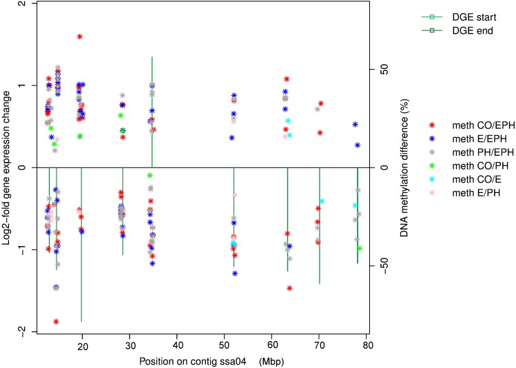
RRBS sequencing yielded on average 42.2 million read pairs, 11.9 million unique CpGs with mapping efficiency of 49%, 6 X coverage and 99% bisulphite conversion rate. Of the 223 genes responding to acute stress in EPH treated animals (log-fold change >1 and Padj < 0.01), 156 were found within 500 kb of regions displaying significant differences in methylation associated with treatment. Several interesting DNA methylation and gene expression effects were found to map to ssa04 in particular (Figure 7). Most of these DNA methylation differences (9849 or 55%) involved either increased or reduced methylation with EPH treatment (37%, 34% and 29% compared to PH, E and CO respectively), 45% of the methylation differences occurred in intergenic, 30% in gene, 10% in promoter, 6% in CDS, 6% in five-prime UTR and 3% in three-prime UTR.
Figure 7.
DNA methylation differences between treatments 0 hr pre-acute stress (stars) and gene expression response (from 0 to 3 hr after acute stress, shown as green bars) mapped across chromosome ssa04 for EPH treated fish.
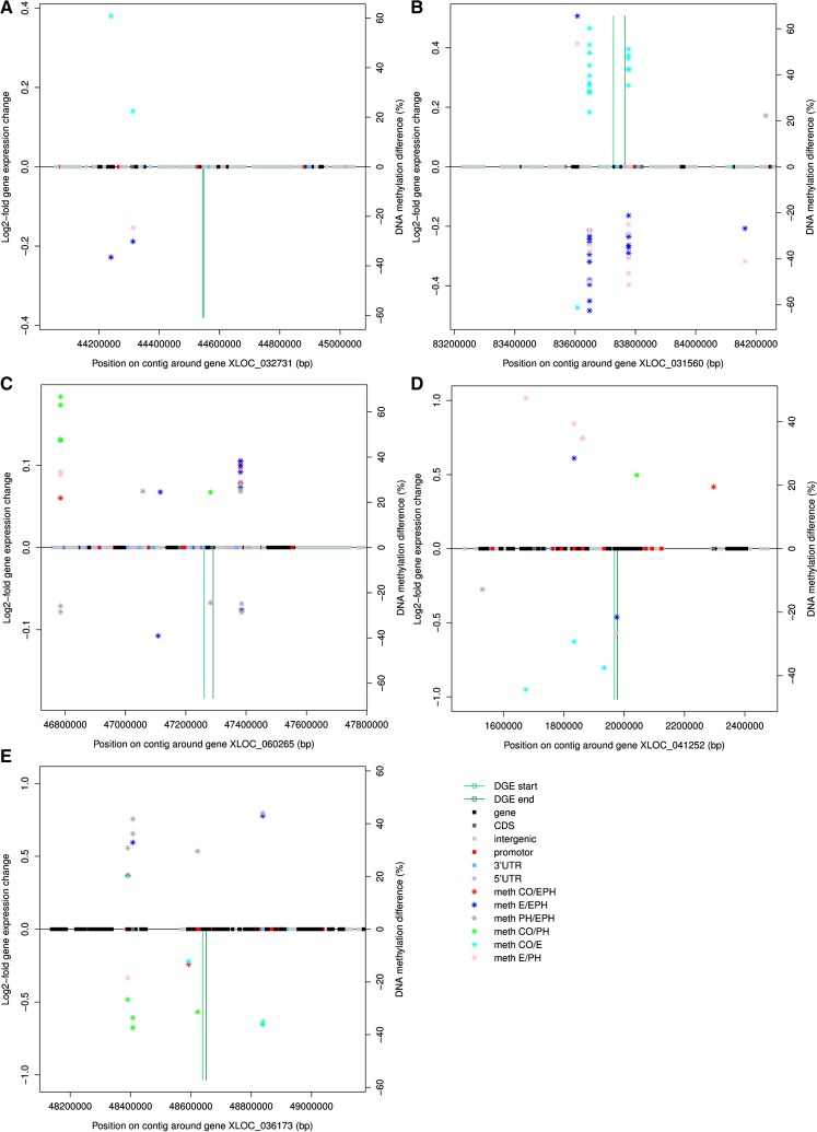
Of the differentially expressed genes in the HPI axis, the down-regulated expression of pomc1a in the CO, E and EPH fish, but up-regulation in PH fish, might be influenced by DNA methylation in the dyactin subunit (dctn1) and Wolf-Hirschhorn syndrome candidate (whsc1) located down-stream on ssa09. The methylation levels were significantly reduced with EPH or PH treatments relative to E treatment, but significantly enhanced with E treatment relative to controls (Figure 8A). gr2 is positioned ∼400 kb from pomc-a1 and showed up-regulated expression in all groups. This might be associated with the DNA methylation in up- and down-stream regions of the promoter and intergenic region of the heat shock 70 protein 4 (hsp70A4) gene, which is located 22 kb from gr2 on ssa09. DNA methylation was significantly reduced with EPH or PH treatments relevant to E treatment and methylation was significantly enhanced with E treatment relative to controls (Figure 8B). It therefore seems unlikely that the enhanced DNA methylation for the E treated fish is connected to the differential gene expression detected for gr2. Other factors are possibly influencing the regulation of gr2 expression. DNA methylation was found to be significantly higher in a region upstream of the serotonin transporter solute carrier family 6 member 4 (slc6a4) on ssa20 in EPH treated fish compared to CO, E and PH treated fish (Figure 8C), while the methylation of a down-stream region was significantly increased by PH treatment compared to E, C and EPH.
Figure 8.
Methylation differences between treatments 0 hr pre-acute stress (stars) and gene expression response (from 0 to 3 hr after acute stress, shown as green bars) for mapped to positions in the genome for EPH treated fish. Significant gene expression changes with acute stress and DNA methylation differences with treatment are shown for pomc-a1 (XLOC_032731) (A), gr2 (XLOC_031560) (B), serotonin transporter solute carrier family 6 member 4 (XLOC_060265) (C), gh2 (XLOC_041252) (D) and around the hsp70 gene (XLOC_036173) (E).
Of the differentially expressed genes in the somatotrophic axis, significant differential DNA-methylation with treatment (q-value < 0.1) was also found in various gene and promoter regions up- and down-stream of gh2 on ssa12 (Figures 6A and 8D). These differentially methylated genic regions code for zinc finger proteins (zf879-like, zf 34-like, zf 558-like, zf 501-like and zf 239-like) and one uncharacterized transcript. A tendency for higher methylation for PH treated than E treated animals (pink stars) and less methylation for E compared to CO treated animals (light blue stars) was detected at sites upstream of the gene (Figure 8D). A single base position in the gh2 gene showed higher methylation in E treated animals (E/EPH blue and E/PH pink stars, Figure 8D). A similar strong pattern of methylation change between treatments was detected for gr2, but this time with the E treatment having significantly reduced methylation relative to CO, and the EPH treatment having significantly higher methylation in this position than E and PH treatments. However, points with methylation differences between treatments were scattered around gh2 and gr2, with no distinct CpG islands of methylation (e.g., gh2, Figure 8D).
The hsp70 on ssa10 is another gene whose expression was significantly down-regulated in response to acute stress in EPH treated fish, but was relatively unresponsive for fish of other treatment groups. Two areas surrounding the gene showed distinct patterns of changed methylation with treatment (q-value < 0.1, Figure 8E). One of these areas was located in a 3′ UTR for transferrin-like domain ∼200 kb up-stream and one in a 3′ UTR for lamin nuclear inner membrane filament B2 ∼200 kb down-stream. Methylation in the upstream 3′ UTR and gene was reduced and increased with PH and EPH treatment respectively.
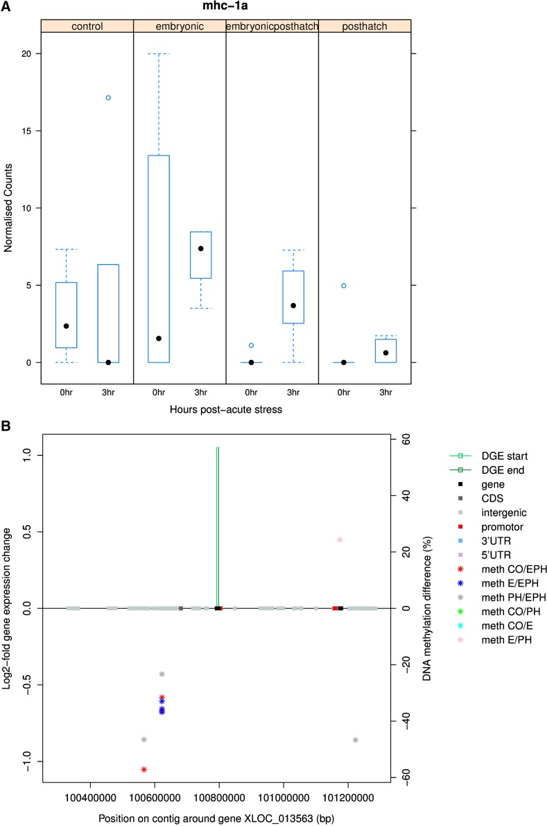
DNA methylation in proximity to several differentially expressed immune genes was also noteworthy. MHC class IA core region is up-regulated with acute stress in EPH treated animals (padj < 0.05) and intergenic regions ∼200kb upstream show higher methylation with EPH treatment (Figure 9). T-Cell Receptor gamma (tcrγ) was significantly down-regulated after acute stress in EPH treated animals (padj < 0.01, unchanged for CO, E and PH) and an uncharacterized gene region ∼100kb downstream was found to contain higher DNA methylation with PH and EPH treatment (Supplementary Figure 3). Toll-like receptor 3 (tlr3) was more than onefold down-regulated with acute stress in EPH treated animals (padj < 0.01) and various genes up- and down-stream showed patterns of increased or decreased methylation depending on treatment comparison (Supplementary Figure 4). Immunoglobulin H (igh) locus B (one transcript) and locus A (two transcripts) were more than onefold down-regulated with acute stress in EPH (padj < 0.01) and E treated animals (padj < 0.05). Differences in methylation in proximity to the igh locus B gene mapped to intergenic, promoter, CDS and gene regions (Supplementary Figure 5). The ATP-binding cassette (abc) transporters were up-regulated with acute stress in EPH treated animals (padj < 0.01). Intergenic regions showing higher methylation with PH treatment and lower levels of methylation with EPH treatment were identified up- and down-stream respectively from the gene (Supplementary Figure 6).
Figure 9.
Box plots of changes in the expression of MHC class 1A (transcript XLOC_013563) for control (CO), embryonic (E), post-hatch (PH) and embryonic post-hatch (EPH) treated fish (A) and map of methylation differences between treatments 0 hr pre-acute stress (stars) (B). The gene expression response for EPH treated fish (from 0 to 3 hr after acute stress) is shown as green bars positioned at the start and end of the gene transcript on the methylation map.
Discussion
Response of the HPI axis to acute stress
The present study documented a relatively weak cortisol response to acute stress in Atlantic salmon at the start feeding stage compared to the ten times greater response that was recently shown in the fully formed adult (Höglund et al. 2017). The tendency for lower cortisol levels throughout the experiment in EPH treated fish, and the reduction of cortisol to below pre-stress levels by 3 hr post-acute stress in EPH treated fish, is an indication that the EPH treatment has modified the overall coping style of the fish relative to other treatments. The suppression of cortisol expression in EPH treated fish, and the long-term effects of this modified response as the fish encounters other stresses in the culture environment, might also be linked to the enhanced growth rate of EPH treated fish detected by our associated study when the fish were transferred to sea (Moghadam et al. 2017).
One possible way that EPH treatment might be modifying the coping style of the fish and suppressing the cortisol response to acute stress is by promoting the expression of genes that inactivate or metabolize cortisol. One such gene is srd5a2, of which two isoforms were detected in salmon, one of which is significantly up-regulated in response to acute stress in EPH treated fish but not in fish receiving other treatments (Figure 4). srd5a2 plays a role in the metabolism and inactivation of cortisol via 5α-reduction of the cortisol A ring double bond and is also responsible for 5α-reduction of testosterone (Fisher et al. 1978). Inhibition of the gene has been shown to augment cortisol action (Nasiri et al. 2015).
Down-regulation of avpi1, crf 1, crf binding protein 1a and 1b transcripts in response to acute stress in all treatment and control groups suggests that the brain hypothalamus-pituitary connection was functioning at the start feeding stage of development and that a negative feedback loop was activated by cortisol at 3 hr post-stress. This was further evidenced by the down-regulated expression of both pomc a1 and a2 in response to acute stress in E and EPH treated and control fish. pomc-b was previously found to respond in the same way in juvenile salmon at the parr stage one hour after acute stress (Madaro et al. 2015).
Modulation of the growth and stress axes in response to repeated stress During early development
In terms of the overall number of genes responding to later life acute stress, PH treated fish responded the least, E the most and EPH gave an intermediate response. This suggests that the duration and timing of the stress treatments effectively modulates the epigenetic programming of the fish and primes the subsequent acute stress response. Cortisol binds and acts on both the GR and MR, which play important roles in the feedback and regulation of cortisol secretion. The gr expression was shown to be a consistent indicator of stress axis activity in sea bass (Terova et al. 2005). Many of the genes that are exclusively responding in EPH treated individuals were found to have putative functions that could contribute to an adaptive response in later life. These subtle differences in the response to acute stress between treatments, as detected for crfr1 and gr, could be affecting how the fish copes and recovers from the stress event, and influencing the hormesis with the EPH treatment that was observed in our associated study (Moghadam et al. 2017). The gr gene tended to be up-regulated in response to stress in CO and EPH treated fish but unchanged in E and PH treated fish (Figure 5B). Life-long stressors such as repeated handling stress are known to lead to larger numbers of GR receptors in the rodent brain and increased methylation of the promoter region of this gene (Anacker et al. 2014). We found that DNA methylation in areas mapping around these genes was associated with the timing and duration of the chronic stress experienced during early development (Figure 8 A and B). Vindas et al. (2016) also showed that early life exposure to chronic stress reduces the response to acute stress and proposed that this could be inducing habituation to the aquaculture environment.
Gh1 transgenic Atlantic salmon not only demonstrated substantially faster growth rates than non-transgenic animals (Du et al. 1992), but also displayed elevated metabolic rate and improved oxygen delivery (Don Stevens and Sutterlin 1999; Cook et al. 2000). The salmonid gh1 and gh2 genes have distinct regulatory modes and are thought to function differently or at different times of development (von Schalburg et al. 2008). The suppression of gh2 and igf1 expression in response to acute stress in EPH treated animals might be contributing to the enhanced growth rate of the EPH treated fish detected in our associated study (Moghadam et al. 2017). The up- and down-stream DNA methylation differences that were detected in EPH fish suggest that epigenetic effects could be steering this differential response. The precise influence of reduced or raised DNA methylation in areas around these genes should be cautiously interpreted. The levels of methylation were measured at 0 hr immediately before animals were subjected to the acute stress. Differences in the extent of DNA methylation in these areas could possibly affect the baseline level of gene expression before the acute stress, the extent of the gene expression response to the acute stress, the direction of expression change in response to the acute stress or might not have any effect at all on the response to acute stress. We restricted our analysis to <500kb up- or down-stream of differentially expressed genes, but it could be that there are other CpG regions outside of these windows where levels of DNA methylation are associated with differential gene expression (either causative or type I error associations). Further experimentation, for example involving DNA methylation editing (Liu et al. 2016), would be needed to test whether DNA methylation at these <500bp up- or down-stream positions are actually influencing the response of these genes to acute stress. The contrasting responses of the stress and growth response axes in EPH treated fish is noteworthy: gh2 and igf1 are more strongly down-regulated at 3hrs post-stress (Figures 6 A and B), whereas, expression of the gr gene is more strongly up-regulated following acute stress in EPH treated fish than fish of other treatments (Figure 5B). This result could indicate a stronger switching away from growth and toward coping with stress following stressful events in EPH treated fish, a strategy that might benefit the recovery of the fish and ultimately provide a better long-term growth response, but more data on temporal changes in the expression of genes in the somatotropic and HPI axes in EPH treated fish is needed to test this hypothesis. A similar pattern has been observed before in response to chronic stress in salmon and sea bass (Moghadam et al. 2017; Fokos et al. 2017; Vindas et al. 2016). Up-stream positive and negative feedback regulators of gh expression, such as gonadotrophin-releasing hormone and preproghrelin-1 and 2 (Lim et al. 2014), tended to show reduced expression in all treatment and control groups. Three transcripts annotated as igf-1 were down-regulated in EPH treated animals and up-regulated in PH treated animals (n.s., like the response of gh2). igf-1 can act as a negative or positive feedback regulator of gh expression (Romero et al. 2012) and is an important mediator of the metabolic response to acute stress (Wiseman et al. 2007).
Serotonin is believed to play an important role in coordinating the stress response of fish and other animals (Backstrom and Winberg 2017) and has been found to be involved in switching the allocation of energy during acute stress from functions affecting growth and reproduction to functions affecting the animal’s ability to cope with stress (Andrews et al. 2015), inhibiting the release of growth hormone (Yu et al. 2008) and regulating the production of ATP from glucose by stimulating glycogen breakdown (Conde-Sieira et al. 2010). Glucosensing systems are deregulated under stress and unable to respond to changes in glucose levels (Conde-Sieira et al. 2010). The expression of the serotonin receptors was generally down-regulated under all treatments in response to the acute stress, but EPH treated fish showed suppressed 5-htr1a expression relative to the other groups before the acute stress was encountered. In comparison, Vindas et al. (2016) reported increased 5-htr transcript levels in the brain stem and hypothalamus of salmon one hour after acute stress, and the catabolism of serotonin was significantly higher in control fish than in fish previously exposed to unpredictable chronic stress. Consistently, the serotonin degrading enzyme monoamine oxidase did not respond to acute stress in the CO, E and PH treatment groups, but was down-regulated in expression in the EPH treatment groups. Previous studies showing that chronic stress treatment during early life influences growth rate as fish enter the sea (Moghadam et al. 2017; Vindas et al. 2016) could be partly explained by some form of mitigated monoamine response.
Repeated stress During early development programs the acute stress response of heat shock protein 70
Heat shock protein (hsp) expression is up-regulated when organisms come under stress and the HSP’s are known to play critical roles in the folding and assembly of proteins, functioning of the immune system, apoptosis and inflammation, all of which help reduce trauma and physical stress (Roberts et al. 2010). This defense mechanism is activated in the earliest developmental stages of fish (e.g., Wu et al. 2018). Expression of hsp70 was more than onefold down-regulated 3 hr after acute shock in EPH treated fish (but not in fish of the other treatments) and 3′UTR regions 200 kb up- and down-stream showed increased DNA methylation with EPH treatment. The response of hsp70 expression to stress differs depending on the timing, duration, type of stress and species (Takle et al. 2005; e.g., Moniruzzaman et al. 2017). Repeated embryonic thermal stress has also been found to reduce or down-regulate subsequent hsp70 responses to acute heat shock in whitefish (Whitehouse et al. 2017). Short-term heat exposure of the early Atlantic cod blastula, and long-term heat exposure to the embryo, both result in up-regulation of hsp70 (Skjærven et al. 2011). hsp70 is upregulated during and after metamorphosis in Senegalese sole and embryos incubated at lower temperatures contained higher levels of hsp70 (Campos et al. 2013). Both cold- and heat-shock induces expression of hsp90 in zebrafish larvae, while hsp70 and hsp70-like expression gradually increases in cold-exposed zebrafish larvae (Long et al. 2012). hsp70 is transiently elevated in trout plasma after heat shock (Faught et al. 2017). The modulated response of hsp70 gene expression to acute stress is another indication that the stress coping style has been modified by the EPH treatment.
Changes in immune system genes
Hormesis elicited by repeated stress, as possibly experienced by EPH treated fish (Moghadam et al. 2017), could in some instances suppress the response of the immune system and make the animal more vulnerable to disease (Tort 2011). Equally, such exposure to mild repeated stress could pre-prepare or enhance the response of the immune system and allow the animals to cope and perform better in the face of infectious disease. Decreased gr responsiveness with stress-related disorders reduces the ability of mammals to resist disease (Raison and Miller 2003). We observed that the response of gr to acute stress was dampened by the E and PH treatments, but that the EPH treated fish responded in a similar fashion to the CO fish. This difference in the responsiveness of the stress axis could therefore be influencing the immune response of the fish.
Various immune responsive genes were differentially expressed in response to acute stress in EPH treated fish. Significant differential DNA methylation associated with treatment was also detected up- and/or down-stream of these loci. Specific alleles of mhc I and II have been found to be associated with resistance to infectious salmon anemia in Atlantic salmon (Kjøglum et al. 2006; Grimholt et al. 2003) and salmon resistant to ISAV infection show differential expression of mhc IIB in the spleen (Dettleff et al. 2017). The tcr recognizes antigens bound to mhc (Guan et al. 2016). Other pentaxin type proteins like tcr (e.g., C-reactive protein, which responds to interleukin-6 secretion by the T-cells and binds to dead or dying cells to activate the complement system; Scheid et al. 1994) were also found to be down-regulated in response to acute stress in EPH treated fish but unchanged in CO, E and PH fish. The tlr3 gene plays an important role in the recognition of viral particles (e.g., salmonid alphavirus subtype 3), and along with the RIG-I-like receptors, uses interferon regulatory factors (3 and 7) to produce interferon-a2 (Xu et al. 2016). Methylation of abc and differential expression of tlr signaling pathway genes have been detected in cold-shocked zebrafish larvae and embryos (Long et al. 2012; Han et al. 2016). Programming of immune response genes could have important implications for the robustness of the fish when exposed to disease in aquaculture, and the effect of the EPH treatment on the gene expression response and effect of these genes therefore warrants further investigation.
Conclusions and relevance for aquaculture
A functional HPI axis was found to exist in Atlantic salmon at 880-odays after fertilization. EPH treatment was found to influence the responses of the HPI and somatotropic neuroendocrine axes to acute stress. Our results suggest that EPH fish could be better able to switch between stress response and growth, and that immune function may also be differentially affected in EPH treated fish. Levels of DNA methylation measured immediately before the acute stress were found to be associated at closely mapping positions to genes that were up- or down-regulated in expression in response to the acute stress in some instances.
The results highlight that the history of the fish is important. It is often assumed that fish return to normal resting condition 24 hr after a stress, whereas our evidence suggests that the fish may never fully recover. It is probably not a good strategy to expend energy on growth in times of stress and the strategy adopted by EPH treated fish when acutely stressed might lead to better recovery and growth in the long-term. However, it must be remembered that these are dynamic responses and the situation could change over time.
The unique immune gene response to acute stress detected in EPH treated fish suggests that the EPH treatment could also have implications for robustness and warrants further investigation. Treatments that are able to modify stress sensitivity therefore could have potential for boosting survivability and productivity at critical stages of production, such as when Atlantic salmon are transferred to cages in the sea when they are particularly stressed and vulnerable to disease.
Acknowledgments
This work was supported by the Research Council of Norway under Grant 194050 and the Norwegian Institute of Food, Fisheries and Aquaculture (Nofima) Strategic Internal Project under Grant 10878. We would like to acknowledge Audny Johansen for excellent assistance in the lab and Dr. Erik Burgerhout for taking the time to read and comment on the manuscript.
H.T., H.J. and Ø.A. conceived and designed the experiment. H.T. and H.J. conducted the challenges and collected samples for transcriptome and methylome sequencing. H.J. coordinated the RNA and DNA extractions and quantifications. H.T. was responsible for the cortisol analyses. N.R. and H.K.M performed the bioinformatics and statistical analyses. N.R. drafted the manuscript. All authors contributed to the interpretation of the data and improvement of the manuscript.
We have followed the ethical guidelines provided by the Norwegian Food and Health authority.
Footnotes
Supplemental material available at Figshare: https://doi.org/10.25387/g3.7586393.
Communicating editor: D. Macqueen
Literature Cited
- Akalin A., Kormaksson M., Li S., Garrett-Bakelman F. E., Figueroa M. E., et al. , 2012. methylKit: a comprehensive R package for the analysis of genome-wide DNA methylation profiles. Genome Biol. 13: R87 10.1186/gb-2012-13-10-r87 [DOI] [PMC free article] [PubMed] [Google Scholar]
- Anacker C., O’Donnell K. J., Meaney M. J., 2014. Early life adversity and the epigenetic programming of hypothalamic-pituitary-adrenal function. Dialogues Clin. Neurosci. 16: 321–333. [DOI] [PMC free article] [PubMed] [Google Scholar]
- Anders S., Pyl P. T., Huber W., 2015. HTSeq–a Python framework to work with high-throughput sequencing data. Bioinformatics 31: 166–169. 10.1093/bioinformatics/btu638 [DOI] [PMC free article] [PubMed] [Google Scholar]
- Andrews P. W., Bharwani A., Lee K. R., Fox M., Thomson J. A., 2015. Is serotonin an upper or a downer? The evolution of the serotonergic system and its role in depression and the antidepressant response. Neurosci. Biobehav. Rev. 51: 164–188. 10.1016/j.neubiorev.2015.01.018 [DOI] [PubMed] [Google Scholar]
- Auperin B., Geslin M., 2008. Plasma cortisol response to stress in juvenile rainbow trout is influenced by their life history during early development and by egg cortisol content. Gen. Comp. Endocrinol. 158: 234–239. 10.1016/j.ygcen.2008.07.002 [DOI] [PubMed] [Google Scholar]
- Backstrom T., Winberg S., 2017. Serotonin Coordinates Responses to Social Stress-What We Can Learn from Fish. Front. Neurosci. 11: 595 10.3389/fnins.2017.00595 [DOI] [PMC free article] [PubMed] [Google Scholar]
- Baker M. E., Funder J. W., Kattoula S. R., 2013a Evolution of hormone selectivity in glucocorticoid and mineralocorticoid receptors. J. Steroid Biochem. Mol. Biol. 137: 57–70. Erratum: 139: 104. 10.1016/j.jsbmb.2013.07.009 [DOI] [PubMed] [Google Scholar]
- Baker M. R., Gobush K. S., Vynne C. H., 2013b Review of factors influencing stress hormones in fish and wildlife. J. Nat. Conserv. 21: 309–318. 10.1016/j.jnc.2013.03.003 [DOI] [Google Scholar]
- Besseau L., Fuentes M., Sauzet S., Beauchaud M., Chatain B., et al. , 2013. Somatotropic axis genes are expressed before pituitary onset during zebrafish and sea bass development. Gen. Comp. Endocrinol. 194: 133–141. 10.1016/j.ygcen.2013.08.018 [DOI] [PubMed] [Google Scholar]
- Best C., Kurrasch D. M., Vijayan M. M., 2017. Maternal cortisol stimulates neurogenesis and affects larval behaviour in zebrafish. Sci. Rep. 7: 40905 10.1038/srep40905 [DOI] [PMC free article] [PubMed] [Google Scholar]
- Bolger A. M., Lohse M., Usadel B., 2014. Trimmomatic: a flexible trimmer for Illumina sequence data. Bioinformatics 30: 2114–2120. 10.1093/bioinformatics/btu170 [DOI] [PMC free article] [PubMed] [Google Scholar]
- Boschen K. E., Keller S. M., Roth T. L., Klintsova A. Y., 2018. Epigenetic mechanisms in alcohol- and adversity-induced developmental origins of neurobehavioral functioning. Neurotoxicol. Teratol. 66: 63–79. 10.1016/j.ntt.2017.12.009 [DOI] [PMC free article] [PubMed] [Google Scholar]
- Campos C., Valente L. M. P., Conceicao L. E. C., Engrola S., Sousa V., et al. , 2013. Incubation temperature induces changes in muscle cellularity and gene expression in Senegalese sole (Solea senegalensis). Gene 516: 209–217. 10.1016/j.gene.2012.12.074 [DOI] [PubMed] [Google Scholar]
- Chatzopoulou A., Heijmans J. P. M., Burgerhout E., Oskam N., Spaink H. P., et al. , 2016. Glucocorticoid-Induced Attenuation of the Inflammatory Response in Zebrafish. Endocrinology 157: 2772–2784. 10.1210/en.2015-2050 [DOI] [PubMed] [Google Scholar]
- Conde-Sieira M., Aguilar A. J., Lopez-Patino M. A., Miguez J. M., Soengas J. L., 2010. Stress alters food intake and glucosensing response in hypothalamus, hindbrain, liver, and Brockmann bodies of rainbow trout. Physiol. Behav. 101: 483–493. 10.1016/j.physbeh.2010.07.016 [DOI] [PubMed] [Google Scholar]
- Cook J. T., Sutterlin A. M., McNiven M. A., 2000. Effect of food deprivation on oxygen consumption and body composition of growth-enhanced transgenic Atlantic salmon (Salmo salar). Aquaculture 188: 47–63. 10.1016/S0044-8486(00)00333-1 [DOI] [Google Scholar]
- de Jesus E. G., Hirano T., Inui Y., 1991. Changes in cortisol and thyroid-hormone concentrations during early development and metamorphosis in the Japanese flounder, Paralichthys olivaceus. Gen. Comp. Endocrinol. 82: 369–376. 10.1016/0016-6480(91)90312-T [DOI] [PubMed] [Google Scholar]
- Dettleff P., Moen T., Santi N., Martinez V., 2017. Transcriptomic analysis of spleen infected with infectious salmon anemia virus reveals distinct pattern of viral replication on resistant and susceptible Atlantic salmon (Salmo salar). Fish Shellfish Immunol. 61: 187–193. 10.1016/j.fsi.2017.01.005 [DOI] [PubMed] [Google Scholar]
- Don Stevens E., Sutterlin A., 1999. Gill morphometry in growth hormone transgenic Atlantic salmon. Environ. Biol. Fishes 54: 405–411. 10.1023/A:1007574308129 [DOI] [Google Scholar]
- Du S. J., Gong Z. Y., Fletcher G. L., Shears M. A., King M. J., et al. , 1992. Growth Enhancement in Transgenic Atlantic Salmon by the Use of an All Fish Chimeric Growth-Hormone Gene Construct. Bio-Technology 10: 176–181. [DOI] [PubMed] [Google Scholar]
- Faught E., Henrickson L., Vijayan M. M., 2017. Plasma exosomes are enriched in Hsp70 and modulated by stress and cortisol in rainbow trout. J. Endocrinol. 232: 237–246. 10.1530/JOE-16-0427 [DOI] [PubMed] [Google Scholar]
- Fisher L. K., Kogut M. D., Moore R. J., Goebelsmann U., Weitman J. J., et al. , 1978. Clinical, endocrinological, and enzymatic characterization of two patients with 5a-reductase deficiency: evidence that a single enzyme is responsible for the 5a reduction of cortisol and testosterone. J. Clin. Endocrinol. Metab. 47: 653–664. 10.1210/jcem-47-3-653 [DOI] [PubMed] [Google Scholar]
- Fokos S., Pavlidis M., Yiotis T., Tsalafouta A., Papandroulakis N., et al. , 2017. Early life low intensity stress experience modifies acute stress effects on juvenile brain cell proliferation of European sea bass (D-Labrax). Behav. Brain Res. 317: 109–121. 10.1016/j.bbr.2016.09.026 [DOI] [PubMed] [Google Scholar]
- Grimholt U., Larsen S., Nordmo R., Midtlyng P., Kjoeglum S., et al. , 2003. MHC polymorphism and disease resistance in Atlantic salmon (Salmo salar); facing pathogens with single expressed major histocompatibility class I and class II loci. Immunogenetics 55: 210–219. 10.1007/s00251-003-0567-8 [DOI] [PubMed] [Google Scholar]
- Guan J., Gonzalez F., Yin Y., Shastri N., 2016. Antigen processing in the ER is monitored by unusually conserved CD8+T cell antigen receptors specific for non-classical Qa-1 MHC molecules. Eur. J. Immunol. 46: 590. [Google Scholar]
- Hagen H., Hansen L. P., Kjelsaas D., Røed H., Schandy T., et al. , 2002. Laksen, elvas konge, Tm and Tom, Vestfossen, Norway. [Google Scholar]
- Han B. S., Li W. H., Chen Z. Z., Xu Q. Q., Luo J. T., et al. , 2016. Variation of DNA Methylome of Zebrafish Cells under Cold Pressure. PLoS One 11: e0160358 10.1371/journal.pone.0160358 [DOI] [PMC free article] [PubMed] [Google Scholar]
- Höglund E., Overli O., Andersson M. A., Silva P., Laursen D. C., et al. , 2017. Dietary L-tryptophan leaves a lasting impression on the brain and the stress response. Br. J. Nutr. 117: 1351–1357. 10.1017/S0007114517001428 [DOI] [PubMed] [Google Scholar]
- Hu F., Crespi E. J., Denver R. J., 2008. Programming neuroendocrine stress axis activity by exposure to glucocorticoids during postembryonic development of the frog, Xenopus laevis. Endocrinology 149: 5470–5481. 10.1210/en.2008-0767 [DOI] [PubMed] [Google Scholar]
- Kapoor A., Dunn E., Kostaki A., Andrews M. H., Matthews S. G., 2006. Fetal programming of hypothalamo-pituitary-adrenal function: prenatal stress and glucocorticoids. J. Physiol. 572: 31–44. 10.1113/jphysiol.2006.105254 [DOI] [PMC free article] [PubMed] [Google Scholar]
- Kjøglum S., Larsen S., Bakke H. G., Grimholt U., 2006. How specific MHC class I and class II combinations affect disease resistance against infectious salmon anaemia in Atlantic salmon (Salmo salar). Fish Shellfish Immunol. 21: 431–441. 10.1016/j.fsi.2006.02.001 [DOI] [PubMed] [Google Scholar]
- Korosi A., Baram T. Z., 2010. Plasticity of the stress response early in life: mechanisms and significance. Dev. Psychobiol. 52: 661–670. 10.1002/dev.20490 [DOI] [PMC free article] [PubMed] [Google Scholar]
- Krueger F., Andrews S. R., 2011. Bismark: a flexible aligner and methylation caller for Bisulfite-Seq applications. Bioinformatics 27: 1571–1572. 10.1093/bioinformatics/btr167 [DOI] [PMC free article] [PubMed] [Google Scholar]
- Langmead B., Trapnell C., Pop M., Salzberg S. L., 2009. Ultrafast and memory-efficient alignment of short DNA sequences to the human genome. Genome Biol. 10: R25 10.1186/gb-2009-10-3-r25 [DOI] [PMC free article] [PubMed] [Google Scholar]
- Li M., Leatherland J. F., 2013. The implications for aquaculture practice of epigenomic programming of components of the endocrine system of teleostean embryos: lessons learned from mammalian studies. Fish Fish. 14: 528–553. 10.1111/j.1467-2979.2012.00486.x [DOI] [Google Scholar]
- Lien S., Koop B. F., Sandve S. R., Miller J. R., Kent M. P., et al. , 2016. The Atlantic salmon genome provides insights into rediploidization. Nature 533: 200–205. 10.1038/nature17164 [DOI] [PMC free article] [PubMed] [Google Scholar]
- Lim, C. T., A. Grossman, and K. B., 2014 Normal Physiology of ACTH and GH Release in the Hypothalamus and Anterior Pituitary in Man in Endotext [Internet], edited by L. J. De Groot, G. Chrousos, K. Dungan and et al. MDText.com, Inc., South Dartmouth (MA).
- Liu X. S., Wu H., Ji X., Stelzer Y., Wu X. B., et al. , 2016. Editing DNA Methylation in the Mammalian Genome. Cell 167: 233–247.e17. 10.1016/j.cell.2016.08.056 [DOI] [PMC free article] [PubMed] [Google Scholar]
- Long Y., Li L. C., Li Q., He X. Z., Cui Z. B., 2012. Transcriptomic Characterization of Temperature Stress Responses in Larval Zebrafish. PLoS One 7: e37209 10.1371/journal.pone.0037209 [DOI] [PMC free article] [PubMed] [Google Scholar]
- Love M. I., Huber W., Anders S., 2014. Moderated estimation of fold change and dispersion for RNA-seq data with DESeq2. Genome Biol. 15: 550 10.1186/s13059-014-0550-8 [DOI] [PMC free article] [PubMed] [Google Scholar]
- Love O. P., Williams T. D., 2008. Plasticity in the adrenocortical response of a free-living vertebrate: the role of pre- and post-natal developmental stress. Horm. Behav. 54: 496–505. 10.1016/j.yhbeh.2008.01.006 [DOI] [PubMed] [Google Scholar]
- Madaro A., Olsen R. E., Kristiansen T. S., Ebbesson L. O. E., Nilsen T. O., et al. , 2015. Stress in Atlantic salmon: response to unpredictable chronic stress. J. Exp. Biol. 218: 2538–2550. 10.1242/jeb.120535 [DOI] [PubMed] [Google Scholar]
- Mateus A. P., Power D. M., Canario A. V. M., 2017. Stress and disease in fish, pp. 187–220 in Fish diseases - Prevention and control strategies, edited by Jeney G. Academic Press, London. [Google Scholar]
- McCormick S. D., Regish A., O’Dea M. F., Shrimpton J. M., 2008. Are we missing a mineralocorticoid in teleost fish? Effects of cortisol, deoxycorticosterone and aldosterone on osmoregulation, gill Na+,K+-ATPase activity and isoform mRNA levels in Atlantic salmon. Gen. Comp. Endocrinol. 157: 35–40. 10.1016/j.ygcen.2008.03.024 [DOI] [PubMed] [Google Scholar]
- Meaney M. J., Szyf M., Seckl J. R., 2007. Epigenetic mechanisms of perinatal programming of hypothalamic-pituitary-adrenal function and health. Trends Mol. Med. 13: 269–277. 10.1016/j.molmed.2007.05.003 [DOI] [PubMed] [Google Scholar]
- Metzger D. C. H., Schulte P. M., 2016. Maternal stress has divergent effects on gene expression patterns in the brains of male and female threespine stickleback. Proceedings of the Royal Society B-Biological Sciences 283 10.1098/rspb.2016.1734 [DOI] [PMC free article] [PubMed] [Google Scholar]
- Moghadam H., Johnsen H., Robinson N., Andersen Ø., Tveiten H., 2017. Impacts of early life stress on the methylome and transcriptome of Atlantic salmon. Sci. Rep. 7: 5023 10.1038/s41598-017-05222-2 [DOI] [PMC free article] [PubMed] [Google Scholar]
- Moniruzzaman M., Midday P., Dhara A., Das D., Ghosal I., et al. , 2017. Change in redox state and heat shock protein expression in an Indian major carp Cirrhinus cirrhosus exposed to zinc and lead. J. Toxicol. Sci. 42: 731–740. 10.2131/jts.42.731 [DOI] [PubMed] [Google Scholar]
- Nasiri M., Nikolaou N., Parajes S., Krone N. P., Valsamakis G., et al. , 2015. 5 alpha-Reductase Type 2 Regulates Glucocorticoid Action and Metabolic Phenotype in Human Hepatocytes. Endocrinology 156: 2863–2871. 10.1210/en.2015-1149 [DOI] [PMC free article] [PubMed] [Google Scholar]
- Nesan D., Kamkar M., Burrows J., Scott I. C., Marsden M., et al. , 2012. Glucocorticoid receptor signaling is essential for mesoderm formation and muscle development in zebrafish. Endocrinology 153: 1288–1300. 10.1210/en.2011-1559 [DOI] [PubMed] [Google Scholar]
- Nesan D., Vijayan M. M., 2012. Embryo exposure to elevated cortisol level leads to cardiac performance dysfunction in zebrafish. Mol. Cell. Endocrinol. 363: 85–91. 10.1016/j.mce.2012.07.010 [DOI] [PubMed] [Google Scholar]
- Palma-Gudiel H., Cordova-Palomera A., Leza J. C., Fananas L., 2015. Glucocorticoid receptor gene (NR3C1) methylation processes as mediators of early adversity in stress-related disorders causality: A critical review. Neurosci. Biobehav. Rev. 55: 520–535. 10.1016/j.neubiorev.2015.05.016 [DOI] [PubMed] [Google Scholar]
- Paragliola R. M., Papi G., Pontecorvi A., Corsello S. M., 2017. Treatment with Synthetic Glucocorticoids and the Hypothalamus-Pituitary-Adrenal Axis. Int. J. Mol. Sci. 18: E2201 10.3390/ijms18102201 [DOI] [PMC free article] [PubMed] [Google Scholar]
- Pavlidis M., Sundvik M., Chen Y. C., Panula P., 2011. Adaptive changes in zebrafish brain in dominant-subordinate behavioral context. Behav. Brain Res. 225: 529–537. 10.1016/j.bbr.2011.08.022 [DOI] [PubMed] [Google Scholar]
- Pikulkaew S., Benato F., Celeghin A., Zucal C., Skobo T., et al. , 2011. The Knockdown of Maternal Glucocorticoid Receptor mRNA Alters Embryo Development in Zebrafish. Dev. Dyn. 240: 874–889. 10.1002/dvdy.22586 [DOI] [PubMed] [Google Scholar]
- Raison C. L., Miller A. H., 2003. When Not Enough Is Too Much: The Role of Insufficient Glucocorticoid Signaling in the Pathophysiology of Stress-Related Disorders. Am. J. Psychiatry 160: 1554–1565. 10.1176/appi.ajp.160.9.1554 [DOI] [PubMed] [Google Scholar]
- Reul J. M. H. M., Collins A., Saliba R. S., Mifsud K. R., Carter S. D., et al. , 2015. Glucocorticoids, epigenetic control and stress resilience. Neurobiol. Stress 1: 44–59. 10.1016/j.ynstr.2014.10.001 [DOI] [PMC free article] [PubMed] [Google Scholar]
- Roberts R. J., Agius C., Saliba C., Bossier P., Sung Y. Y., 2010. Heat shock proteins (chaperones) in fish and shellfish and their potential role in relation to fish health: a review. J. Fish Dis. 33: 789–801. 10.1111/j.1365-2761.2010.01183.x [DOI] [PubMed] [Google Scholar]
- Romero C. J., Pine-Twaddell E., Sima D. I., Miller R. S., He L., et al. , 2012. Insulin-Like Growth Factor 1 Mediates Negative Feedback to Somatotroph GH Expression via POU1F1/CREB Binding Protein Interactions. Mol. Cell. Biol. 32: 4258–4269. 10.1128/MCB.00171-12 [DOI] [PMC free article] [PubMed] [Google Scholar]
- Rose A. J., Herzig S., 2013. Metabolic control through glucocorticoid hormones: An update. Mol. Cell. Endocrinol. 380: 65–78. 10.1016/j.mce.2013.03.007 [DOI] [PubMed] [Google Scholar]
- Scheid C., Young R., McDermott R., Fitzsimmons L., Scarffe J. H., et al. , 1994. Immune function of patients receiving recombinant human interleukin-6 (IL-6) in a phase-I clinical study – Induction of C-reactive protein and IGE and inhibition of natural-killer and lymphokine-activated killer-cell activity. Cancer Immunol. Immunother. 38: 119–126. [DOI] [PMC free article] [PubMed] [Google Scholar]
- Seckl J. R., 2008. Glucocorticoids, developmental ‘programming’ and the risk of affective dysfunction. Prog. Brain Res. 167: 17–34. 10.1016/S0079-6123(07)67002-2 [DOI] [PubMed] [Google Scholar]
- Skjærven K. H., Olsvik P. A., Finn R. N., Holen E., Hamre K., 2011. Ontogenetic expression of maternal and zygotic genes in Atlantic cod embryos under ambient and thermally stressed conditions. Comp. Biochem. Physiol. A Mol. Integr. Physiol. 159: 196–205. 10.1016/j.cbpa.2011.02.026 [DOI] [PubMed] [Google Scholar]
- Stouthart A., Lucassen E., van Strien F. J. C., Balm P. H. M., Lock R. A. C., et al. , 1998. Stress responsiveness of the pituitary-interrenal axis during early life stages of common carp (Cyprinus carpio). J. Endocrinol. 157: 127–137. 10.1677/joe.0.1570127 [DOI] [PubMed] [Google Scholar]
- Szyf M., Weaver I., Meaney M., 2007. Maternal care, the epigenome and phenotypic differences in behavior. Reprod. Toxicol. 24: 9–19. 10.1016/j.reprotox.2007.05.001 [DOI] [PubMed] [Google Scholar]
- Takle H., Baeverfjord G., Lunde M., Kolstad K., Andersen O., 2005. The effect of heat and cold exposure on HSP70 expression and development of deformities during embryogenesis of Atlantic salmon (Salmo salar). Aquaculture 249: 515–524. 10.1016/j.aquaculture.2005.04.043 [DOI] [Google Scholar]
- Terova G., Gornati R., Rimoldi S., Bernardini G., Saroglia M., 2005. Quantification of a glucocorticoid receptor in sea bass (Dicentrarchus labrax, L.) reared at high stocking density. Gene 357: 144–151. 10.1016/j.gene.2005.06.016 [DOI] [PubMed] [Google Scholar]
- Tort L., 2011. Stress and immune modulation in fish. Dev. Comp. Immunol. 35: 1366–1375. 10.1016/j.dci.2011.07.002 [DOI] [PubMed] [Google Scholar]
- Trapnell C., Pachter L., Salzberg S. L., 2009. TopHat: discovering splice junctions with RNA-Seq. Bioinformatics 25: 1105–1111. 10.1093/bioinformatics/btp120 [DOI] [PMC free article] [PubMed] [Google Scholar]
- Tsalafouta A., Papandroulakis N., Gorissen M., Katharios P., Flik G., et al. , 2014. Ontogenesis of the HPI axis and molecular regulation of the cortisol stress response during early development in Dicentrarchus labrax. Sci. Rep. 4: 5525 10.1038/srep05525 [DOI] [PMC free article] [PubMed] [Google Scholar]
- Vander A. J., Sherman J. H., Luciano D. S., 1970. Human physiology- The mechanisums of body function, McGraw-Hill Book Company, New York. [Google Scholar]
- Vindas M. A., Madaro A., Fraser T. K., Hoglund E., Olsen R. E., et al. , 2016. Coping with a changing environment: the effects of early life stress. R. Soc. Open Sci. 3: 160382 10.1098/rsos.160382 [DOI] [PMC free article] [PubMed] [Google Scholar]
- von Schalburg K. R., Yazawa R., de Boer J., Lubieniecki K. P., Goh B., et al. , 2008. Isolation, characterization and comparison of Atlantic and Chinook salmon growth hormone 1 and 2. BMC Genomics 9: 522 10.1186/1471-2164-9-522 [DOI] [PMC free article] [PubMed] [Google Scholar]
- Weinstock M., 2008. The long-term behavioural consequences of prenatal stress. Neurosci. Biobehav. Rev. 32: 1073–1086. 10.1016/j.neubiorev.2008.03.002 [DOI] [PubMed] [Google Scholar]
- Whitehouse L. M., McDougall C. S., Stefanovic D. I., Boreham D. R., Somers C. M., et al. , 2017. Development of the embryonic heat shock response and the impact of repeated thermal stress in early stage lake whitefish (Coregonus clupeaformis) embryos. J. Therm. Biol. 69: 294–301. 10.1016/j.jtherbio.2017.08.013 [DOI] [PubMed] [Google Scholar]
- Wilson K. S., Matrone G., Livingstone D. E. W., Al-Dujaili E. A. S., Mullins J. J., et al. , 2013. Physiological roles of glucocorticoids during early embryonic development of the zebrafish (Danio rerio). J. Physiol. 591: 6209–6220. 10.1113/jphysiol.2013.256826 [DOI] [PMC free article] [PubMed] [Google Scholar]
- Wiseman S., Osachoff H., Bassett E., Malhotra J., Bruno J., et al. , 2007. Gene expression pattern in the liver during recovery from an acute stressor in rainbow trout. Comp. Biochem. Physiol. Part D Genomics Proteomics 2: 234–244. 10.1016/j.cbd.2007.04.005 [DOI] [PubMed] [Google Scholar]
- Wu S. G., Hu G. J., Zhao X. P., Wang Q., Jiang J. H., 2018. Synergistic potential of fenvalerate and triadimefon on endocrine disruption and oxidative stress during rare minnow embryo development. Environ. Toxicol. 33: 759–769. 10.1002/tox.22563 [DOI] [PubMed] [Google Scholar]
- Wu Y. H., Patchev A. V., Daniel G., Almeida O. F. X., Spengler D., 2014. Early-Life Stress Reduces DNA Methylation of the Pomc Gene in Male Mice. Endocrinology 155: 1751–1762. 10.1210/en.2013-1868 [DOI] [PubMed] [Google Scholar]
- Xu, C., O. Evensen, and H.M. Munang’andu, 2016 De Novo Transcriptome Analysis Shows That SAV-3 Infection Upregulates Pattern Recognition Receptors of the Endosomal Toll-Like and RIG-I-Like Receptor Signaling Pathways in Macrophage/Dendritic Like TO-Cells. Viruses-Basel 8 (4). [DOI] [PMC free article] [PubMed]
- Yehuda R., Flory J. D., Bierer L. M., Henn-Haase C., Lehrner A., et al. , 2015. Lower Methylation of Glucocorticoid Receptor Gene Promoter 1(F) in Peripheral Blood of Veterans with Posttraumatic Stress Disorder. Biol. Psychiatry 77: 356–364. 10.1016/j.biopsych.2014.02.006 [DOI] [PubMed] [Google Scholar]
- Yu Y., Wong A. O. L., Chang J. P., 2008. Serotonin interferes with Ca2+ and PKC signaling to reduce gonadotropin-releasing hormone-stimulated GH secretion in goldfish pituitary cells. Gen. Comp. Endocrinol. 159: 58–66. 10.1016/j.ygcen.2008.07.021 [DOI] [PubMed] [Google Scholar]
Associated Data
This section collects any data citations, data availability statements, or supplementary materials included in this article.
Data Availability Statement
The GSA Figshare portal was used to upload supplemental files (captions below). All raw sequences were deposited to the NCBI Short Read Archive under the BioProject IDs PRJNA388534 and PRJNA510456. Supplemental material available at Figshare: https://doi.org/10.25387/g3.7586393.


