

Description
A 13-year-old girl presented with a 4-month history of diminution of vision in both eyes. The best corrected distance visual acuity was 20/30 OU. Diffuse light examination on slit lamp revealed bluish white tiny opacities throughout the lens forming concentric layers in both eyes (figure 1A). Detailed slit examination highlighted opacities of various colours: white, blue, greenish-blue, grey and red (figure 1B). The posterior segment was unremarkable in both eyes. Her brother also had similar lenticular opacities suggestive of cerulean cataract. Though the child did not complain of other visual disturbances, glare and contrast sensitivity might also be tested. A regular follow-up was advised.
Figure 1.
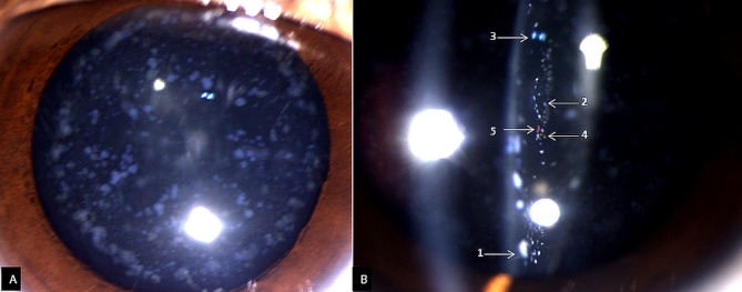
(A) Diffuse light slit lamp photograph of the right eye shows bluish-white opacities throughout the lens forming concentric layers. (B) Slit examination of the same eye with multicoloured opacities marked with numbers: white (1), blue (2), greenish-blue (3), grey (4) and red (5).
Cerulean or blue-dot cataracts are autosomal dominant early onset bilateral cataracts with concentric layers of bluish-white opacities which may form large cuneiform shapes in the mid-periphery.1–5 These appear from birth to early childhood but may not be diagnosed until adulthood. It may be caused by mutations in certain genes: crystallin β-B2 (CRYBB2), crystallin γ-D (CRYGD), musculoaponeurotic fibrosarcoma and major intrinsic protein genes.1–3 The mutations in these genes are responsible for early onset pulverulent cataract and cerulean cataract.3
The progression of cataract is slow and may not become visually significant till the third to fourth decades when surgery may be helpful.4 5 Initially the opacities appear at the edge of the fetal lens nucleus. With time the opacities involve the adult nucleus and the cortex in the form of concentric circles. The central lesions are arranged radially. They may also have associated central sutural cataract.5 The multicoloured cortical opacities were perhaps dense and present in the visual axis in the present case and may be the reason for the slight visual disturbance. Patients with cerulean cataract do not have any associated systemic comorbidities and need not be evaluated.
Learning points.
Cerulean or blue-dot cataract is an early onset familial cataract with predominant bluish-white lenticular opacities.
Although cerulean cataract may be observed in childhood, it is usually visually insignificant.
The visual acuity usually gets affected in adulthood, when cataract extraction may be needed.
Footnotes
Contributors: DK: acquisition of data, analysis and interpretation of data, drafting the article. NJ: acquisition of data, drafting the article. PS: acquisition of data, revising the article critically for important intellectual content. PC: conception and design, revising the article critically for important intellectual content. All authors: final approval of the version published, agreement to be accountable for the article and to ensure that all questions regarding the accuracy or integrity of the article are investigated and resolved.
Funding: The authors have not declared a specific grant for this research from any funding agency in the public, commercial or not-for-profit sectors.
Competing interests: None declared.
Provenance and peer review: Not commissioned; externally peer reviewed.
Patient consent for publication: Parental/guardian consent obtained.
References
- 1. Litt M, Carrero-Valenzuela R, LaMorticella DM, et al. Autosomal dominant cerulean cataract is associated with a chain termination mutation in the human beta-crystallin gene CRYBB2. Hum Mol Genet 1997;6:665–8. 10.1093/hmg/6.5.665 [DOI] [PubMed] [Google Scholar]
- 2. Armitage MM, Kivlin JD, Ferrell RE. A progressive early onset cataract gene maps to human chromosome 17q24. Nat Genet 1995;9:37–40. 10.1038/ng0195-37 [DOI] [PubMed] [Google Scholar]
- 3. Xiao X, Li W, Wang P, et al. Cerulean cataract mapped to 12q13 and associated with a novel initiation codon mutation in MIP. Mol Vis 2011;17:2049–55. [PMC free article] [PubMed] [Google Scholar]
- 4. Provencher LM, Critser B, Johnson AT. Cerulean cataract. JAMA Ophthalmol 2018;136:e183150 10.1001/jamaophthalmol.2018.3150 [DOI] [PubMed] [Google Scholar]
- 5. Ram J, Singh A. Cerulean cataract. QJM 2019;37 10.1093/qjmed/hcz038 [DOI] [PubMed] [Google Scholar]