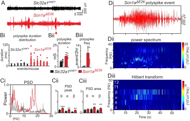
Figure 2. Scn1aΔE26 exhibit frequent spontaneous seizures.
(A) traces of raw EcoG activity show that Scn1aΔE26 mice but not Slc32a1cre/+ mice exhibit frequent spontaneous burst of high amplitude poly-spike activity. The arrow identifies a typical seizure-like poly-spike event that was analyzed further by power spectral analysis in panel D. Polyspike events with a minimum duration of 14 ms were accompanied by seizure-like behavior and so were considered epileptic activity. (B), Scn1aΔE26 mice showed more frequent epileptic poly-spike bursts of activity (Bi, dotted line designates duration threshold for epileptic activity); poly-spike bursts (>14 ms) occurred more frequently in Scn1aΔE26 mice (Bi-Bii) (control 0.13 ± 0.1 events/2 hr, n = 6; Scn1aΔE26 0.37 ± 0.05 events/2 hr, n = 6; T10 = 3.009, p<0.01) and lasted for a longer duration (Biii) (control 7.6 ± 0.6 ms, n = 6; Scn1aΔE26 15.6 ± 0.8 ms, n = 6, T10 = 2.268, p<0.05) compared to control animals. Ci, representative power spectrum density (PSD) plots of spontaneous poly-spike burst events show typical strong activity in the theta-, alpha and beta frequency range in Scn1aΔE26 but not control mice. Cii-Ciii (Figure 2—source data 1), summary data (normalized to the maximum value at each event) show PSD peak (Cii) and PSD area under the curve (Ciii) of each frequency range for each genotype. Note that poly-spike burst events measured in Scn1aΔE26 mice show increased activity in the theta, alpha and beta range. Di-iii, poly-spike burst events recorded from a Scn1aΔE26 mouse (arrow in panel A) plotted on an expanded time scale (Di) and corresponding time frequency distribution (Dii) and deconstructed spectrum into its various frequency domains (Diii). These results were compared using a two-way ANOVA and the Sidak multiple comparison test. *, p<0.05; **, p<0.01; ***p<0.001.

