Figure 1.
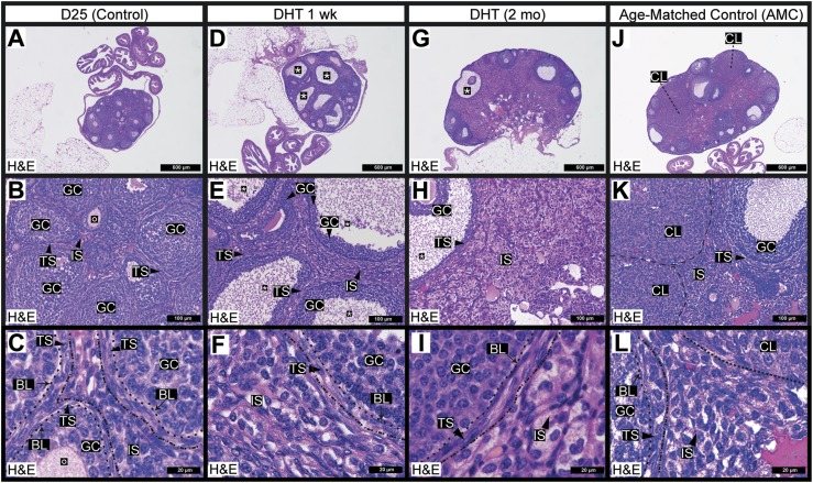
Ovarian follicular and stromal morphology was markedly altered in a mouse model of androgen excess. (A) Histological [hematoxylin and eosin (H&E) stain] examination of the D25 mouse ovary revealed healthy growing follicles and served as a nonluteinized, noncycling control. (B, C) Many follicles in the D25 ovaries contained oocytes surrounded by granulosa cells and theca cells. Theca cells are polarized spindle-shaped mesenchymal cells (generally two to three layers thick) that were found outside the follicle-associated basal lamina. Outside the theca layer was an immature interstitial compartment, the cells of which were derived from steroidogenic theca cells remaining from atretic follicles. This compartment was sparse in D25 control mice. (D–F) DHT treatment of 1 wk led to the formation of many cystic follicles (annotated with asterisks). (E) DHT-treated animals developed an abnormal thickening of the ovarian interstitial layer. (G–I) DHT treatment of 2 mo prevented the generation of corpora lutea and promoted the formation of cystic follicles but did not prevent the formation of theca stroma or the expansion of the interstitial stroma. (I) The expanded interstitial stroma exhibited an abnormal, hyperplastic, and fatty appearance. (J–L) The ovary of a representative AMC mouse contained multiple corpora lutea, which were absent in D25 mouse ovary. (K) Healthy luteal and granulosa cells in the ovary were evident on morphologic comparison. (L) Theca cells were well polarized in the AMC control ovaries, and the interstitial compartment bore a distinct, presumably healthy appearance. N ≥ 3 for all conditions. Dashed lines outline specific ovarian structures that are labeled individually. BL, basal lamina; CL, corpora lutea; GC, granulosa cell; IS, interstitial stroma; O, oocyte; TS, theca stroma.

