Abstract
Introduction
The aim of our study was to assess the efficacy of Phyllanthus niruri standardized extract, combined with magnesium and B6 vitamin, used to treat uncomplicated nephrolithiasis.
Methods
We included in the present study 48 patients with uncomplicated nephrolithiasis, with the maximum calculi diameter of up to 15 mm, confirmed by non-contrast-enhanced computer tomography. Each patient followed a three-month therapeutic regimen with the above mentioned combination, with imaging assessment of the calculi after treatment.
Results
Per patient
The mean age of the patients was 48 years. The median number of calculi was 1 and the mean dimension was 5.5 mm. The stone-free status after treatment was not correlated with gender (p=0.7), side location (p=0.8) or with the number of calculi (p=0.3), but we found a correlation with the location in the upper or middle calyx (54.5% vs 13.8%, p=0.008) and with the maximum diameter (p=0.001).
Per stone
60 calculi were analyzed, 8.3% being located in the upper calyx, 36.7% in the middle and 55% in the lower one. After treatment, 40% were absent, 21.7% showed lower dimensions and 38.3% remained unchanged, with the mean reduction of 1.7 mm. We identified a cut-off value of ≤ 3 mm (AUC 0.9, CI:0.8–0.9, p<0.0001) for the prediction of stone-free status after treatment.
Conclusions
The current treatment had the highest efficacy in achieving stone-free status for patients with calculi ≤ 3 mm, located in the middle or upper calyx. A higher duration of the treatment might show improved results.
Keywords: antilithogenic, Phyllanthus, kidney calculi, triterpenes
Introduction
Nephrolithiasis is one of the most frequent urological benign pathology, being encountered in 1–15% of the patients [1]. This condition is 2–3 times more frequent in women compared to men, with a peak incidence being reported between 30 and 40 years [2]. The vast majority of renal calculi are composed of calcium oxalate, followed by uric acid and struvite [3].
Treatment options include extracorporeal shock wave lithotripsy, percutaneous nephrolithotomy, flexible or rigid ureteroscopy with laser stone fragmentation and retrieval and, in selected cases, the laparoscopic approach might be advised [4].
Even with adequate treatment, the recurrence rate varies between 22 and 51% at a mean follow-up of 2 to 7 years. The main reason for this finding is the imbalance between promoters and inhibitors of the lithogenic process. Patients with hypercalciuria, hyperuricosuria and hyperoxaluria have a higher incidence of recurrence, as well as those with hypocitraturia and hypomagesuria, both of them binding free calcium, thus inhibiting calcium oxalate nucleation [5].
Phyllanthus niruri, commonly known as “stone-breaker”, is a member of Euphorbiceae family, being used for over 2000 years in treating urolithiasis [6]. P. niruri contains triterpenes, which are considered to be the main antilithogenic factor. It has been observed that the afore mentioned compound decreases the urinary excretion of oxalate and calcium near their normal concentration, while interfering with the glycosaminoglycans found in the matrix of the precipitating crystals, making them smoother and more fragile [7–9]. Another noticeable effect is that calcium oxalate crystals remain equidistantly dispersed in urine treated with P. niruri extract [10].
The aim of this study was to assess the efficacy of Phyllanthus niruri standardized extract, combined with magnesium and B6 vitamin, for the treatment of uncomplicated nephrolithiasis.
Patients and methods
Patient selection
The current study included 48 patients who presented to our department between September and December 2017 with nephrolithiasis, initially diagnosed by abdominal ultrasound and confirmed by non-contrast-enhanced computer tomography (CT). All patients signed the written informed consent, approved by our hospital’s ethics committee.
Inclusion criteria were defined as asymptomatic nephrolithiasis, with the maximum diameter of the calculus up to 15 mm and with serum and urinary assessment within the biological reference interval.
Exclusion criteria were considered hypersensitivity to any of the active principles or excipients of the compound, pregnancy and lactation, imaging evidence of ureterohydronephrosis, renal colic at admission or during the study period, liver or kidney impaired function, altered coagulation, positive urine culture, concomitant use of other antilithogenic substances, urological and non-urological malignant diseases.
Out of 48 patients, 8 of them met one of the exclusion criteria before completing 3 months of treatment: 5 stopped the treatment prior to the established due date and 3 developed renal colic. Forty patients were included in the final study database.
Study layout
All patients underwent non-contrast-enhanced CT before initializing the treatment. The identified calculi were classified according to their number, location (superior, middle, inferior calyx) and size.
The treatment consisted of administration of capsules that combined three active principles, as follows: 225 mg dried Phyllanthus niruri leaf extract, 152 mg (40.5% of dietary reference intake) magnesium stearate and 2 mg (142.9% of dietary reference intake) pyridoxine hydrochloride (B6 vitamin). Used excipients were magnesium hydroxide, lactose monohydrate and talcum.
Each patient followed the same therapeutic regimen: 1 capsule twice a day, for 3 months. After treatment, the patients were reassessed by another non-contrast-enhanced CT, in order to evaluate the changes in number, location and size of the calculi.
The statistical analysis was performed using Medcalc v.12.4 (MedCalc Software bvba, Ostend, Belgium; https://www.medcalc.org). P<0.05 was considered statistically significant.
Results
Per patient analysis
The study included a number of 40 patients, with the mean age of 48 years (range: 19–85 years), of whom 55% were men and 45% were female. The mean number of calculi was 1 (range: 1–4) and the mean dimension was 5.5 mm (range: 2–14 mm).
In 40% of cases, the calculi were located in the right kidney, 45% of patients had left kidney lithiasis, whereas 15% of patients had bilateral kidney lithiasis.
After treatment, a quarter of the patients were stone free. The stone free status was not correlated with the gender of the patients (p=0.7), with the side location (p=0.8) or the number of the calculi (p=0.3). A percentage of 13.8% of the patients with inferior calyx lithiasis achieved stone-free status after treatment, in comparison with 54.5% of the patients with calculi in the middle or superior calyx (p=0.008).
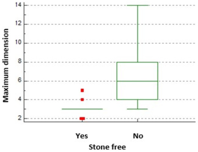
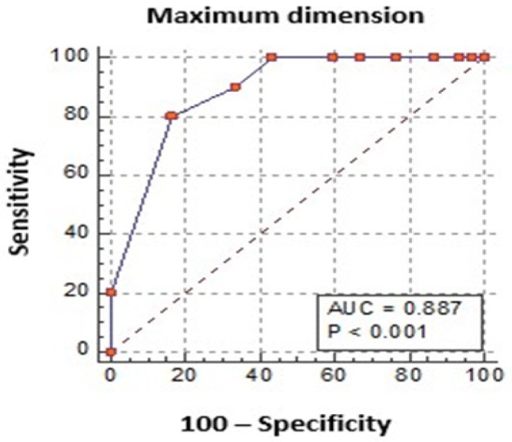
The maximum dimension of the calculi was correlated with achieving stone-free status (p=0.001) (Figure 1). The presence of a calculi equal or smaller than 3 mm had a Se of 80%, a Sp of 83.3% and an AUC of 0.887 (CI: 0.746–0.965, p<0.0001) for stone-free status after treatment (Figure 2).
Figure 1.
Stone-free status after treatment.
Figure 2.
A cut-off value of 3 mm had a high accuracy for the prediction of stone-free status after treatment.
Per stone analysis
We analyzed a number of 60 calculi in the 40 patients included in the study. The mean dimension before treatment was 4.93 mm (range 2–14 mm). Half of the calculi were located on the right side and half on the left side. The inferior calyx harbored 55% of the calculi, whereas 36.7% were located in the middle and 8.3% in the superior calyx.
A percentage of 40% of calculi were absent on the post-treatment imaging, while 21.7% were shown with smaller dimensions. A number of 23 calculi (38.3%) had the same dimension as before the treatment (Table I).
Table I.
Response to treatment stratified by the initial dimension of calculi.
| Response to treatment/dimension of calculi | ≤ 3 mm | > 3 mm and < 6 mm | ≥ 6 mm |
|---|---|---|---|
| Absent (No of patients) | 21 | 3 | 0 |
| Reduced dimension (No of patients) | 2 | 7 | 4 |
| Identical dimension (No of patients) | 4 | 6 | 13 |
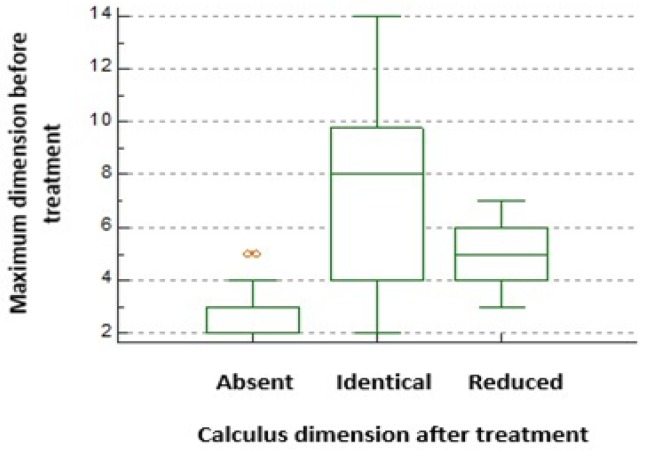
The mean dimension of the remaining calculi after the treatment was 5.7 mm (range: 2–14 mm) and the mean reduction of the dimension was 1.7 mm (range: 1–2 mm) (Figure 3).
Figure 3.
Calculus dimension change after treatment.
Using ROC curve analysis, we identified a cut-off of ≤ 3 mm (AUC 0.9, CI:0.8–0.9, p<0.0001) for the prediction of stone-free status after treatment (Figure 4). Calculi with a dimension >3 mm and <6 mm were most likely to reduce their dimension after treatment (AUC 0.6, CI: 0.4–0.7, p=0.08), whereas calculi of 6 mm or bigger would remain at the same dimension (AUC 0.83, CI: 0.7–0.9, p<0.0001). Using these cut-off values for the pre-treatment dimension of calculi, we observed a significant correlation between the dimension and response to treatment, p<0.0001.
Figure 4.
ROC curve analysis of the accuracy for the prediction of stone-free status after treatment depending on the initial dimension of calculi: a) ≤ 3 mm, b) 3–6mm, c) ≥ 6 mm.
The caliceal location of the calculi was significantly correlated with the response to treatment, as 68.18% of the calculi located in the middle calyx were absent on post-treatment imaging, in comparison with only 18% from the inferior calyx, p=0.003 (Table II).
Table II.
Change in calculi diameter stratified by their caliceal location.
| Response to treatment/location of calculi | Inferior calyx | Middle calyx | Superior calyx |
|---|---|---|---|
| Absent (No of patients) | 6 | 15 | 3 |
| Reduced dimension (No of patients) | 10 | 3 | 0 |
| Identical dimension (No of patients) | 17 | 4 | 2 |
Discussion
Urolithiasis remains one of the most frequent benign urological pathology, second after benign prostatic hyperplasia, with average size of calculi at diagnosis between 5 and 10 mm [11]. In our study, the mean diameter of the calculi was 4.93 mm, for which the risk of requiring a surgical intervention in the next 3 years is considered to be approximately 20%, the risk of developing renal colic is 40% and of increasing in size is 50%, as reported by Burgher et al. [12].
Phytotherapy is one of the first choices regarding pharmaceutical treatment. Micali et al. [13] analyzed the stone-free status after 3 to 6 months of treatment with dried extract of P. niruri in 78 patients with a history of multiple sessions of extracorporeal shock-wave lithotripsy. Average calculi diameter before treatment was 12.2 mm. The imaging assessment was done by performing abdominal X-ray and ultrasound monthly. After 6 months, 93.5% of the patients achieved stone-free status (calculi <3 mm). Response to treatment was statistically significant for calculi located in the lower calyx (p=0.03 for stone-free status and p<0.001 for fragments <3 mm), contrasting to our study, where the upper and mid calyx were the elected locations for calculi to reduce their size or to be dissolved (p=0.003).
Another study, conducted by Pucci et al. [14] used 4.5g of sun-dried P. niruri leaves, 2 times a day, for 3 months, followed by 3 months wash-out period. The study included 56 patients, with average stone size of 15.6 mm. Imaging evaluation was carried out using renal ultrasonography. After the wash-out period, mean diameter of stone fragments was 11.2 mm (p=0.0002). The size of the calculi had decreased in 67.8% of patients, remained unchanged in 17.8% and increased in 14.3%, in comparison with 40% of calculi being absent in our group and 21.7% showing lower dimensions. The difference in the dimension might be as a result of the imaging used, CT being the most accurate for urinary lithiasis in comparison with ultrasound or abdominal X-ray.
Other plants have proven their efficacy for the treatment of urolithiasis. Singh et al. [15] studied the effects of Dolichous biflorus, Fabaceae family, commonly known as horse gram. Forty-five patients were included. First group, 24 patients, with mean calculus size of 5.42 mm, identified by abdominal X-ray or ultrasound, were given 1–2 g/day of dried extract for 6 months. Mean diameter measured 4.26 mm. The second group comprised 21 patients, with mean calculus size of 6.46 mm, which was given 10 to 15 ml of potassium citrate six hourly, for 6 months. After treatment, the stone size dropped to 4.64 mm, but it was not considered a statistically significant change (p>0.05).
Celosia argentea, Amaranthaceae family (silver cock’s comb), has ruled out potassium citrate in a clinical study performed by Rana Singh et al. [16]. Twenty-one patients received 10 mg/kg dried seed extract three times a day, compared to 23 patients receiving 0.25 mL/kg potassium citrate, 4 times a day for 6 months. The calculi were assessed using abdominal X-ray or renal ultrasound every three months. Intragroup analysis showed a significant reduction of the lithiasis in the first group (6.47 vs 3.9 mm, p<0.05), but not for the second one (6.46 vs 4.04, p>0.05). Intergroup evaluation was also statistically significant (p>0.05).
Although previous studies did not favor potassium citrate therapy, results may come with a longer period of time. Lojanapiwat et al. [17] studied potassium citrate therapy vs. placebo in 76 treated patients (39 and 37, respectively). All of them underwent extracorporeal shock-wave lithotripsy or percutaneous nephrolithotomy, being stone-free or with fragments <4 mm at the beginning of the study. The average stone size was 2.54 and 2.65 mm, respectively. The treated group received 27mEq potassium-sodium citrate, three times a day, for 1 year. Plain abdominal X-ray was used for evaluation. In the treated group, 92.3% of stone-free patients remained stone-free, compared to 57.7% in control one.
Soygur et al. [18] performed a similar clinical trial, targeting especially lower caliceal calculi. During 12 months, 90 patients who underwent at least one session of extracorporeal shock wave lithotripsy prior to this study were given 60 mEq potassium citrate daily. Mean reported calculus size was 9.2 mm. The stone recurrence, assessed with abdominal X-ray, was zero in the stone-free treated group, compared to 28.6% in stone-free control group. In the group with residual fragments, 45.5% of treated patients achieved the stone-free status, while the rest remained the same size, compared to untreated ones, where 12.5% were stone-free, 25% of stones remained the same and 62.5% increased in size.
In our study, the mean diameter of calculi was 4.93 mm (range 2–14 mm), similar to the studies taken into consideration. For these patients, extracorporeal shock wave lithotripsy and flexible ureteroscopy may represent an option. However, each technique associates specific morbidity rate.
Even though some self-limited side effects have been described by previously mentioned studies during phytotherapy, these were mild and did not necessitate discontinuation of treatment. Singh et al. [16] reported abdominal pain, hematuria and dysuria after using 1–2g/day Dolichous biflorus extract, symptoms which improved by the end of the 6-month period. Pucci et al. [14] listed similar symptoms after 6 months of 4.5g/day sun-dried Phyllantus niruri leaves. Micali et al. [13] used 2g/day of Phyllantus niruri extract, with no adverse effects to declare. A possible explanation would be the chosen pharmaceutical form, used for a lengthier period of time. In our study, no side-effects were noticed, so we consider that a lower, standardized dose might be safer and just as efficient.
Conclusion
The Phyllanthus niruri standardized extract administered for a 3-month period had the highest efficacy in achieving stone-free status for patients with calculi ≤ 3 mm, located in the middle or upper calyx. In patients with calculi between 3–6 mm, a 1.7 mm reduction was observed after 3 months of treatment. A higher duration of the treatment might show improved results. Other treatment options should be indicated in patients with calculi > 6 mm.
Acknowledgements
This paper was supported by grant no. 5120/2017 for the project entitled “Phyllanthus niruri standardized extract combined with magnesium and B6 vitamin treatment efficacy evaluation in patients with uncomplicated nephrolithiasis”, financed by Fiterman Pharma and Iuliu Hatieganu University of Medicine and Pharmacy, Cluj-Napoca, Romania.
References
- 1.Khan SR, Pearle MS, Robertson WG, Gambaro G, Canales BK, Doizi S, et al. Kidney stones. Nat Rev Dis Primers. 2016;2:16008. doi: 10.1038/nrdp.2016.8. [DOI] [PMC free article] [PubMed] [Google Scholar]
- 2.Romero V, Akpinar H, Assimos DG. Kidney stones: a global picture of prevalence, incidence, and associated risk factors. Rev Urol. 2010;12:e86–e96. [PMC free article] [PubMed] [Google Scholar]
- 3.Spivacow FR, Del Valle EE, Lores E, Rey PG. Kidney stones: Composition, frequency and relation to metabolic diagnosis. Medicina (B Aires) 2016;76:343–348. [PubMed] [Google Scholar]
- 4.Nambirajan T, Jeschke S, Albqami N, Abukora F, Leeb K, Janetschek G. Role of laparoscopy in management of renal stones: single-center experience and review of literature. J Endourol. 2005;19:353–359. doi: 10.1089/end.2005.19.353. [DOI] [PubMed] [Google Scholar]
- 5.Kang HW, Seo SP, Kwon WA, Woo SH, Kim WT, Kim YJ, et al. Distinct metabolic characteristics and risk of stone recurrence in patients with multiple stones at the first-time presentation. Urology. 2014;84:274–278. doi: 10.1016/j.urology.2014.02.029. [DOI] [PubMed] [Google Scholar]
- 6.Kieley S, Dwivedi R, Monga M. Ayurvedic medicine and renal calculi. J Endourol. 2008;22:1613–1616. doi: 10.1089/end.2008.0020. [DOI] [PubMed] [Google Scholar]
- 7.Boim MA, Heilberg IP, Schor N. Phyllanthus niruri as a promising alternative treatment for nephrolithiasis. Int Braz J Urol. 2010;36:657–664. doi: 10.1590/s1677-55382010000600002. [DOI] [PubMed] [Google Scholar]
- 8.Freitas AM, Schor N, Boim MA. The effect of Phyllanthus niruri on urinary inhibitors of calcium oxalate crystallization and other factors associated with renal stone formation. BJU Int. 2002;89:829–834. doi: 10.1046/j.1464-410x.2002.02794.x. [DOI] [PubMed] [Google Scholar]
- 9.Nishiura JL, Campos AH, Boim MA, Heilberg IP, Schor N. Phyllanthus niruri normalizes elevated urinary calcium levels in calcium stone forming (CSF) patients. Urol Res. 2004;32:362–366. doi: 10.1007/s00240-004-0432-8. [DOI] [PubMed] [Google Scholar]
- 10.Barros ME, Lima R, Mercuri LP, Matos JR, Schor N, Boim MA. Effect of extract of Phyllanthus niruri on crystal deposition in experimental urolithiasis. Urol Res. 2006;34:351–357. doi: 10.1007/s00240-006-0065-1. [DOI] [PubMed] [Google Scholar]
- 11.Fisang C, Anding R, Müller SC, Latz S, Laube N. Urolithiasis--an interdisciplinary diagnostic, therapeutic and secondary preventive challenge. Dtsch Arztebl Int. 2015;112:83–91. doi: 10.3238/arztebl.2015.0083. [DOI] [PMC free article] [PubMed] [Google Scholar]
- 12.Burgher A, Beman M, Holtzman JL, Monga M. Progression of nephrolithiasis: long-term outcomes with observation of asymptomatic calculi. J Endourol. 2004;18:534–539. doi: 10.1089/end.2004.18.534. [DOI] [PubMed] [Google Scholar]
- 13.Micali S, Sighinolfi MC, Celia A, De Stefani S, Grande M, Cicero AF, et al. Can Phyllanthus niruri affect the efficacy of extracorporeal shock wave lithotripsy for renal stones? A randomized, prospective, long-term study. J Urol. 2006;176:1020–1022. doi: 10.1016/j.juro.2006.04.010. [DOI] [PubMed] [Google Scholar]
- 14.Pucci ND, Marchini GS, Mazzucchi E, Reis ST, Srougi M, Evazian D, et al. Effect of phyllanthus niruri on metabolic parameters of patients with kidney stone: a perspective for disease prevention. Int Braz J Urol. 2018;44:758–764. doi: 10.1590/S1677-5538.IBJU.2017.0521. [DOI] [PMC free article] [PubMed] [Google Scholar]
- 15.Singh RG, Behura SK, Kumar R. Litholytic property of Kulattha (Dolichous biflorus) vs potassium citrate in renal calculus disease: a comparative study. J Assoc Physicians India. 2010;58:286–289. [PubMed] [Google Scholar]
- 16.Singh RG, Singh TB, Kumar R, Dwivedi US, Moorthy KN, Kumar N. A comparative pilot study of litholytic properties of Celosia argental (Sitivaraka) versus potassium citrate in renal calculus disease. J Altern Complement Med. 2012;18:427–428. doi: 10.1089/acm.2011.0431. [DOI] [PubMed] [Google Scholar]
- 17.Lojanapiwat B, Tanthanuch M, Pripathanont C, Ratchanon S, Srinualnad S, Taweemonkongsap T, et al. Alkaline citrate reduces stone recurrence and regrowth after shockwave lithotripsy and percutaneous nephrolithotomy. Int Braz J Urol. 2011;37:611–616. doi: 10.1590/s1677-55382011000500007. [DOI] [PubMed] [Google Scholar]
- 18.Soygür T, Akbay A, Küpeli S. Effect of potassium citrate therapy on stone recurrence and residual fragments after shockwave lithotripsy in lower caliceal calcium oxalate urolithiasis: a randomized controlled trial. J Endourol. 2002;16:149–152. doi: 10.1089/089277902753716098. [DOI] [PubMed] [Google Scholar]


