Abstract
C-Peptide (“connecting” peptide – molecular formula C112H179N35O46) is a peptide made of 31 aminoacids, making the bond between A and B chains of insulin from the pro-insulin molecule. Pro-insulin is the precursor of the insulin that is synthesized in the beta-pancreatic cells. After its discovery in 1967 by Steiner et al, together with the discovery of insulin biosynthesis, C-peptide seemed to bring new benefits, having similar effects as those of insulin. Unfortunately, the subsequent studies have classified C-peptide as a biologically inactive peptide. After the ‘90s, however, both studies on animals and those on human subjects with type 1 diabetes where C-peptide had been administered showed that it played important biological parts in improving kidney function and nerve conduction velocity, as well as in increasing blood flow in muscles, skin, kidneys, thus being seen as a possible treatment for chronic complications of type 1 diabetes. Although for a long time C-peptide has been considered to be an inert biological product, recent research has emphasized its active biological function. C-peptide bonds to the membrane of certain types of cells (neuronal, endothelial, renal tubular cells, fibroblasts) through a surface receptor coupled with a G protein, and it determines multiple effects at the cellular level: it improves the quality of red cells, generating a better oxygenation of tissues; it has a vasodilator effect for muscles, skin, kidneys; it generates blood flow increase in skeletal muscles and at the skin level; it decreases glomerular hyper-filtering; it reduces albumin urinary excretion; it improves the function and structure of nerves in patients with type 1 diabetes and C-peptide deficiency, but not in healthy subjects. Therefore, C-peptide could have a therapeutic potential in preventing some of the late complications of diabetes mellitus.
Keywords:C-Peptide, G protein, diabetes mellitus, insulin secretion.
INTRODUCTION
C-Peptide (“connecting” peptide – molecular formula C112H179N35O46) is a peptide made of 31 aminoacids, making the bond between A and B chains of insulin from the pro-insulin molecule (Fig. 1). Pro-insulin is the precursor of the insulin that is synthesized in the beta-pancreatic cells.It is transported to the Golgi complex and embedded in secretory grains covered by a coating called clathrin. C-peptide plays an essential role in the correct folding of pro-insulin through the two disulfide bridges. After maturation, the secretory grains lose their clathrin covering, and pro-insulin will be submitted to the action of two types of endopeptidases (2 and 3 convertases), generating the forming of two intermediate products: 31,32-split pro-insulin, and 64-, 65-split pro-insulin, respectively. Of these two molecules, four aminoacids (31, 32 and 64, 65) will be excluded, resulting in insulin and C-peptide that will be discharged in echimolar ratio in the portal circulation (1). The C-peptide fragment resulting from the splitting of pro-insulin during the secretor process releases insulin as a complex of two united peptide chains. Echimolar quantities ofinsulin and C-peptide are subsequently released into circulation. C-peptide has a longer halving time (25-30 minutes) compared to insulin (5-10 minutes), it is not metabolized by the liver but in the proximal renal tubes, from where it is secreted by urine in proportion of 5-10%. C-peptide may be measured in serum, plasma or urine, being much more exact than insulin in appraising the endogen secretion of insulin. As it is not present in injectable insulin preparations, C-peptide may be measured in patients treated with insulin.
After its discovery in 1967 by Steiner et al, together with the discovery of biosynthesis of insulin (2), C-peptide seemed to bring new benefits, having effects similar to those of insulin. Unfortunately, subsequent studies have classified C-peptide as a biologically inactive peptide. After the ‘90s, however, both studies on animals and human subjects with type 1 diabetes where C-peptide had been administered showed that it played important biological parts in improving kidney function and nerve conduction velocity as well as in increasing blood flow in muscles, skin, kidneys, thus being seen as a possible treatment for chronic complications of type 1 diabetes (3-5).
BIOLOGICAL PART AND CELLULAR EFFECTS OF C-PEPTIDE
Although for a long time C-peptide has been considered to be an inert biological product, recent research has emphasized its active biological function. C-peptide bonds to the membrane of certain types of cells (neuronal, endothelial, renal tubular cells, fibroblasts) through a surface receptor coupled with a G protein, and it determines multiple effects at the cellular level:
- it stimulates the activity of Na+-K +ATPase (6, 7): it is well known that, in diabetic patients, the activity of Na-K-ATPase pump is reduced at the level of several types of tissues, which determines the occurrence of chronic complications of diabetes. Studies have shown that the administration of C-peptide in rats stimulated the activity of Na-K-ATPase in renal tubular cells and in nerve cells (8, 9). Also, at the erythrocyte level, there is a decrease in Na-K-ATPase activity, which determines the alteration of red cell deformability and a growth of blood viscosity with role in the occurrence of micro-vascular complications of type 1 diabetes. The administering of C-peptide leads to an improvement of red cell deformability and better tissue oxygenation (10, 11).
- effects on eNOS: studies have shown that the administering of C-peptide activates the release of nitric oxide (NO) by stimulating endothelial synthetase (eNOS), with vasodilator effect on muscles, skin and kidneys (7, 12);
- it stimulates the activity of phosphoinositide 3-kinase, triggering the growth of cellular, tubular renal and neuronal proliferation (13);
- MAPK stimulation (mitogen activated protein kinases): C-peptide significantly stimulates cellular proliferation by activating C phospholipase, increasing the intracellular concentration of Ca2+, followed by phosphorylation and activation of MAPK (14).
Consequently, the administration of C-peptide triggers the growth of blood flow in skeletal muscles and at the skin level, decreases glomerular hyper-filtering, reduces the urinary secretion of albumin, and improves the function and structure of nerves in patients with type 1 diabetes and deficit of C-peptide, but not in healthy subjects. Thus, C-peptide could play a potential therapeutic part in preventing some of the late complications of diabetes mellitus (15).
DOSING OF C-PEPTIDE/NORMAL VALUES PER AGE GROUPS
The values of C-peptide depend on several factors: sampling moment (basal or stimulated), patient's age, the dosing method used. Yet, there is no optimal standardization regarding the dosing of C-peptide, which requires caution in interpreting the values of C-peptide, taking into account the fact that they differ depending of the methods that are used and on the laboratory (16)
From a practical point of view, the dosing of C-peptide is of clinical importance in evaluating cases of hypoglycemia, measuring the reserve of beta-pancreatic cells in patients with type 1 diabetes mellitus (DM), making differential diagnosis in case of type 1 DM/type 2 DM/MODY diabetes/ latent diabetes of autoimmune type in adults (LADA) or when evaluating insulin resistance in obese patients and in those with polycystic ovaries.
Low values of C-peptide may appear in case of: abusive administering of insulin; pancreatectomy; type 1 DM and in the latent autoimmune diabetes of the adult (LADA).
High values of C-peptide may appear in: type 2 DM and MODY; endogenous hyperinsulinism; ingestion of oral antidiabetic drugs; insulinoma; transplant of pancreas.
Measuring of C-peptide in the blood. We may determine basal C-peptide/C-peptide a jeun or stimulated C-peptide. When we want to assess the resistance to insulin in non-insulin treated patients, the dosing à jeun is preferred. The dosing of the stimulated C-peptide (postprandial: dosing of C-peptide at 90-120 minutes after meal or stimulated by glucagon – dosing à jeun of C-peptide at six minutes after adm IV of 1 mL of glucagon) would be indicated to be used when testing the residual function of beta insular cells in insulin-treated patients (16).
Measuring of C-peptide in urine. The total quantity of C-peptide excreted in urine/day represents approximately 5% of the pancreatic secretion. The concentration in urine is usually 10-20 times higher than in plasma, and the absence of proteases in urine (unlike plasma) gives C-peptide a greater stability at room temperature. Apparently, the dosing of C-peptide in urine is more attractive, being totally non-invasive, which may be very useful, particularly in children. It may be dosed in the collected urine/24 hours or in spontaneous sample, dosing the ratio C-peptide/urinary creatinine (nmol/mmol).
EVOLUTION OF C-PEPTIDE IN TYPE 1 DIABETES
Type 1 DM supposes the destruction of the pancreatic beta cells by an autoimmune process that begins way before setting the clinical diagnosis and it continues for a long time in the evolution of the disease, as shown by research (19). The first data describing the modifications at the level of pancreatic beta cells immediately after the diagnosis were published in 1973 (New England Journal of Medicine), mentioning a growth of the C-peptide level in the remission interval, respectively a drop of its concentrations when coming out of remission. The decline of pancreatic beta cells residual function correlated with the drop of C-peptide values occurs in the period of time before diagnosis (even up to six months) as well as immediately afterwards (20). Most patients with type 1 DM have an endogenous secretion of insulin at diagnosis and in the following 1-2 years (21, 22) and, according to some authors, even five years from onset (23).
On the other hand, some studies have proven the existence of unexpectedly large concentrations in subjects with type 1 DM diagnosed for several years (24). The DCCT study shows that 48% of all patients with type 1 DM screened for 1-5 years after diagnosis have registered a value of the stimulated C-peptide of . 0.2 nmol/L. This is the level where patients obviously have a better glycemic control, fewer micro-vascular complications and a lower risk of hypoglycemia (25). Several factors influencing the rate of decrease of the insular reserve in patients with type 1 SD have been identified: age on onset, quality of glycemic control, level of autoimmune markers, genetic factors such as HLA, insulin gene or simply individual variations (26-28). Patients diagnosed in the pre-puberty period show lower levels on onset (<0.2 nmol/L) as well as a more accelerated rate of C-peptide decrease in the evolution of the disease, comparatively with those diagnosed at puberty or as adults (0.3-0.9 nmol/L basal C-peptide, respectively: 0.6-1.3 nmol/L stimulated C-peptide) (29). The same DCCT concluded that patients with intensive treatment since the onset had a lower decline rate of C-peptide (30). It was noticed that the smaller the age at onset, the smaller the values of C-peptide. If C-peptide is high at onset, then its values will drop faster in the first year of evolution. Those with high BMI usually show higher values of C-peptide, but the rate of C-peptide decrease in the first year is also high. Asymptomatic patients with a low HbA1c at onset associate a high C-peptide at onset, but they have a fast drop in the first year (31).
BRIEFLY:
- most patients with type 1 diabetes show an obvious decline in the reserve of insular beta cells in five years since onset, due to their destruction by an autoimmune mechanism;
- the level of C-peptide in children is generally lower than in adults, with a much faster drop in its rate (particularly in children under five);
- studies have shown that patients with type 1 diabetes continue to secrete C-peptide in a low quantity, sometimes in 10 years after onset, and these beta cells that are still secretor may be reactivated by prandial stimulation (32);
- C-peptide persistence has been demonstrated to be advantageous for patients: the DCCT study has shown that a stimulated C-peptide . 0.2 nmol/L (200 pmol/L) was associated with a favorable prognosis of clinical evolution: lower frequency of chronic complications, less severe hypoglycemia.
CLINICAL UTILITY OF C-PEPTIDE
From a practical point of view, the dosing of C-peptide is of clinical importance in the following situations: measurement of insulin endogenous secretion in patients with diabetes mellitus; differentiation between type 1 DM/type 2 DM/MODY/latent diabetes of autoimmune type in adults (LADA) and initiating the suitable treatment; suspicion of insulin-resistance in obese patients or in those with polycystic ovary syndrome (high C-peptide); evaluation of hypoglycemia due to hyperinsulin (high values of C peptide) versus hypoglycemia of other causes than hyperinsulinism (C-peptide with low values).
C-PEPTIDE – AS MEASURE OF INSULIN SECRETION
Knowing that C-peptide is secreted in an echimolar ratio as the insulin in pancreatic beta cells, its level provides data on insulin endogenous secretion in patients with type 1 DM. Given that approximately 50% of insulin produced by the pancreas is metabolized in the liver, the serum level of insulin does not accurately show the level of insulin secretion (in both insulin-treated and non-insulin treated patients).
The values of C-peptide must be carefully interpreted in renal insufficiency (~ 50% is metabolized in urine and only 5% is eliminated in urine). Therefore, in renal impairment, the values of C-peptide may be falsely high (16, 33). In insulin-resistant obese patients, C-peptide values may be either normal or high (16).
DIFFERENTIATION FROM SD t1/SD2
The dosing of C-peptide is the key element for a correct diagnosis of SD (34). C-peptide allows for the differentiation between DM 1 and DM 2, particularly when long-term diabetes is concerned, because C-peptide values may be superposable in both types of diabetes at onset. In DM 1, the level of C-peptide drops quickly, so that the utility of C-peptide testing is obvious after 3-5 years since onset, when most patients have low values of C-peptide. The low values of C-peptide in the first years after diagnosis may confirm the presence of type 1 DM (values < 0.2 nmol/L show a severe insulin deficiency). The high values must be carefully interpreted – they may reveal the presence of an endogenous secretion of insulin during remission (16).
IDENTIFICATION OF PATIENTS WITH MODY TYPE DIABETES
The persistence of C-peptide values within the normal limits after the end of the remission pleads for MODY type diabetes. It is important to identify such situations, when it may be opportune to change the treatment (stop administering insulin and introduce sulphonyl urea derivatives) (16, 35). However, C-peptide is not useful in differentiating DM 2 from MODY-type diabetes (36).
DETERMINATION OF PROGNOSIS
In type 1 DM, the preservation of residual insular secretion (highlighted by the optimal level of stimulated C-peptide: >0.2 nmol/L) is associated with a better glycemic control, fewer cases of hypoglycemia and a lower rate of micro-vascular complications (37). In type 2 DM, high values of C-peptide are associated with a high risk of macro- vascular complications (38-40). The relation between C-peptide and occurrence of microvascular complications in type 2 DM is not fully elucidated.
THERAPEUTIC PERSPECTIVES
In the last 20 years, increasing information has been emerging on the physiology of C-peptide and treatment possibilities that it offers in type 1 diabetes. It is now known that C-peptide bonds specifically to the cellular membrane by a G-protein receptor, and it activates the Na-K-ATPase system, increases the expression of NO-synthetase as well as certain transcription factors that play a part in the anti-inflammatory and antioxidant mechanisms. Studies on animals and recent research on patients with DM 1 have demonstrated that administering of C-peptide in substitution doses has favourable effects on the structural and functional modifications induced by hyperglycemia of DM in peripheral nerves, kidneys and at retina level (41). Therefore, C-peptide may be taken into account as a potential therapeutic agent in chronic complications of DM: neuropathy, nephropathy and retinopathy. The discovery of C-peptide intracellular effects provides a new perspective over the relation between C-peptide and the occurrence of microvascular complications in DM (42).
Conflicts of interest: none declared.
Financial support: none declared.
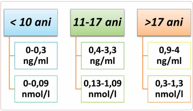
FIGURE 1.
Normal values of basal C-peptide in children (18)
TABLE 1.
Normal values of basal C-peptide in children (18)
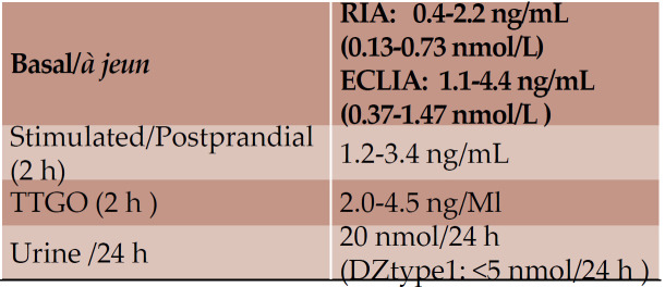
TABLE 2.
Normal values of C-peptide in adults (18)
Contributor Information
Carmen NOVAC, M. S. Curie” Children Emergency Hospital, Bucharest, Romania.
Gabriela RADULIAN, ”Carol Davila” University of Medicine and Pharmacy, Bucharest, Romania; „N. C. Paulescu” National Institute of Diabetes, Nutrition and Metabolic Diseases, Bucharest, Romania.
Anca ORZAN, ”M. S. Curie” Children Emergency Hospital, Bucharest, Romania; ”Carol Davila” University of Medicine and Pharmacy, Bucharest, Romania.
Mihaela BALGRADEAN, ”M. S. Curie” Children Emergency Hospital, Bucharest, Romania; ”Carol Davila” University of Medicine and Pharmacy, Bucharest, Romania.
REFERENCES
- 1.Rhodes CJ, Shoelson S, Halban PA. Insulin Biosynthesis, Processing, and Chemistry, in: Joslin’s Diabetes Mellitus, 14th edition. 2005.
- 2.Steiner DF, Cunningham D, Spigelman L, Aten B. Insulin biosynthesis:evidence for a precursor. Science. 1967;157:697–700. doi: 10.1126/science.157.3789.697. [DOI] [PubMed] [Google Scholar]
- 3.Luppi P, Cifarelli V, Wahren J. C-peptide and long-term complications of diabetes. Pediatr Diabetes. 2011;12:276–292. doi: 10.1111/j.1399-5448.2010.00729.x. [DOI] [PubMed] [Google Scholar]
- 4.Sima AA, Zhang W, Muzik O, et al. Sequential abnormalities in type 1 diabetic encephalopathy and the effects of C-peptide. Rev Diabet Stud. 2009;6:211–222. doi: 10.1900/RDS.2009.6.211. [DOI] [PMC free article] [PubMed] [Google Scholar]
- 5.Wahren J, Ekberg K, Jörnvall H. C-peptide is a bioactive peptide. Diabetologia. 2007;50:503–509. doi: 10.1007/s00125-006-0559-y. [DOI] [PubMed] [Google Scholar]
- 6.Vague P, Coste TC, Jannot MF, et al. C-peptide, Na+,K+-ATPase, and Diabetes. Experimental Diab Res. 2004;5:37–50. doi: 10.1080/15438600490424514. [DOI] [PMC free article] [PubMed] [Google Scholar]
- 7.Wahren J, Ekberg K, Johansson J, et al. Role of C-peptide in human physiology. Am J Physiol Endocrinol Metab. 2000;5:E759–E768. doi: 10.1152/ajpendo.2000.278.5.E759. [DOI] [PubMed] [Google Scholar]
- 8.Ido Y, Vindigni A, Chang K, Stramm L, et al. Prevention of vascular and neural dysfunction in diabetic rats by C-peptide. Science. 1997;277:563–566. doi: 10.1126/science.277.5325.563. [DOI] [PubMed] [Google Scholar]
- 9.Ohtomo Y, Bergman T, Johansson BL, et al. Differential effects of proinsulin C-peptide fragments on Na+, K+-ATPase activity of renal tubule segments. . Diabetologia. 1998;41:287–291. doi: 10.1007/s001250050905. [DOI] [PubMed] [Google Scholar]
- 10.Johansson BL, Linde B, Wahren J. Effects of C-peptide on blood flow, capillary diffusion capacity and glucose utilization in the exercising forearm of type 1 (insulindependent) diabetic patients. Diabetologia. 1992;35:1511–1158. doi: 10.1007/BF00401369. [DOI] [PubMed] [Google Scholar]
- 11.Kunt T, Schneider S, Pfutzner A, et al. The effect of human proinsulin C-peptide on erythrocyte deformability in patients with Type I diabetes mellitus. Diabetologia. 1999;42:465–471. doi: 10.1007/s001250051180. [DOI] [PubMed] [Google Scholar]
- 12.Johansson BL, Wahren J, Pernow J. C-peptide increases forearm blood flow in patients with type 1 diabetes via a nitric oxide-dependent mechanism. Am J Physiol Endocrinol Metab. 2003;285:E864–E870. doi: 10.1152/ajpendo.00001.2003. [DOI] [PubMed] [Google Scholar]
- 13.Johansson BL, Borg K, Fernqvist-Forbes E. Beneficial effects of C-peptide on incipient nephropathy and neuropathy in patients with Type 1 diabetes mellitus. Diabet Med. 2000;17:181–189. doi: 10.1046/j.1464-5491.2000.00274.x. [DOI] [PubMed] [Google Scholar]
- 14.Zhong Z, Davidescu A, Ehren I, et al. C peptide stimulates ERK 1/2 and JNK MAP kinases via activation of protein kinase C in human renal tubular cells. Diabetologia. 2005;48:187–197. doi: 10.1007/s00125-004-1602-5. [DOI] [PubMed] [Google Scholar]
- 15.Wahren J, et al. The clinical potential of C-peptide replacement in type 1 diabetes. Diabetes. 2012;61:761–772. doi: 10.2337/db11-1423. [DOI] [PMC free article] [PubMed] [Google Scholar]
- 16.Jones AG, Hattersley AT. The clinical utility of C-peptide measurement in the care of patients with diabetes. Diabet Med. 2013;7:803–817. doi: 10.1111/dme.12159. [DOI] [PMC free article] [PubMed] [Google Scholar]
- 17.Laborator Synevo. Referinþele specifice tehnologiei de lucru utilizate. Ref Type: Catalog. 2010.
- 18.Constantin C, Cheta D. Proinsulina si peptidul C. Tratat roman de boli metabolice, Edit. Brumar, Timisoara. 2010. pp. 119–123.
- 19.Palmer JP. C-peptide in the natural history of type 1 diabetes. Diabetes Metab Res Rev. 2009;25:325–328. doi: 10.1002/dmrr.943. [DOI] [PMC free article] [PubMed] [Google Scholar]
- 20.Black MB, Rosenfield RL, Mako ME, et al. Sequential changes in beta-cell function in insulintreated diabetic patients assessed by c-peptide immunoreactivity. N Engl J Med. 1973;288:1144–1148. doi: 10.1056/NEJM197305312882202. [DOI] [PubMed] [Google Scholar]
- 21.Decline of C-peptide during the first year after diagnosis of Type 1 diabetes in children and adolescents. Diabetes Res Clin Pract. Proteins Laboratory Testing and Clinical Use, Media Print Taunusdruck GmbH, Frankfurt am Main. 2013;100:203–209. doi: 10.1016/j.diabres.2013.03.003. [DOI] [PubMed] [Google Scholar]
- 22.Greenbaum CJ, Beam CA, Boulware D, et al. Type 1 Diabetes TrialNet Study Group. Fall in C-peptide during first 2 years from diagnosis: evidence of at least two distinct phases from composite Type 1 Diabetes TrialNet data. Diabetes. 2012;61:2066–2073. doi: 10.2337/db11-1538. [DOI] [PMC free article] [PubMed] [Google Scholar]
- 23.Sørensen JS, Johannesen J, Pociot F, et al. Residual β-Cell Function 3–6 Years After Onset of Type 1 Diabetes Reduces Risk of Severe Hypoglycemia in Children and Adolescents. Diabetes Care. 2013;11:3454–3459. doi: 10.2337/dc13-0418. [DOI] [PMC free article] [PubMed] [Google Scholar]
- 24.Keenan HA, Sun JK, Levine J, et al. Residual insulin production and pancreatic β-cell turnover after 50 years of diabetes: Joslin Medalist Study. Diabetes. 2010;59:2846–2853. doi: 10.2337/db10-0676. [DOI] [PMC free article] [PubMed] [Google Scholar]
- 25.The Diabetes Control and Complications Trial Research Group. Effect of intensive therapy on residual β-cell function in patients with type 1 diabetes in the diabetes control and complications trial. Ann Intern Med. 1998;128:517–523. doi: 10.7326/0003-4819-128-7-199804010-00001. [DOI] [PubMed] [Google Scholar]
- 26.Törn C, Landin-Olsson M, Lernmark Å, et al. Prognostic factors for the course of β cell function in autoimmune diabetes. . J Clin Endocrinol Metab. 2000;85:4619–4623. doi: 10.1210/jcem.85.12.7065. [DOI] [PubMed] [Google Scholar]
- 27.Petrone A, Galgani A, Spoletini M, et al. Residual insulin secretion at diagnosis of type 1 diabetes is independently associated with both, age of onset and HLA genotype. Diabetes Metab Res Rev. 2005;21:271–275. doi: 10.1002/dmrr.549. [DOI] [PubMed] [Google Scholar]
- 28.Petrone A, Spoletini M, Zampetti S, et al. Buzz reduced residual β-cell function and worse metabolic control. . Diabetes Care. 2008;31:1214–1218. doi: 10.2337/dc07-1158. [DOI] [PubMed] [Google Scholar]
- 29.Palmer JP, Fleming GA, Greenbaum CJ, et al. C-peptide is the appropriate outcome measure for type 1 diabetes clinical trials to preserve β-cell function. Diabetes. 2004;53:250–264. doi: 10.2337/diabetes.53.1.250. [DOI] [PubMed] [Google Scholar]
- 30.The Diabetes Control and Complications Trial Research Group. The effect of intensive treatment of diabetes on the development and progression of long-term complications in insulin-dependent diabetes mellitus. N Engl J Med. 1993;329:977–986. doi: 10.1056/NEJM199309303291401. [DOI] [PubMed] [Google Scholar]
- 31.Ludvigsson J, Carlsson A, Deli A, et al. Decline of C-peptide during the first year after diagnosis of Type 1 diabetes in children and adolescents. Media Print Taunusdruck GmbH, Frankfurt am Main. 2013;100:203–209. doi: 10.1016/j.diabres.2013.03.003. [DOI] [PubMed] [Google Scholar]
- 32.Oram RA, et al. The majority of patients with long-duration type 1 diabetes are insulin microsecretors and have functioning beta cells. Diabetologia. 2014;1:187–191. doi: 10.1007/s00125-013-3067-x. [DOI] [PMC free article] [PubMed] [Google Scholar]
- 33.Covic AM, Schelling JR, Constantiner M, et al. Serum C-peptide concentrations poorly phenotype type 2 diabetic end-stage renal disease patients. Kidney Int. 2000;58:1742–1750. doi: 10.1046/j.1523-1755.2000.00335.x. [DOI] [PubMed] [Google Scholar]
- 34.Ludvigsson J, et al. C-peptide in the classification of of diabetes in children and adolescents. Pediatric Diabetes. 2012;1:45–50. doi: 10.1111/j.1399-5448.2011.00807.x. [DOI] [PubMed] [Google Scholar]
- 35.ISPAD. guidelines. 2014.
- 36.Besser RE, Shepherd MH, McDonald TJ, et al. Urinary C-peptide creatinine ratio is a practical outpatient tool for identifying hepatocyte nuclear factor 1-á/hepatocyte nuclear factor 1-α maturity-onset diabetes of the young from long-duration type 1 diabetes. Diabetes Care. 2011;34:286–291. doi: 10.2337/dc10-1293. [DOI] [PMC free article] [PubMed] [Google Scholar]
- 37.Steffes MW, Sibley S, Jackson M, Thomas W. Beta-cell function and the development of diabetes-related complications in the Diabetes Control and Complications Trial. Diabetes Care. 2003;26:832–836. doi: 10.2337/diacare.26.3.832. [DOI] [PubMed] [Google Scholar]
- 38.Bo S, Cavallo-Perin P, Gentile L, et al. Relationship of residual beta-cell function, metabolic control and chronic complications in type 2 diabetes mellitus. Acta Diabetol. 2000;37:125–129. doi: 10.1007/s005920070014. [DOI] [PubMed] [Google Scholar]
- 39.Sari R, Balci MK. Relationship between C peptide and chronic complications in type-2 diabetes mellitus. J Natl Med Assoc. 2005;97:1113–1118. [PMC free article] [PubMed] [Google Scholar]
- 40.Inukai T, Matsutomo R, Tayama K, et al. Relation between the serum level of C-peptide and risk factors for coronary heart disease and diabetic microangiopathy in patients with type-2 diabetes mellitus. Exp Clin Endocrinol Diabetes. 1999;107:40–45. doi: 10.1055/s-0029-1212071. [DOI] [PubMed] [Google Scholar]
- 41.Wahren J, Larsson C. C-peptide: new findings and therapeutic possibilities. https://www.ncbi.nlm.nih.gov/pubmed/25648391. 2015. [DOI] [PubMed]
- 42.Yosten GL, Maric-Bilkan C, Luppi P, Wahren J. Physiological effects and therapeutic potential of proinsulin C-peptide. Am J Physiol Endocrinol Metab. 2014;307:E955–E968. doi: 10.1152/ajpendo.00130.2014. [DOI] [PMC free article] [PubMed] [Google Scholar]


