Key Points
Question
What is the long-term association of low birth weight with ocular geometry in adulthood?
Findings
This population-based, observational cohort study of 7120 participants with birth weight and follow-up data found an association of low birth weight with steeper corneal curvature, thinner central cornea, and shorter axial length among adults aged 40 to 80 years.
Meaning
Although the association of low birth weight with ocular geometry in childhood persisted in adulthood, the findings could not be controlled for retinopathy of prematurity and its treatment, which may affect the outcomes.
This population-based cohort study evaluates whether low birth weight is associated with anterior segment anatomy and axial length in adulthood among participants in the Gutenberg Health Study.
Abstract
Importance
Low birth weight is associated with altered ocular organ development in childhood, including the morphology of the eye. However, no population-based data exist about this association in adulthood.
Objective
To evaluate whether low birth weight has a long-term association with anterior segment anatomy and axial length in adulthood.
Design, Setting, and Participants
The Gutenberg Health Study is a population-based, observational cohort study in Germany. All participants underwent ocular biometry. Among the participants with follow-up and self-reported birth weight available, associations were assessed between low birth weight and anterior segment anatomy and axial length using multivariable linear regression analysis with adjustment for age and sex. In patients with phakia, anterior chamber depth and lens thickness were also examined. Data for this study were collected from April 27, 2012, through April 28, 2017, and analyzed from January through April 2018.
Exposures
Low birth weight.
Main Outcomes and Measures
Corneal curvature, central corneal thickness, white-to-white distance, anterior chamber depth, lens thickness, and axial length.
Results
Overall, 11 294 eyes of 7120 participants were included (52.4% female; mean [SD] age, 56.2 [10.3] years). Most of the participants were white (98.6%). After adjustment for age and sex, an association was found between a lower birth weight and steeper corneal curvature (β = 0.005 mm/100 g; 95% CI, 0.005-0.006 mm/100 g; P < .001), smaller white-to-white distance (β = 0.006 mm/100 g; 95% CI, 0.005-0.007 mm/100 g; P < .001), thinner central corneal thickness (β = 0.327 μm/100 g; 95% CI, 0.229-0.425 μm/100 g; P < .001), and shorter axial length (β = 0.006 mm/100 g; 95% CI, 0.003-0.010 mm/100 g; P < .001). However, anterior chamber depth and lens thickness were not associated with low birth weight in participants with phakia (10 510 eyes of 5279 participants).
Conclusions and Relevance
These analyses demonstrate an association between low birth weight and altered ocular geometry in adults aged 40 to 80 years, suggesting that birth weight and associated factors are crucial in anatomical ocular morphologic development. Retinopathy of prematurity and its treatment may affect ocular anatomy but could not be further analyzed in this study.
Introduction
Low birth weight is an indicator of impaired prenatal growth or preterm birth and is a risk factor for systemic comorbidities in adulthood such as arterial hypertension and diabetes.1,2 Recent reports3,4 show that altered prenatal growth development affects ocular morphology in childhood and adolescence. However, few population-based studies have reported associations between low birth weight and corneal curvature,3 corneal power,4 and axial length3 in childhood and adolescence. Therefore, impaired prenatal growth development is thought to affect morphologic organ development of the eye in childhood.3,4,5 We expect that these changes persist in adulthood, but the long-term effects of prenatal growth restriction on ocular morphology in adults are rarely investigated, and results differ. Although some authors6 hypothesize that morphologic differences due to low birth weight and prematurity diminish within the first decade of life, an Australian study7 reported associations of low birth weight with shorter axial length and steeper curvature of the cornea in participants ranging from 5 to 80 years of age.
Ocular geometry is linked to the prevalence and incidence of age-related macular degeneration8,9 and the prevalence of open-angle glaucoma10 and diabetic retinopathy,11 the main eye diseases leading to visual impairment and blindness in industrialized countries.12 Therefore, determining whether low birth weight affects ocular geometry and contributes to an increased risk for ocular comorbidities is imperative.
The aim of this study was to evaluate the association of low birth weight with ocular geometry in adults aged 40 to 80 years. Our hypothesis was that low birth weight is linked to altered ocular geometry in adulthood, resulting in steeper corneal curvatures and shorter axial lengths.
Methods
Study Population
The Gutenberg Health Study (GHS) is an observational, population-based, prospective, single-center cohort study at the University Medical Center of the Johannes Gutenberg University Mainz, Mainz, Germany.13 The population was sampled for each decade of age with equal amounts of individuals of both sexes and residences (urban and rural). The recruitment efficacy proportion was 61.2%. For this analysis, participants of the 5-year follow-up from April 27, 2012, through April 28, 2017, were included because only in the follow-up examination did every participant undergo ocular biometry. Overall, 12 423 participants (82.8%) of the GHS cohort had a follow-up examination after 5 years. All participants underwent an ophthalmological examination, including ocular biometry and Scheimpflug imaging, anterior segment photography, and a medical history interview. Written informed consent was obtained from all study participants before their entry into the study, and the GHS complies with Good Clinical Practice, Good Epidemiological Practice, and the ethical principles of the Declaration of Helsinki. The study protocol and study documents were approved by the local ethics committee of the Medical Chamber of Rhineland-Palatinate, Germany.
Birth Weight
In the present analysis, only participants with self-reported birth weight were included. The participants were asked on invitation to the GHS study center to review their records or family albums for a documented birth weight, and respondents were divided into the following birth weight groups: a low birth weight less than 2500 g (group 1), a normal birth weight of 2500 to 4000 g (group 2), and a high birth weight greater than 4000 g (group 3). In addition, participants with birth weights of less than 1000 g and greater than 6000 g were excluded because these self-reported data are suspected to be unreliable.
Ophthalmological Examination
A detailed description of the ophthalmological examinations is reported elsewhere.14 In brief, ocular biometry was performed (LenStar 900 device; Haag Streit). For the measurements, the participants were instructed to fixate on the center of the ocular biometer’s internal fixation target. Directional arrows of the device instruct the investigator to obtain an optimal focus, which is achieved when the smallest circles possible are displayed. This procedure was performed to ensure the highest measurement accuracy and to minimize measurement errors. Overall, the device performs 3 single measurements per examination and then computes the mean value. The following measurements were assessed with ocular biometry: corneal radius, white-to-white distance as a measurement of corneal diameter (measurement of the horizontal diameter of a best-fitted circle to the outer border of the iris), central corneal thickness, anterior chamber depth, lens thickness, and axial length. Every variable was checked for outliers. We compared these measurements with anterior segment photographs and Scheimpflug images recorded with a high-resolution rotating camera system (Pentacam HR; Oculus). Ocular biometric measurements were excluded when they were likely to be invalid compared with these other imaging modalities.
Covariates
Factors that may affect our main outcome measures, including age, sex, socioeconomic status (SES) based on income, educational level, occupation, and ocular comorbidities, were considered covariates. We defined SES according to the SES index used within the German Health Update 2009,15 which ranges from 3 to 21 (3 indicates the lowest SES; 21, the highest SES). Ocular comorbidities were assessed as self-reported corneal disease, age-related macular degeneration, and glaucoma. Participants with a history of corneal disease were excluded owing to possible altered ocular anatomy.
Statistical Analysis
Data were analyzed from January through April 2018. The main outcome measures were corneal radius, white-to-white distance, central corneal thickness, anterior chamber depth, lens thickness, and axial length. In a nonresponder analysis, we analyzed the difference between participants who reported birth weight and those with missing birth weight information. We compared the birth weight data distribution of our cohort with the distributions reported in the medical literature and governmental data.16,17 Descriptive statistics were computed for the main outcome measures. Absolute and relative frequencies were calculated for dichotomous variables; mean (SD), for approximately normally distributed data; and median (interquartile range), for the remaining variables. Linear regression models with general estimating equations were used to assess associations and to account for correlations between corresponding eyes. In model 1, the main outcome measures were tested in a univariate analysis with birth weight as an independent variable; in model 2, the associations were adjusted for age and sex. Furthermore, sensitivity analyses were performed with age, sex, SES, and ocular comorbidities, such as self-reported, age-related macular degeneration and glaucoma, as potential confounders. All models were computed in a first step including birth weight as a continuous variable and in a second step including birth weight as a categorical variable (<2500, 2500-4000, and >4000 g). The nonstandardized beta coefficients (β) are reported in this study. Pearson correlation coefficients were computed for the univariate association between birth weight (continuous) and the main outcome measures. The data were analyzed with R (version 3.3.1; R Foundation for Statistical Computing).18 This explorative study had no adjustment for multiple testing. The P value reflects the level of evidence, and no significance level was defined.
Results
Participant Characteristics
The 5-year follow-up examination of the GHS included 12 423 participants, of whom 7183 reported their birth weight. Forty-one participants were excluded because their self-reported birth weights were less than 1000 g or greater than 6000 g, and 22 persons were excluded due to a history of corneal disease, leaving a birth weight population of 7120 participants. Participant characteristics such as sex, age, ocular comorbidities, and ocular geometry, including corneal radius, white-to-white distance, central corneal thickness, anterior chamber depth, lens thickness, and axial length are reported in Table 1. The mean (SD) age of all participants was 56.2 (10.3) years; 3728 were female (52.4%) and 3392 (47.6%) were male. Most of the participants were white (98.6%). Furthermore, 10 510 eyes of 5279 participants were phakic. Overall, 382 participants reported a birth weight less than 2500 g (group 1); 5837, from 2500 to 4000 g (group 2); and 901, greater than 4000 g (group 3).
Table 1. Characteristics of the Study Sample and Ocular Geometric Variables of the Gutenberg Health Study by Birth Weight Groupsa.
| Birth Weight Group | |||
|---|---|---|---|
| Variable | Low (<2500 g) (n = 382) | Normal (2500-4000 g) (n = 5837) | High (>4000 kg) (n = 901) |
| Female, No. (%) | 253 (66.2) | 3167 (54.2) | 308 (34.2) |
| Age, mean (SD), y | 57.7 (10.8) | 55.8 (10.2) | 58.0 (10.5) |
| SES index, mean (SD)b | 13.11 (4.27) | 13.76 (4.24) | 13.62 (4.33) |
| Birth weight, median (IQR), g | 2100 (1750-2300) | 3360 (3000-3600) | 4500 (4200-4700) |
| Eye disease present, No. (%) | |||
| Glaucoma | 17 (4.4) | 191 (3.3) | 36 (4.0) |
| AMD | 9 (2.4) | 75 (1.3) | 14 (1.6) |
| Right eye measurements | |||
| Mean corneal radius, mean (SD), mm | 7.67 (0.26) | 7.77 (0.28) | 7.85 (0.27) |
| White-to-white distance, mean (SD), mm | 12.1 (0.4) | 12.2 (0.4) | 12.3 (0.4) |
| Central corneal thickness, mean (SD), μm | 545 (34) | 550 (35) | 555 (35) |
| Anterior chamber depth, mean (SD), mm | 3.29 (0.39) | 3.28 (0.35) | 3.26 (0.37) |
| Lens thickness, mean (SD), mm | 4.30 (0.35) | 4.30 (0.36) | 4.34 (0.39) |
| Axial length, mean (SD), mm | 23.7 (1.4) | 23.8 (1.3) | 23.9 (1.3) |
| Left eye measurements | |||
| Mean corneal radius, mean (SD), mm | 7.67 (0.27) | 7.76 (0.28) | 7.84 (0.26) |
| White-to-white distance, mean (SD), mm | 12.1 (0.4) | 12.2 (0.4) | 12.3 (0.4) |
| Central corneal thickness, mean (SD), μm | 545 (33) | 550 (35) | 556 (34) |
| Anterior chamber depth, mean (SD), mm | 3.27 (0.38) | 3.26 (0.34) | 3.24 (0.36) |
| Lens thickness, mean (SD), mm | 4.36 (0.34) | 4.36 (0.35) | 4.41 (0.38) |
| Axial length, mean (SD), mm | 23.4 (1.4) | 23.8 (1.3) | 23.9 (1.2) |
Abbreviations: AMD, age-related macular degeneration; IQR, interquartile range; SES, socioeconomic status.
Includes 7120 participants.
Calculated using the German Health Update 2009,15 which ranges from 3 (lowest SES) to 21 (highest SES).
Nonresponder Analysis
Overall, 5240 of the follow-up participants (42.2%) did not report birth weight data. In the nonresponder analysis, participants with self-reported birth weights were 7.8 years younger and more likely female (3751 of 7183 [52.2%] vs 2314 of 5240 [44.2%]) than those without self-reported data on birth weight (eTable 1 in the Supplement). Ocular variables only revealed small differences adjusted for sex and age (eTable 1 in the Supplement), except for axial length; eyes of participants with self-reported birth weight were longer by 0.14 mm in the right eyes and 0.13 mm in the left eyes.
Birth Weight as a Continuous Variable
Anterior Segment of the Eye
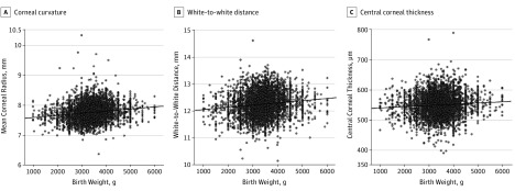
Linear associations were observed between birth weight and corneal curvature (Figure 1A), white-to-white distance (Figure 1B), and central corneal thickness (Figure 1C). Pearson correlation coefficient between birth weight and corneal curvature was 0.17 (P < .001); white-to-white distance, 0.13 (P < .001); and central corneal thickness, 0.08 (P < .001).
Figure 1. Scatterplot of Birth Weight With Corneal Radius, White-to-White Distance, and Central Corneal Thickness.
Includes 7120 participants in the Gutenberg Health Study. Participants with lower birth weights showed a steeper corneal curvature, smaller white-to-white distance, and a thinner central corneal thickness.
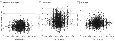
Corneal curvature was steeper in the adults with low birth weights. Multivariable analysis with adjustments for age and sex revealed that low birth weight was linked to a steeper corneal radius (β = 0.005 mm/100 g; 95% CI, 0.005-0.006 mm/100 g; P < .001). A shorter white-to-white distance was observed in adults with low birth weights in the multivariable analysis adjusted for age and sex (β = 0.006 mm/100 g; 95% CI, 0.005-0.007 mm/100 g; P < .001). In the multivariable analysis, an association was found between birth weight and central corneal thickness, showing thinner central corneal thickness measurements in participants with low birth weights (β = 0.327 μm/100 g; 95% CI, 0.229-0.425 μm/100 g; P < .001) (Table 2). For the analysis of anterior chamber depth and lens thickness, the data of 10 510 eyes of 5279 participants were included. Anterior chamber depth (r = 0.003; P = .047) and lens thickness (r = 0.002; P = .74) did not show an association with birth weight after adjustment for age and sex (Table 2; Figure 2A and B).
Table 2. Linear Associations of Ocular Geometric Variables With Birth Weighta.
| Variable by Birth Weight | Model 1b | Model 2c | ||
|---|---|---|---|---|
| β Coefficient (95% CI) | P Value | β Coefficient (95% CI) | P Value | |
| Mean corneal radius, mm/100 g | 0.007 (0.006 to 0.008) | <.001 | 0.005 (0.005 to 0.006) | <.001 |
| White-to-white distance, mm/100 g | 0.008 (0.007 to 0.009) | <.001 | 0.006 (0.005 to 0.007) | <.001 |
| Central corneal thickness, μm/100 g | 0.406 (0.309 to 0.502) | <.001 | 0.327 (0.229 to 0.425) | <.001 |
| Anterior chamber depth, mm/100 g | 0.001 (0.0001 to 0.002) | .03 | −0.0001 (−0.001 to 0.001) | .83 |
| Lens thickness, mm/100 g | 0.0003 (−0.001 to 0.001) | .60 | −0.001 (−0.002 to 0.0002) | .14 |
| Axial length, mm/100 g | 0.014 (0.011 to 0.018) | <.001 | 0.006 (0.003 to 0.010) | <.001 |
Includes 7120 participants from the Gutenberg Health Study. Linear regression analysis used generalized estimating equations to control for correlations between right and left eyes.
Indicates crude model without adjustment.
Adjusted for sex and age.
Figure 2. Scatterplot of Birth Weight With Anterior Chamber Depth, Lens Thickness, and Axial Length.
Includes 7120 participants in the Gutenberg Health Study. Participants with lower birth weights showed a shorter axial length.
Axial Length
Pearson correlation analysis revealed a univariate association between low birth weight and axial length (r = 0.08; P < .001) (Figure 2C) and in the multivariable analysis after adjustment for age and sex (β = 0.006 mm/100 g; 95% CI, 0.003-0.010 mm/100 g; P < .001), indicating that the participants with a low birth weight were more likely to have a shorter axial length.
Birth Weight Categorized Into Low, Normal, and High Groups
Anterior Segment
When analyzing associations with birth weight categorized into low, normal, and high groups, the corneal radius was smaller in the low birth weight group compared with that in the normal birth weight group in the multivariable analysis after adjustment for age and sex (β = −0.08 mm; 95% CI, −0.11 to −0.06 mm; P < .001). Furthermore, the participants with high birth weights had a flatter corneal radius after adjustment for sex and age (β = 0.06 mm; 95% CI, 0.04-0.07 mm; P < .001) (Table 3).
Table 3. Associations of Ocular Geometric Variables With Birth Weight Groupsa .
| Variable by Birth Weight | Model 1b | Model 2c | ||
|---|---|---|---|---|
| β Coefficient (95% CI) | P Value | β Coefficient (95% CI) | P Value | |
| Mean corneal radius, mm | ||||
| <2500 g | −0.10 (−0.12 to −0.08) | <.001 | −0.08 (−0.11 to −0.06) | <.001 |
| 2500 g-4000 g | 1 [Reference] | NA | 1 [Reference] | NA |
| >4000 g | 0.07 (0.06 to 0.09) | <.001 | 0.06 (0.04 to 0.07) | <.001 |
| White-to-white distance, mm | ||||
| <2500 g | −0.12 (−0.16 to −0.09) | <.001 | −0.09 (−0.13 to −0.06) | <.001 |
| 2500-4000 g | 1 [Reference] | NA | 1 [Reference] | NA |
| >4000 g | 0.08 (0.06 to 0.11) | <.001 | 0.06 (0.04 to 0.09) | <.001 |
| Central corneal thickness, μm | ||||
| <2500 g | −4.76 (−7.54 to −1.97) | <.001 | −4.06 (−6.85 to −1.26) | .004 |
| 2500-4000 g | 1 [Reference] | NA | 1 [Reference] | NA |
| >4000 g | 5.97 (4.10 to 7.90) | <.001 | 5.08 (3.15 to 7.00) | <.001 |
| Anterior chamber depth, mm | ||||
| <2500 g | 0.01 (−0.02 to 0.04) | .58 | 0.03 (−0.0002 to 0.06) | .052 |
| 2500-4000 g | 1 [Reference] | NA | 1 [Reference] | NA |
| >4000 g | −0.02 (−0.04 to −0.0003) | .047 | −0.03 (−0.05 to −0.01) | .008 |
| Lens thickness, mm | ||||
| <2500 g | −0.003 (−0.033 to 0.03) | .85 | −0.02 (−0.05 to 0.002) | .07 |
| 2500-4000 g | 1 [Reference] | NA | 1 [Reference] | NA |
| >4000 g | 0.04 (0.02 to 0.07) | <.001 | 0.01 (−0.01 to 0.02) | .60 |
| Axial length, mm | ||||
| <2500 g | −0.15 (−0.26 to −0.04) | .01 | −0.07 (−0.18 to 0.04) | .20 |
| 2500-4000 g | 1 [Reference] | NA | 1 [Reference] | NA |
| >4000 g | 0.10 (0.02 to 0.16) | .008 | 0.01 (−0.06 to 0.08) | .71 |
Abbreviation: NA, not applicable.
Includes 7120 participants from the Gutenberg Health Study (382 with low birth weight, 5837 with normal birth weight, and 901 with high birth weight). Linear regression analysis using generalized estimating equations controlled for correlations between right and left eyes.
Indicates crude model without adjustment.
Adjusted for sex and age.
White-to-white distance as a surrogate for corneal diameter was shorter in the low birth weight group in the multivariable analysis after adjustment for sex and age (β = −0.09 mm; 95% CI, −0.13 to −0.06 mm; P < .001). The participants with high birth weights showed greater white-to-white distance in the multivariable analysis adjusted for age and sex (β = 0.06 mm; 95% CI, 0.04-0.09; P < .001).
The low birth weight group showed thinner central corneal thickness compared with that in the normal birth weight group in the multivariable analysis adjusted for age and sex (β = −4.06 μm; 95% CI, −6.85 to −1.26 μm; P = .004). The participants with high birth weights showed greater central corneal thickness compared with that in the participants with normal birth weights after adjustment for sex and age (β = 5.08 μm; 95% CI, 3.15-7.00 μm; P < .001) (Table 3).
Regarding anterior chamber depth and lens thickness, no differences were observed between the low and normal birth weight groups after adjustment for age and sex. The participants with high birth weights showed shallower anterior chamber depth in the age- and sex-adjusted analysis (β = −0.027 mm; 95% CI, −0.05 to −0.01 mm; P = .008). Lens thickness did not differ between participants with high and normal birth weights in the multivariable analysis.
Axial Length
Axial length did not differ between the low and normal birth weight groups in the multivariable analysis. Similarly, the participants with high birth weights showed comparable axial length to that of the normal birth weight group in the multivariable analysis after adjusting for age and sex (Table 3).
Sensitivity Analysis
A sensitivity analysis including adjustments for sex, age, SES, age-related macular degeneration, and glaucoma in different models revealed similar associations between birth weight and ocular geometry (eTable 2 in the Supplement). Analyses of the 3 birth weight groups with adjustments for sex, age, SES, and ocular comorbidities yielded similar findings (eTable 3 in the Supplement).
Discussion
In our analysis, we investigated associations between low birth weight and eye anatomy in adults from a population-based study sample. Adults aged 40 to 80 years with low birth weights were more likely to have a thinner central cornea, a steeper corneal curvature, a shorter corneal diameter, and a shorter axial length but not an altered anterior chamber depth or lens thickness. These data provide new insight into the long-term effects of low birth weight, suggesting that prenatal growth development and associated factors are linked to alterations in ocular geometry that persist beyond childhood.
Our data show that the corneal radius and the white-to-white distance as a surrogate marker for corneal diameter were smaller in adults with low birth weights, leading to a steeper corneal curvature. This association is already known in children5,6,19,20 but is rarely investigated in adults. Some authors21,22 postulated that the steeper corneal curvature diminishes step by step in infancy. In contrast, other groups have shown that differences in the corneal curvature due to low birth weight persist until 8 to 10 years of age6 and are still present at 12 to 15 years of age.4 Similarly, the Australian Twins Eye Study7 showed that participants with low birth weight have a steeper corneal curvature at 5 to 80 years of age. Fielder et al23 suggested that lower environmental temperature after preterm delivery may lead to developmental delay of the cornea, resulting in less flattening of the corneal curvature in former preterm infants.
We observed an association between low birth weight and a thinner central corneal thickness in adulthood. Previous studies in children have shown that preterm infants have a thicker central corneal thickness compared with full-term infants.19,24 Some authors have suggested that these differences diminish in preterm infants until full-term age is reached.25 This hypothesis is consistent with studies that did not find any differences in central corneal thickness between former preterm infants and full-term infants assessed in childhood,6,26 which is in contrast to our results in low birth weight adults. Whether a thinner central cornea may be a risk factor for glaucoma development remains under investigation,27 and whether low birth weight may also be linked to glaucoma warrants further exploration. In contrast to corneal characteristics, birth weight was not associated with anterior chamber depth or lens thickness after adjusting for age and sex, which is congruent to population-based studies in schoolchildren3,5 and the work of Sun et al7 conducted within the Australian Twins Eye Study.
Although our analysis of birth weight as a continuous variable showed an association with axial length, this small effect was not present in the secondary analyses when birth weight was categorized into low, normal, and high groups. Previous studies in children revealed that low birth weight is associated with a shorter axial length.6,26,28 Similarly, Sun and colleagues7 found an association between shorter axial length and low birth weight in the Australian Twins Eye Study at an age range from 5 to 80 years.
Overall, altered ocular geometry, such as hyperopia and a short axial length, is linked to the prevalence and incidence of age-related macular degeneration8,9 and the prevalence of diabetic retinopathy,11 whereas myopia and a longer axial length are linked to open-angle glaucoma,10 all of which are the main eye diseases leading to visual impairment and blindness in industrialized countries.12 Future work is planned to evaluate whether participants with low birth weight have an increased risk for these diseases.
Strengths and Limitations
The strengths of our study include the large sample size and the population-based study design. Furthermore, strict standardized examinations reduce examiner-dependent variations, and all investigators were masked to participants’ birth weight data.
However, only 7183 participants (57.8%) remembered their birth weights, reflecting a severe limitation of our study. Participants who reported their birth weights were in general younger and more often female. A nonresponder analysis revealed that, with consideration of the age and sex structure of the study sample, differences in ocular variables between the 2 groups were small. Thus, we assume that, having adjusted for age and sex, our analyses can be generalized to the German population.
In addition, a further limitation is that birth weight was self-reported and could not be proved by birth reports. However, to ensure validity of the self-reported birth weight data, every participant was asked to review his or her records or family album for documented birth weight at invitation to the GHS study. Unfortunately, misclassification must be considered when interpreting our results. Previous reports found high reliability between self-reported birth weight and documented birth weight in medical birth reports.7 In addition, the distribution of self-reported birth weight in our study was similar to that in governmental data in the early 1970s as reported previously.16,17
Forty-one participants reported birth weights less than 1000 g or greater than 6000 g and were excluded from our analysis because these results were suspected to be unreliable. However, a sensitivity analysis with inclusion of these excluded participants showed similar results. Another limitation is the lack of data regarding gestational age and the status of potential retinopathy of prematurity and its treatment among the study participants. Consequently, we cannot distinguish whether birth weight was low, appropriate, or high relative to gestational age and whether the participants were born preterm or full term. Because both factors may affect ocular geometry, other factors in addition to low birth weight may have contributed to our findings. Our data could not be controlled for retinopathy of prematurity and its treatment, which may affect these findings. In addition, previous studies recruited participants with far more extreme low birth weight than our population-based approach. Consequently, our approach reflects the effect of birth weight in the general population, whereas the effects of extreme low birth weight may differ.
Conclusions
Our results highlight the contention that the association of low birth weight with ocular geometry persists into adulthood, suggesting that birth weight is a decisive factor associated with altered anatomical, ocular morphologic development. Low birth weight is associated with a steeper corneal curvature, a smaller corneal diameter (as estimated by the white-to-white distance as a surrogate), and a thinner central cornea. These findings suggest that restricted prenatal growth development as a fetus-specific factor is associated with long-lasting morphologic ocular alterations in adulthood.
eTable 1. Characteristics of the Study Sample (n = 12 423) and Ocular Geometric Variables of the Gutenberg Health Study by Reported and Missed Birth Weight Groups Adjusted for Sex and Age
eTable 2. Associations of Ocular Geometry with Birth Weight (n = 7120) Adjusted for Age, Sex, Socioeconomic Status, and Ocular Comorbidities
eTable 3. Associations of Ocular Morphologic Variables With Birth Weight Groups in the Gutenberg Health Study Adjusted for Sex, Age, Socioeconomic Status, and Ocular Comorbidities
References
- 1.Barker DJ. Maternal nutrition, fetal nutrition, and disease in later life. Nutrition. 1997;13(9):807-813. doi: 10.1016/S0899-9007(97)00193-7 [DOI] [PubMed] [Google Scholar]
- 2.Barker DJ. The developmental origins of adult disease. J Am Coll Nutr. 2004;23(6)(suppl):588S-595S. doi: 10.1080/07315724.2004.10719428 [DOI] [PubMed] [Google Scholar]
- 3.Ojaimi E, Robaei D, Rochtchina E, Rose KA, Morgan IG, Mitchell P. Impact of birth parameters on eye size in a population-based study of 6-year-old Australian children. Am J Ophthalmol. 2005;140(3):535-537. doi: 10.1016/j.ajo.2005.02.048 [DOI] [PubMed] [Google Scholar]
- 4.Fieß A, Schuster AK, Pfeiffer N, Nickels S. Association of birth weight with corneal power in early adolescence: results from the National Health and Nutrition Examination Survey (NHANES) 1999-2008. PLoS One. 2017;12(10):e0186723. doi: 10.1371/journal.pone.0186723 [DOI] [PMC free article] [PubMed] [Google Scholar]
- 5.Saw SM, Tong L, Chia KS, et al. The relation between birth size and the results of refractive error and biometry measurements in children. Br J Ophthalmol. 2004;88(4):538-542. doi: 10.1136/bjo.2003.025411 [DOI] [PMC free article] [PubMed] [Google Scholar]
- 6.Fieß A, Kölb-Keerl R, Knuf M, et al. Axial length and anterior segment alterations in former preterm infants and full-term neonates analyzed with Scheimpflug imaging. Cornea. 2017;36(7):821-827. doi: 10.1097/ICO.0000000000001186 [DOI] [PubMed] [Google Scholar]
- 7.Sun C, Ponsonby AL, Brown SA, et al. Associations of birth weight with ocular biometry, refraction, and glaucomatous endophenotypes: the Australian Twins Eye Study. Am J Ophthalmol. 2010;150(6):909-916. doi: 10.1016/j.ajo.2010.06.028 [DOI] [PubMed] [Google Scholar]
- 8.Fisher DE, Klein BE, Wong TY, et al. Incidence of age-related macular degeneration in a multi-ethnic United States population: the Multi-Ethnic Study of Atherosclerosis. Ophthalmology. 2016;123(6):1297-1308. doi: 10.1016/j.ophtha.2015.12.026 [DOI] [PMC free article] [PubMed] [Google Scholar]
- 9.Pan CW, Ikram MK, Cheung CY, et al. Refractive errors and age-related macular degeneration: a systematic review and meta-analysis. Ophthalmology. 2013;120(10):2058-2065. doi: 10.1016/j.ophtha.2013.03.028 [DOI] [PubMed] [Google Scholar]
- 10.Marcus MW, de Vries MM, Junoy Montolio FG, Jansonius NM. Myopia as a risk factor for open-angle glaucoma: a systematic review and meta-analysis. Ophthalmology. 2011;118(10):1941-1992.e1. doi: 10.1016/j.ophtha.2011.03.012 [DOI] [PubMed] [Google Scholar]
- 11.Fu Y, Geng D, Liu H, Che H. Myopia and/or longer axial length are protective against diabetic retinopathy: a meta-analysis. Acta Ophthalmol. 2016;94(4):346-352. doi: 10.1111/aos.12908 [DOI] [PubMed] [Google Scholar]
- 12.Bourne RR, Jonas JB, Flaxman SR, et al. ; Vision Loss Expert Group of the Global Burden of Disease Study . Prevalence and causes of vision loss in high-income countries and in Eastern and Central Europe: 1990-2010. Br J Ophthalmol. 2014;98(5):629-638. doi: 10.1136/bjophthalmol-2013-304033 [DOI] [PubMed] [Google Scholar]
- 13.Wild PS, Zeller T, Beutel M, et al. The Gutenberg Health Study [in German]. Bundesgesundheitsblatt Gesundheitsforschung Gesundheitsschutz. 2012;55(6-7):824-829. doi: 10.1007/s00103-012-1502-7 [DOI] [PubMed] [Google Scholar]
- 14.Höhn R, Kottler U, Peto T, et al. The ophthalmic branch of the Gutenberg Health Study: study design, cohort profile and self-reported diseases. PLoS One. 2015;10(3):e0120476. doi: 10.1371/journal.pone.0120476 [DOI] [PMC free article] [PubMed] [Google Scholar]
- 15.Lampert T, Kroll LE, Müters S, Stolzenberg H. Measurement of the socioeconomic status within the German Health Update 2009 (GEDA) [in German]. Bundesgesundheitsblatt Gesundheitsforschung Gesundheitsschutz. 2013;56(1):131-143. doi: 10.1007/s00103-012-1583-3 [DOI] [PubMed] [Google Scholar]
- 16.Fieß A, Schuster AK, Nickels S, et al. Association of low birth weight with myopic refractive error and lower visual acuity in adulthood: results from the population-based Gutenberg Health Study (GHS). Br J Ophthalmol. 2019;103(1):99-105. doi: 10.1136/bjophthamol-2017-311774 [DOI] [PubMed] [Google Scholar]
- 17.Deutsches statistisches Bundesamt. Bevölkerung und Erwerbstätigkeit, Bevölkerungsbewegung. In: Statistisches Bundesamt, Fachserie 1, Reihe 2, 1972-1980. [Google Scholar]
- 18.R: A Language and Environment for Statistical Computing [computer program]. Vienna, Austria: R Foundation for Statistical Computing; 2016.
- 19.Kirwan C, O’Keefe M, Fitzsimon S. Central corneal thickness and corneal diameter in premature infants. Acta Ophthalmol Scand. 2005;83(6):751-753. doi: 10.1111/j.1600-0420.2005.00559.x [DOI] [PubMed] [Google Scholar]
- 20.Hittner HM, Rhodes LM, McPherson AR. Anterior segment abnormalities in cicatricial retinopathy of prematurity. Ophthalmology. 1979;86(5):803-816. doi: 10.1016/S0161-6420(79)35437-9 [DOI] [PubMed] [Google Scholar]
- 21.Inagaki Y. The rapid change of corneal curvature in the neonatal period and infancy. Arch Ophthalmol. 1986;104(7):1026-1027. [DOI] [PubMed] [Google Scholar]
- 22.Friling R, Weinberger D, Kremer I, Avisar R, Sirota L, Snir M. Keratometry measurements in preterm and full term newborn infants. Br J Ophthalmol. 2004;88(1):8-10. doi: 10.1136/bjo.88.1.8 [DOI] [PMC free article] [PubMed] [Google Scholar]
- 23.Fielder AR, Levene MI, Russell-Eggitt IM, Weale RA. Temperature—a factor in ocular development? Dev Med Child Neurol. 1986;28(3):279-284. doi: 10.1111/j.1469-8749.1986.tb03873.x [DOI] [PubMed] [Google Scholar]
- 24.Autzen T, Bjørnstrøm L. Central corneal thickness in premature babies. Acta Ophthalmol (Copenh). 1991;69(2):251-252. doi: 10.1111/j.1755-3768.1991.tb02720.x [DOI] [PubMed] [Google Scholar]
- 25.Portellinha W, Belfort R Jr. Central and peripheral corneal thickness in newborns. Acta Ophthalmol (Copenh). 1991;69(2):247-250. doi: 10.1111/j.1755-3768.1991.tb02719.x [DOI] [PubMed] [Google Scholar]
- 26.Ecsedy M, Kovacs I, Mihaltz K, et al. Scheimpflug imaging for long-term evaluation of optical components in Hungarian children with a history of preterm birth. J Pediatr Ophthalmol Strabismus. 2014;51(4):235-241. doi: 10.3928/01913913-20140521-04 [DOI] [PubMed] [Google Scholar]
- 27.Medeiros FA, Weinreb RN. Is corneal thickness an independent risk factor for glaucoma? Ophthalmology. 2012;119(3):435-436. doi: 10.1016/j.ophtha.2012.01.018 [DOI] [PMC free article] [PubMed] [Google Scholar]
- 28.Hirano S, Yamamoto Y, Takayama H, Sugata Y, Matsuo K. Ultrasonic observation of eyes in premature babies, part 6: growth curves of ocular axial length and its components (author’s transl) [in Japanese]. Nippon Ganka Gakkai Zasshi. 1979;83(9):1679-1693. [PubMed] [Google Scholar]
Associated Data
This section collects any data citations, data availability statements, or supplementary materials included in this article.
Supplementary Materials
eTable 1. Characteristics of the Study Sample (n = 12 423) and Ocular Geometric Variables of the Gutenberg Health Study by Reported and Missed Birth Weight Groups Adjusted for Sex and Age
eTable 2. Associations of Ocular Geometry with Birth Weight (n = 7120) Adjusted for Age, Sex, Socioeconomic Status, and Ocular Comorbidities
eTable 3. Associations of Ocular Morphologic Variables With Birth Weight Groups in the Gutenberg Health Study Adjusted for Sex, Age, Socioeconomic Status, and Ocular Comorbidities


