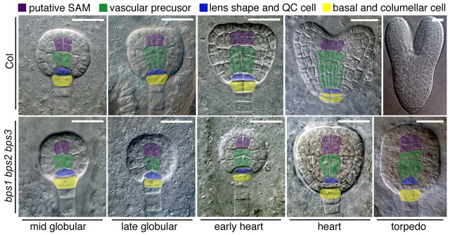
Fig. 4.
Embryonic cell division defects in vascular and root meristem precursors of bps triple mutants. Top row: wild-type (Col-0) embryos. Bottom row: bps1-2 bps2-2 bps3-2 triple mutant embryos. Embryo staging was based on phenotypically wild-type siblings (bps2 bps3), and is indicated below the images. DIC images were false colored to facilitate comparisons of cell types between the wild type and the bps triple mutant. The major defects identified in this analysis were provascular/procambium development and cell divisions at the root apex. Scale bars: 25 μm in all images except the wild-type torpedo embryo (50 μm).

