Abstract
Sensory perception in the inner ear relies on the hair bundle, the highly polarized brush of movement detectors that crowns hair cells. We previously showed that, in the mouse cochlea, the edge of the forming bundle is defined by the ‘bare zone’, a microvilli-free sub-region of apical membrane specified by the Insc-LGN-Gαi protein complex. We now report that LGN and Gαi also occupy the very tip of stereocilia that directly abut the bare zone. We demonstrate that LGN and Gαi are both essential for promoting the elongation and differential identity of stereocilia across rows. Interestingly, we also reveal that total LGN-Gαi protein amounts are actively balanced between the bare zone and stereocilia tips, suggesting that early planar asymmetry of protein enrichment at the bare zone confers adjacent stereocilia their tallest identity. We propose that LGN and Gαi participate in a long-inferred signal that originates outside the bundle to model its staircase-like architecture, a property that is essential for direction sensitivity to mechanical deflection and hearing.
KEY WORDS: Hair cell, Hair bundle, Stereocilia, Staircase-like organization, LGN/Gpsm2, Gαi, Mouse
Summary: In the mouse cochlea, LGN and Gai guide planar polarity in hair cells and are essential for stereocilia elongation, the staircase pattern of the hair bundle and hearing.
INTRODUCTION
Spearheading sensory perception in the inner ear, epithelial hair cells (HCs) transform sound waves, gravity or head movements into electrical impulses that are relayed to the brain by peripheral neurons. Hair bundle deflection exerts force on molecular tip-links that connect neighboring shorter and taller stereocilia across rows, which activates the transduction machinery located at the base of each link (Zhao and Müller, 2015). HCs are receptive to bundle displacements along a single planar axis that is indicated by the direction of the gradient of stereocilia height (Pickles et al., 1984; Shotwell et al., 1981). How the bundle adopts its staircase pattern, however, remains largely unclear. Resident bundle proteins such as Myo15a and Myo3a, and their cargo delivered to stereocilia tips, are required to various extents for graded heights and concurrent graded amounts of some proteins at tips across rows (Belyantseva et al., 2003, 2005; Ebrahim et al., 2016; Lelli et al., 2016; Manor et al., 2011; Mburu et al., 2003; Probst et al., 1998; Salles et al., 2009). However, the mechanisms that instruct morphological and molecular asymmetry across the bundle remain unknown. External cues, including a role for the extracellular matrix or the kinocilium, have been proposed but not experimentally validated (Manor and Kachar, 2008).
Tissue and cell polarization during early development lay the structural foundations required for sensory function. Before a marked staircase pattern and tip-links arise postnatally in the mouse cochlea, ‘core’ planar cell polarity (PCP) proteins that are asymmetrically enriched at cell-cell junctions ensure uniform bundle orientation across HCs (May-Simera and Kelley, 2012). Furthermore, recent studies have clarified that planar asymmetry of the cytoskeleton in each HC (which is manifested, for example, by the V-shaped or semi-circular edge of the bundle and the off-center position of the kinocilium) depends on ciliary proteins, small GTPases and the Insc-LGN-Gαi complex (Bhonker et al., 2016; Ezan et al., 2013; Grimsley-Myers et al., 2009; Jones et al., 2008; Sipe and Lu, 2011; Tarchini et al., 2013). As the staircase pattern of the hair bundle is effectively a planar asymmetry in stereocilia height, we investigate here whether cell-intrinsic planar polarity signals could participate in its establishment. We show that following their planar polarized enrichment at the ‘bare zone’, which is the smooth apical sub-region that defines the lateral edge of the bundle (Tarchini et al., 2013), LGN (Gpsm2) and Gαi become specifically enriched at the tips of adjacent stereocilia that will form the tallest row. Stunted postnatal stereocilia and lack of differential identity across rows in absence of LGN and Gαi function suggest a model whereby planar polarity information in the flat region of the apical membrane is used to establish differential stereocilia identity across the bundle.
RESULTS AND DISCUSSION
LGN and Gαi colocalize at the tips of stereocilia in the first row
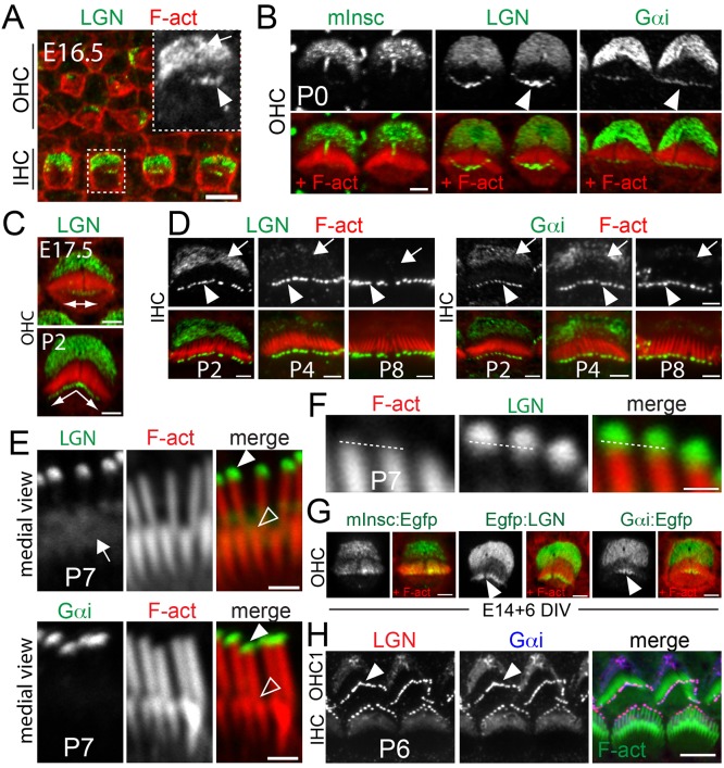
Insc-LGN-Gαi are planar polarized in murine cochlear post-mitotic HCs from embryonic day (E) 15.0 (Bhonker et al., 2016; Ezan et al., 2013; Tarchini et al., 2013), labeling and expanding the bare zone (Tarchini et al., 2013). At E16.5, we found that LGN and Gαi become enriched in a second apical compartment: the tips of developing stereocilia in inner hair cells (IHCs) (Fig. 1A; data not shown). At birth, in both IHCs and outer hair cells (OHCs), LGN and Gαi, but not Insc, are detected at the distal tip of stereocilia adjacent to the bare zone (Fig. 1B). Although initially restricted to central stereocilia in the bundle, LGN-Gαi spread to encompass all stereocilia in the first row (Fig. 1C). Stereocilia enrichment becomes more prominent after birth, whereas protein accumulation at the bare zone, which dominates during embryogenesis, concomitantly decreases (Fig. 1D). LGN-Gαi amounts at the tips are initially comparable in IHCs and OHCs, but decrease during bundle maturation in OHCs (Fig. S1A). LGN-Gαi immunostaining signal extends distally beyond F-actin in the tallest row, but is undetectable in shorter rows (Fig. 1E,F; Fig. S1B), and a similar protein distribution is observed in vestibular HCs (Fig. S1C). Importantly, LGN signal in stereocilia is absent in Lgn mutants (Fig. S1D), and this specific tip enrichment is recapitulated when LGN and Gαi proteins carrying Myc or Egfp tags are transfected into HCs in cochlear cultures (Fig. 1G; Fig. S1E). Overexpressed fusion proteins maintain specificity for the first row (Fig. S1E), suggesting that LGN-Gαi are not able to enter shorter stereocilia located further away from the bare zone. At all stages, LGN and Gαi colocalize at tips in a similar way to their colocalization at the bare zone (Fig. 1H; Fig. S1E), as expected from their well-established direct binding (Du and Macara, 2004). Together, these results uncover a novel and intriguing dual subcellular LGN-Gαi distribution in HCs that suggests a potential role in stereocilia development.
Fig. 1.
LGN and Gαi, but not Insc, localize at the distal tip of stereocilia in the tallest row. (A) LGN immunostaining at E16.5 (one turn position). Inset: LGN channel from the boxed IHC region. (B) Insc, LGN and Gαi immunostaining at P0 (base). (C) LGN at E17.5 and P2. Stereocilia tip enrichment (double-headed arrow) is initially restricted to central stereocilia, but spreads peripherally with time. (D) LGN and Gαi in IHCs at P2, P4 and P8. Bare zone localization decreases with time. (E) Medial view of LGN and Gαi in P7 IHC bundles. No protein enrichment is detected at tips in the second row. See Fig. S1B for a lateral side view. (F) Higher magnification view of LGN at the tips. LGN extends distally beyond the F-actin core of stereocilia (broken lines). (G) Egfp-tagged LGN and Gαi recapitulate endogenous protein distribution, including stereocilia tips. Tagged Insc localizes at the bare zone and can invade the bundle, but is not specifically enriched at stereocilia tips. Cochleas were electroporated at E14.5 and cultured for 6 days. (H) Co-immunostaining for LGN and Gαi at P6 (one turn position). Solid arrowheads, stereocilia tips in the first row; open arrowheads, stereocilia tips in the second row; arrows, bare zone. All images are en face views of the apical surface of the cochlear sensory epithelium where lateral (abneural) is upwards and medial (neural) is downwards. F-actin (F-act) is labeled with phalloidin. Scale bars: 5 µm in A,H; 2 µm in B-E,G; 1 µm in F.
LGN and Gαi are required for stereocilia elongation during bundle maturation
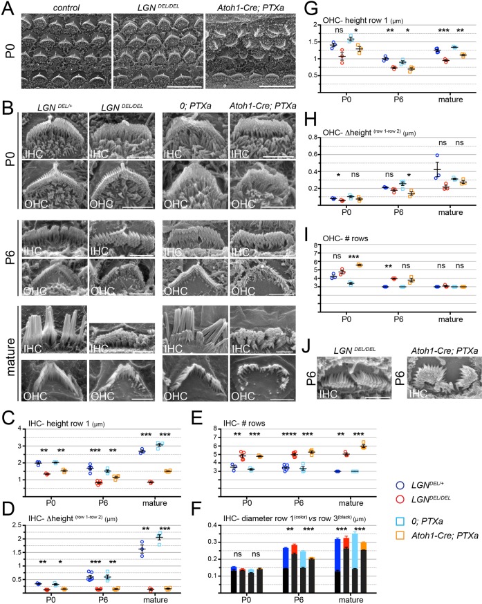
LGN or Gαi inactivation leads to disorganization of stereocilia distribution at the HC apical membrane (Bhonker et al., 2016; Ezan et al., 2013; Tarchini et al., 2013), which we interpreted to be the result of defective bare zone specification and expansion (Tarchini et al., 2013). Prompted by LGN-Gαi localization at their tips, we undertook a detailed analysis of stereocilia morphology during development. Interestingly, in Lgn mutants, IHC stereocilia in the first row are slightly shorter at birth (postnatal day 0, P0) (Fig. 2A-C), a phenotype that increases in severity at P6 and results in drastically stunted stereocilia at maturity (Fig. 2B,C; Fig. S3). While mature IHC stereocilia normally form three rows of markedly different height and girth, the supernumerary rows of similar thickness and a very shallow staircase pattern in Lgn mutants suggest that the bundles remain immature (Fig. 2B,D-F).
Fig. 2.
Hair bundle defects in the absence of LGN and Gαi function. (A) Scanning electron microscopy of the organ of Corti at P0 (half-turn) in a control (LgnDEL/+) and in LgnDEL/DEL and Atoh1-Cre; PTXa mutants. (B) Higher magnification medial views of representative single IHCs (top) and OHCs (bottom) at P0, P6 and mature stages. Mature Lgn and PTXa samples are at P14 and P22, respectively. See lateral views in Fig. S3A. (C-F) Quantification of IHC stereocilia height in the first row (C), the height differential between the first and second row (D), the number of rows across the bundle (E), and the stereocilia thickness in the first (colored) and third (black) rows (F). (G-I) Quantification of OHC stereocilia height in the first row (G), the height differential between the first and second row (H), and the number of rows across the bundle (I). (J) Representative examples of defective stereocilia distribution in LgnDEL/DEL and Atoh1-Cre; PTXa mutants. 0; PTXa controls have the Rosa26-(stop)-PTXa allele, but lack the Atoh1-Cre transgene. Average per animal±s.e.m. is plotted for n≥3 animals. Welch's t-test (ns, P>0.05; *P≤0.05; **P≤0.01; ***P≤0.001; ****P≤0.0001). In F, the t-test reflects thickness differences in row 3. All stereocilia quantifications are detailed in Tables S1 and S2. Cochleas were analyzed halfway in the basal turn (P0, P6) and at the apical turn (mature). Scale bars: 10 µm in A; 2 µm in B,J.
To bypass functional redundancy between Gnai1, Gnai2 and Gnai3, we have previously analyzed transgenic fetuses expressing Pertussis toxin catalytic subunit (PTXa) in HCs (Tarchini et al., 2013). PTXa specifically ADP-ribosylates inhibitory G proteins, as only this class of G proteins carries a conserved C-terminal cysteine target site; PTXa is routinely used to inactivate Gαi signaling (Milligan, 1988; Wise et al., 1997). To extend this approach, we produced a stable mouse line in which PTXa expression is Cre-inducible at the Rosa26 locus (Fig. S2A,B). Using FoxG1-Cre (Hébert and McConnell, 2000) to induce PTXa in early otic progenitors, we observed cochlear defects that are apparently restricted to HC apical differentiation and recapitulate the severe HC planar misorientation reported previously (Fig. S2C) (Tarchini et al., 2013). These animals die shortly after birth, however, so we used Atoh1-Cre (Matei et al., 2005) to restrict PTXa mostly to post-mitotic HCs in order to study postnatal bundle morphogenesis. Atoh1-Cre; PTXa IHCs have bundle defects that are strikingly reminiscent of Lgn mutants, with supernumerary rows of identical-looking stereocilia and a drastically shortened first row (Fig. 2B-F). Thus, LGN and Gαi are both required for postnatal stereocilia elongation and play an essential role in generating graded asymmetric identities across rows, with mutant IHCs harboring an almost flat bundle of ‘generic’ stereocilia. OHC bundle defects are generally similar but less severe, with supernumerary rows largely corrected at maturity and normal stereocilia thickness across rows (Fig. 2B,G-I; data not shown). Milder first row shortening and better preserved staircase-like organization probably reflects the fact that first row elongation and thickening is much less pronounced in OHCs compared with IHCs (compare Fig. 2C,D,G,H). Stereocilia shortening and shallower staircase patterns are also observed in vestibular HCs in the utricle (Fig. S3B-D).
At mature stages, the IHCs of Atoh1-Cre; PTXa mutants have longer stereocilia than do Lgn mutants (Fig. 2B,C). Milder shortening could reflect a knock-down rather than a full inactivation of Gαi function by PTXa. Accordingly, leftover Gαi protein can be detected in less differentiated HCs, where Cre expression occurs later (Fig. S2D). By contrast, disorganized stereocilia distribution at the apical membrane is more pronounced in Atoh1-Cre; PTXa than in Lgn mutants (Fig. 2J). It will be interesting to combine Gnai1-Gnai2-Gnai3 mutants to extend the PTXa results by removing single or multiple endogenous Gαi proteins.
Although loss-of-function approaches cannot resolve subcellular protein function, we propose that the LGN-Gαi complex regulates stereocilia elongation from their stereocilia tip location because: (1) shorter stereocilia are observed postnatally, when LGN-Gαi tip enrichment is more prominent and bare zone accumulation decreases; (2) resident proteins are more likely to influence stereocilia elongation than proteins located outside the bundle; and (3) proteins localized at stereocilia tips and regulating height have been reported previously (Manor and Kachar, 2008) (discussed below).
Stereocilia stunting in absence of LGN-Gαi coincides with profound hearing loss
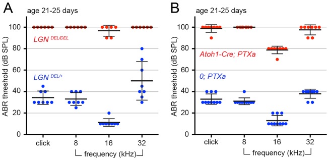
Very limited auditory brainstem responses could be elicited from click sounds and pure tone frequencies (8, 16 and 32 kHz) in young Lgn and Atoh1-Cre; PTXa adult mutants at the highest sound pressure tested (Fig. 3A,B). Stunted stereocilia are a likely cause for near-complete deafness, and could be the etiology of hereditary hearing loss reported in multiple families with recessive mutations in LGN/GPSM2 (Doherty et al., 2012; Walsh et al., 2010; Yariz et al., 2012). In addition, planar polarity defects and abnormal stereocilia distribution could also play a role (Bhonker et al., 2016; Ezan et al., 2013; Tarchini et al., 2013). Direct binding between LGN and Gαi is essential for proper bundle morphogenesis and hearing, as suggested by a recent study using a mutant in which LGN lacks the Goloco motifs that mediate its interaction with Gαi (Bhonker et al., 2016). Of note, Lgn and Atoh1-Cre; PTXa cochleas are still able to incorporate the FM1-43 dye, and the Cdh23 protein can still be detected near stereocilia tips in mature Lgn mutants, suggesting that LGN-Gαi do not directly affect mechanosensory channels or tip-links (Fig. S3E,F). Although Lgn and PTXa mutants do not display obvious balance problems in their cage, their vestibular HC bundle anomalies (Fig. S3B-D) might possibly explain the poor skills observed during free swimming (Movie 1) (Goodyear et al., 2012). Together, these results underscore the importance of LGN-Gαi signaling for HC function.
Fig. 3.
Absence of LGN and Gαi function leads to profound deafness. Auditory brainstem response thresholds for constitutive Lgn mutants (Lgn DEL/DEL) (A) and Gαi inactivation in post-mitotic HCs (Atoh1-Cre; PTXa) (B). LgnDEL/DEL (n=6) and LgnDEL/+ control littermates (n=8), Atoh1-Cre; PTXa (n=8) and 0; PTXa (n=10) control littermates (without the Atoh1-Cre transgene) were tested once at P21-P25 (mean±s.d.).
Evidence for a molecular link between the bare zone and stereocilia tips
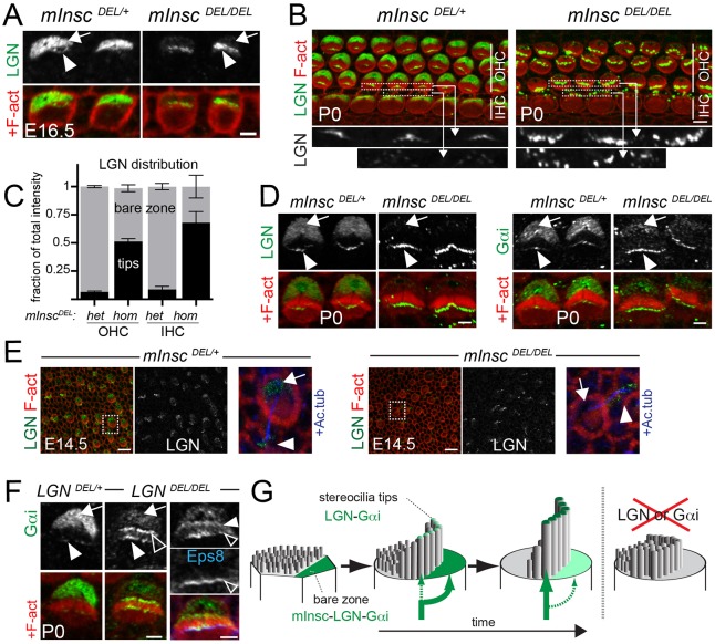
In mitotic cells, LGN-Gαi-mediated regulation of spindle orientation depends on their binding partners (Morin and Bellaïche, 2011). For example, LGN can bind Insc, a protein that directs vertical epithelial divisions and is typically absent during planar divisions (Konno et al., 2008; Kraut et al., 1996; Postiglione et al., 2011; Poulson and Lechler, 2010; Žigman et al., 2005). We reasoned that binding partners could differentially regulate the trafficking of LGN-Gαi to distinct HC compartments. Consistently, we noticed increased amounts of LGN and Gαi before birth at stereocilia tips in absence of Insc, which itself accumulates at the bare zone but not at the tips (Fig. 4A-D and Fig. 1B,G). Interestingly, LGN-Gαi enrichment at the bare zone is conversely reduced in Insc mutants (Fig. 4A-D). The same redistribution across compartments is observed in utricular HCs in Insc mutants (Fig. 4E). Thus, before birth, Insc buffers LGN-Gαi at the bare zone, limiting protein amount at tips. Similarly, LGN is required to keep Gαi at the bare zone, thereby preventing an excess amount of Gαi from accumulating at stereocilia tips and ectopically localizing at their base (Fig. 4F; Fig. S4A). A proper balance of proteins between compartments can be restored by providing Insc or LGN in individual HCs of Insc or Lgn mutant explants, respectively (Fig. S4B,C). These results uncover a cell-intrinsic mechanism that is able to tune relative protein distribution between compartments. Although this mechanism de facto establishes an interesting link between planar polarization and stereocilia tip enrichment in the first row (Fig. 4G), the reason for limiting LGN-Gαi amounts at tips before birth is still unclear. LGN-Gαi excess at tips in mutants is observed only before the normal increase in protein accumulation there after birth, with unchanged LGN-Gαi amounts present at tips postnatally in Insc mutant (Fig. S4D). Consistently, transfected Insc no longer antagonizes LGN at tips in more mature Insc mutant explants (Fig. S4C). Finally, we did not observe stereocilia over-elongation or obvious bundle defects in Insc mutants (not shown). However, transient LGN-Gαi excess could produce subtle changes in the kinetics of stereocilia elongation that are difficult to detect in fixed tissue, and LGN-Gαi at tips might only influence elongation after birth, when stunted stereocilia start to be visible in the corresponding mutants.
Fig. 4.
Balanced LGN-Gαi distribution between the bare zone and stereocilia tips. (A) LGN immunostaining at E16.5 (apical turn) in the indicated genotypes. Mutant IHCs have decreased levels of LGN at the bare zone (arrow) and increased LGN levels in stereocilia (arrowhead). (B) LGN immunostaining at P0. Boxed regions are shown at higher magnification underneath and specifically show LGN stereocilia enrichment in OHCs and IHCs. (C) Fraction of total LGN staining intensity at the bare zone (gray) and stereocilia tips (black) in OHCs and IHCs of P0 InscDEL/DEL and control littermates (mean±s.e.m.). Nine OHCs (three per row) and three IHCs at the cochlea base were analyzed for n≥3 animals for each genotype. (D) LGN (left) and Gαi (right) immunostaining in P0 OHCs. (E) LGN immunostaining in utricular epithelium at E14.5. A single HC (boxed) is magnified in the right-most panels, where acetylated tubulin (Ac. tub) co-staining reveals the kinocilium in blue. (F) Gαi immunostaining at P0 in OHCs. Gαi excess in the bundle is seen at the tips (solid arrowhead) and the base (open arrowhead) of stereocilia. In the right-most panels, stereocilia tips are labeled with Eps8 (blue). Arrows and arrowheads indicate the bare zone and stereocilia tips, respectively. (G) Model depicting the Insc-LGN-Gαi complex distribution (green) during HC differentiation and the mutant phenotype. Scale bars: 2 µm in A,D,F; 5 µm in B; 10 µm in E.
From a top-down perspective, it is tempting to hypothesize that selective trafficking of LGN-Gαi into the first row of stereocilia could be achieved through planar polarized enrichment at the bare zone, which is immediately adjacent along the epithelial plane (Fig. 1B; Fig. 4G). Accordingly, LGN-Gαi are first enriched at the bare zone before becoming detectable at tips, and bare zone-restricted Insc can influence early accumulation of LGN-Gαi at tips. Stereociliary LGN-Gαi would then directly define a tallest row identity, and indirectly implement distinct stereocilia heights and girth across rows in the bundle. The tallest row has indeed been shown to influence stereocilia morphology in the shorter rows via tip-links (Caberlotto et al., 2011a,b; Xiong et al., 2012).
Importantly however, further work is required to establish formally a causal link between the planar polarity machinery and staircase architecture in the bundle. As dual LGN-Gαi localization is observed throughout HC apical differentiation (E16.5 to P8), time-based conditional genetics is unlikely to help, and alternative approaches will be needed to manipulate protein enrichment in a compartment-specific manner. It will also be interesting to ask whether LGN-Gαi are part of a larger protein complex at stereocilia tips. The Myo15a-Whrn-Eps8 complex is essential for shaping the bundle after birth, and mice with mutations in any of these proteins show stunted stereocilia and shallow staircase organization that is very reminiscent of the LGN-Gαi phenotype described here (Manor et al., 2011; Mburu et al., 2003; Probst et al., 1998; Zampini et al., 2011). Interestingly, Myo15a-Whrn-Eps8 are not enriched at the bare zone, or polarized in the plane, and there is currently no explanation for the observation that their graded enrichment at tips is proportional to stereocilia height (Belyantseva et al., 2003, 2005; Delprat et al., 2005; Rzadzinska et al., 2004).
In summary, we report the striking co-occurrence of planar polarity and stereocilia elongation defects in absence of LGN and Gαi function. We propose an innovative model whereby planar polarity signals that originate outside the bundle are used to instruct graded stereocilia heights inside the bundle. We identify the Insc-LGN-Gαi complex as an outstanding candidate for conveying information across compartments, and establishing the staircase-like bundle architecture that is essential for sensory function in HCs.
MATERIALS AND METHODS
Animals
Lgn and Insc mutants have been described previously (Tarchini et al., 2013). The generation of PTXa mice is described in the supplementary Materials and Methods. All animal work was performed in accordance with the Canadian Council on Animal Care guidelines, and reviewed for compliance and approved by the Animal Care and Use Committee of The Jackson Laboratory.
Immunostaining and FM1-43 uptake
Cochleas and utricles were isolated from the temporal bone and processed for immunostaining as previously described (Tarchini et al., 2013). To compare protein distribution between the bare zone and stereocilia tips (Fig. 4), Insc and Lgn mutants and control littermates were processed identically and incubated with a pre-pooled mix to ensure similar antibody concentration. The same cochlear position was imaged in controls and mutants using the same exposure settings, and images were treated identically. For FM1-43 uptake, P4 cochleas were immersed in FM1-43FX (ThermoFisher; 5 µM) for 1 min, fixed for 10 min in 4% PFA, mounted and imaged immediately. Details of the antibodies used, imaging and quantitative analysis can be found in the supplementary Materials and Methods.
Cochlear cultures
Briefly, cochleas from CD1 or B6FVB wild-type mice or mutant mice were collected at E14.5, electroporated with 1-5 µg/µl circular DNA (27 V, 27 ms, 6 pulses at 500 ms intervals) and cultured for 6 days in Matrigel (Corning), as previously described (Tarchini et al., 2013).
Scanning electron microscopy
Temporal bones were fixed for 1 h in 4% PFA before exposing the cochlear sensory epithelium and removing the tectorial membrane. Samples were then incubated overnight in 2.5% glutaraldehyde and 4% PFA in 1 mM MgCl2 and 0.1 M sodium cacodylate buffer. After further dissection, cochlear and utricular samples were progressively dehydrated in ethanol and incubated through a graded isoamyl acetate series, before being dried using critical point drying or hexamethyldisilazane (Electron Microscopy Sciences). After sample sputtering with gold-palladium, images were acquired on a Hitachi 3000N VP at 20 kV using 5000-10,000× magnification. Details of the quantitative analysis can be found in the supplementary Materials and Methods.
Auditory brainstem response and swimming procedure
Briefly, mice were anesthetized with tribromoethanol (2.5 mg per 10 g of body weight), and tested using the Smart EP evoked potential system (Intelligent Hearing Systems) as described previously (Zheng et al., 1999). Thresholds were determined by increasing the sound pressure level (SPL) in 10 dB increments. To observe free swimming behavior (Goodyear et al., 2012), mice were placed in a 38×45 cm plastic tub filled with 10 cm of water at 25°C. Their behavior was filmed for 1 min and videos were evaluated blind to genotype.
Acknowledgements
We thank Wenning Qin and Yingfan Zhang for technical assistance with the PTXa mouse, Cong Tian and Kenneth Johnson for help with Auditory Brainstem Response tests, and Fumio Matsuzaki, Quansheng Du and Jun Yang for LGN and PDZD7 antibodies.
Footnotes
Competing interests
The authors declare no competing or financial interests.
Author contributions
Conceptualization: B.T. and M.C.; Experimentation and data analysis: B.T., A.L.D.T. and N.D.; Manuscript writing: B.T.; Manuscript editing: M.C. and A.L.D.T.; Supervision and funding: B.T. and M.C.
Funding
This work was supported by a research grant from the Canadian Institutes of Health Research [MOP-102584] to M.C. and by a start-up from the Jackson Laboratory to B.T. B.T. was supported by a Human Frontiers Science Program long-term fellowship [LT 00041/2007-L/3] and M.C. is a Senior Fellow of the Fonds de la recherche du Québec–Santé/Fondation Antoine Turmel.
Supplementary information
Supplementary information available online at http://dev.biologists.org/lookup/doi/10.1242/dev.139089.supplemental
References
- Belyantseva I. A., Boger E. T. and Friedman T. B. (2003). Myosin XVa localizes to the tips of inner ear sensory cell stereocilia and is essential for staircase formation of the hair bundle. Proc. Natl. Acad. Sci. USA 100, 13958-13963. 10.1073/pnas.2334417100 [DOI] [PMC free article] [PubMed] [Google Scholar]
- Belyantseva I. A., Boger E. T., Naz S., Frolenkov G. I., Sellers J. R., Ahmed Z. M., Griffith A. J. and Friedman T. B. (2005). Myosin-XVa is required for tip localization of whirlin and differential elongation of hair-cell stereocilia. Nat. Cell Biol. 7, 148-156. 10.1038/ncb1219 [DOI] [PubMed] [Google Scholar]
- Bhonker Y., Abu-Rayyan A., Ushakov K., Amir-Zilberstein L., Shivatzki S., Yizhar-Barnea O., Elkan-Miller T., Tayeb-Fligelman E., Kim S. M., Landau M. et al. (2016). The GPSM2/LGN GoLoco motifs are essential for hearing. Mamm. Genome 27, 29-46. 10.1007/s00335-015-9614-7 [DOI] [PMC free article] [PubMed] [Google Scholar]
- Caberlotto E., Michel V., Boutet de Monvel J. and Petit C. (2011a). Coupling of the mechanotransduction machinery and stereocilia F-actin polymerization in the cochlear hair bundles. Bioarchitecture 1, 169-174. 10.4161/bioa.1.4.17532 [DOI] [PMC free article] [PubMed] [Google Scholar]
- Caberlotto E., Michel V., Foucher I., Bahloul A., Goodyear R. J., Pepermans E., Michalski N., Perfettini I., Alegria-Prevot O., Chardenoux S. et al. (2011b). Usher type 1G protein sans is a critical component of the tip-link complex, a structure controlling actin polymerization in stereocilia. Proc. Natl. Acad. Sci. USA 108, 5825-5830. 10.1073/pnas.1017114108 [DOI] [PMC free article] [PubMed] [Google Scholar]
- Delprat B., Michel V., Goodyear R., Yamasaki Y., Michalski N., El-Amraoui A., Perfettini I., Legrain P., Richardson G., Hardelin J. P. et al. (2005). Myosin XVa and whirlin, two deafness gene products required for hair bundle growth, are located at the stereocilia tips and interact directly. Hum. Mol. Genet. 14, 401-410. 10.1093/hmg/ddi036 [DOI] [PubMed] [Google Scholar]
- Doherty D., Chudley A. E., Coghlan G., Ishak G. E., Innes A. M., Lemire E. G., Rogers R. C., Mhanni A. A., Phelps I. G., Jones S. J. M. et al. (2012). GPSM2 mutations cause the brain malformations and hearing loss in Chudley-McCullough syndrome. Am. J. Hum. Genet. 90, 1088-1093. 10.1016/j.ajhg.2012.04.008 [DOI] [PMC free article] [PubMed] [Google Scholar]
- Du Q. and Macara I. G. (2004). Mammalian Pins is a conformational switch that links NuMA to heterotrimeric G proteins. Cell 119, 503-516. 10.1016/j.cell.2004.10.028 [DOI] [PubMed] [Google Scholar]
- Ebrahim S., Avenarius M. R., Grati M., Krey J. F., Windsor A. M., Sousa A. D., Ballesteros A., Cui R., Millis B. A., Salles F. T. et al. (2016). Stereocilia-staircase spacing is influenced by myosin III motors and their cargos espin-1 and espin-like. Nat. Commun. 7, 10833 10.1038/ncomms10833 [DOI] [PMC free article] [PubMed] [Google Scholar]
- Ezan J., Lasvaux L., Gezer A., Novakovic A., May-Simera H., Belotti E., Lhoumeau A.-C., Birnbaumer L., Beer-Hammer S., Borg J. P. et al. (2013). Primary cilium migration depends on G-protein signalling control of subapical cytoskeleton. Nat. Cell Biol. 15, 1107-1115. 10.1038/ncb2819 [DOI] [PubMed] [Google Scholar]
- Goodyear R. J., Jones S. M., Sharifi L., Forge A. and Richardson G. P. (2012). Hair bundle defects and loss of function in the vestibular end organs of mice lacking the receptor-like inositol lipid phosphatase PTPRQ. J. Neurosci. 32, 2762-2772. 10.1523/JNEUROSCI.3635-11.2012 [DOI] [PMC free article] [PubMed] [Google Scholar]
- Grimsley-Myers C. M., Sipe C. W., Geleoc G. S. G. and Lu X. (2009). The small GTPase Rac1 regulates auditory hair cell morphogenesis. J. Neurosci. 29, 15859-15869. 10.1523/JNEUROSCI.3998-09.2009 [DOI] [PMC free article] [PubMed] [Google Scholar]
- Hébert J. M. and McConnell S. K. (2000). Targeting of cre to the Foxg1 (BF-1) locus mediates loxP recombination in the telencephalon and other developing head structures. Dev. Biol. 222, 296-306. 10.1006/dbio.2000.9732 [DOI] [PubMed] [Google Scholar]
- Jones C., Roper V. C., Foucher I., Qian D., Banizs B., Petit C., Yoder B. K. and Chen P. (2008). Ciliary proteins link basal body polarization to planar cell polarity regulation. Nat. Genet. 40, 69-77. 10.1038/ng.2007.54 [DOI] [PubMed] [Google Scholar]
- Konno D., Shioi G., Shitamukai A., Mori A., Kiyonari H., Miyata T. and Matsuzaki F. (2008). Neuroepithelial progenitors undergo LGN-dependent planar divisions to maintain self-renewability during mammalian neurogenesis. Nat. Cell Biol. 10, 93-101. 10.1038/ncb1673 [DOI] [PubMed] [Google Scholar]
- Kraut R., Chia W., Jan L. Y., Jan Y. N. and Knoblich J. A. (1996). Role of inscuteable in orienting asymmetric cell divisions in Drosophila. Nature 383, 50-55. 10.1038/383050a0 [DOI] [PubMed] [Google Scholar]
- Lelli A., Michel V., Boutet de Monvel J., Cortese M., Bosch-Grau M., Aghaie A., Perfettini I., Dupont T., Avan P., El-Amraoui A. et al. (2016). Class III myosins shape the auditory hair bundles by limiting microvilli and stereocilia growth. J. Cell Biol. 212, 231-244. 10.1083/jcb.201509017 [DOI] [PMC free article] [PubMed] [Google Scholar]
- Manor U. and Kachar B. (2008). Dynamic length regulation of sensory stereocilia. Semin. Cell Dev. Biol. 19, 502-510. 10.1016/j.semcdb.2008.07.006 [DOI] [PMC free article] [PubMed] [Google Scholar]
- Manor U., Disanza A., Grati M. H., Andrade L., Lin H., Di Fiore P. P., Scita G. and Kachar B. (2011). Regulation of stereocilia length by myosin XVa and whirlin depends on the actin-regulatory protein Eps8. Curr. Biol. 21, 167-172. 10.1016/j.cub.2010.12.046 [DOI] [PMC free article] [PubMed] [Google Scholar]
- Matei V., Pauley S., Kaing S., Rowitch D., Beisel K. W., Morris K., Feng F., Jones K., Lee J. and Fritzsch B. (2005). Smaller inner ear sensory epithelia in Neurog1 null mice are related to earlier hair cell cycle exit. Dev. Dyn. 234, 633-650. 10.1002/dvdy.20551 [DOI] [PMC free article] [PubMed] [Google Scholar]
- May-Simera H. and Kelley M. W. (2012). Planar cell polarity in the inner ear. Curr. Top. Dev. Biol. 101, 111-140. 10.1016/B978-0-12-394592-1.00006-5 [DOI] [PubMed] [Google Scholar]
- Mburu P., Mustapha M., Varela A., Weil D., El-Amraoui A., Holme R. H., Rump A., Hardisty R. E., Blanchard S., Coimbra R. S. et al. (2003). Defects in whirlin, a PDZ domain molecule involved in stereocilia elongation, cause deafness in the whirler mouse and families with DFNB31. Nat. Genet. 34, 421-428. 10.1038/ng1208 [DOI] [PubMed] [Google Scholar]
- Milligan G. (1988). Techniques used in the identification and analysis of function of pertussis toxin-sensitive guanine nucleotide binding proteins. Biochem. J. 255, 1-13. 10.1042/bj2550001 [DOI] [PMC free article] [PubMed] [Google Scholar]
- Morin X. and Bellaïche Y. (2011). Mitotic spindle orientation in asymmetric and symmetric cell divisions during animal development. Dev. Cell 21, 102-119. 10.1016/j.devcel.2011.06.012 [DOI] [PubMed] [Google Scholar]
- Pickles J. O., Comis S. D. and Osborne M. P. (1984). Cross-links between stereocilia in the guinea pig organ of Corti, and their possible relation to sensory transduction. Hear. Res. 15, 103-112. 10.1016/0378-5955(84)90041-8 [DOI] [PubMed] [Google Scholar]
- Postiglione M. P., Jüschke C., Xie Y., Haas G. A., Charalambous C. and Knoblich J. A. (2011). Mouse inscuteable induces apical-basal spindle orientation to facilitate intermediate progenitor generation in the developing neocortex. Neuron 72, 269-284. 10.1016/j.neuron.2011.09.022 [DOI] [PMC free article] [PubMed] [Google Scholar]
- Poulson N. D. and Lechler T. (2010). Robust control of mitotic spindle orientation in the developing epidermis. J. Cell Biol. 191, 915-922. 10.1083/jcb.201008001 [DOI] [PMC free article] [PubMed] [Google Scholar]
- Probst F. J., Fridell R. A., Raphael Y., Saunders T. L., Wang A., Liang Y., Morell R. J., Touchman J. W., Lyons R. H., Noben-Trauth K. et al. (1998). Correction of deafness in shaker-2 mice by an unconventional myosin in a BAC transgene. Science 280, 1444-1447. 10.1126/science.280.5368.1444 [DOI] [PubMed] [Google Scholar]
- Rzadzinska A. K., Schneider M. E., Davies C., Riordan G. P. and Kachar B. (2004). An actin molecular treadmill and myosins maintain stereocilia functional architecture and self-renewal. J. Cell Biol. 164, 887-897. 10.1083/jcb.200310055 [DOI] [PMC free article] [PubMed] [Google Scholar]
- Salles F. T., Merritt R. C. Jr., Manor U., Dougherty G. W., Sousa A. D., Moore J. E., Yengo C. M., Dosé A. C. and Kachar B. (2009). Myosin IIIa boosts elongation of stereocilia by transporting espin 1 to the plus ends of actin filaments. Nat. Cell Biol. 11, 443-450. 10.1038/ncb1851 [DOI] [PMC free article] [PubMed] [Google Scholar]
- Shotwell S. L., Jacobs R. and Hudspeth A. J. (1981). Directional sensitivity of individual vertebrate hair cells to controlled deflection of their hair bundles. Ann. N. Y. Acad. Sci. 374, 1-10. 10.1111/j.1749-6632.1981.tb30854.x [DOI] [PubMed] [Google Scholar]
- Sipe C. W. and Lu X. (2011). Kif3a regulates planar polarization of auditory hair cells through both ciliary and non-ciliary mechanisms. Development 138, 3441-3449. 10.1242/dev.065961 [DOI] [PMC free article] [PubMed] [Google Scholar]
- Tarchini B., Jolicoeur C. and Cayouette M. (2013). A molecular blueprint at the apical surface establishes planar asymmetry in cochlear hair cells. Dev. Cell 27, 88-102. 10.1016/j.devcel.2013.09.011 [DOI] [PubMed] [Google Scholar]
- Walsh T., Shahin H., Elkan-Miller T., Lee M. K., Thornton A. M., Roeb W., Abu Rayyan A., Loulus S., Avraham K. B., King M. C. et al. (2010). Whole exome sequencing and homozygosity mapping identify mutation in the cell polarity protein GPSM2 as the cause of nonsyndromic hearing loss DFNB82. Am. J. Hum. Genet. 87, 90-94. 10.1016/j.ajhg.2010.05.010 [DOI] [PMC free article] [PubMed] [Google Scholar]
- Wise A., Watson-Koken M.-A., Rees S., Lee M. and Milligan G. (1997). Interactions of the alpha2A-adrenoceptor with multiple Gi-family G-proteins: studies with pertussis toxin-resistant G-protein mutants. Biochem. J. 321, 721-728. 10.1042/bj3210721 [DOI] [PMC free article] [PubMed] [Google Scholar]
- Xiong W., Grillet N., Elledge H. M., Wagner T. F. J., Zhao B., Johnson K. R., Kazmierczak P. and Müller U. (2012). TMHS is an integral component of the mechanotransduction machinery of cochlear hair cells. Cell 151, 1283-1295. 10.1016/j.cell.2012.10.041 [DOI] [PMC free article] [PubMed] [Google Scholar]
- Yariz K. O., Walsh T., Akay H., Duman D., Akkaynak A. C., King M.-C. and Tekin M. (2012). A truncating mutation in GPSM2 is associated with recessive non-syndromic hearing loss. Clin. Genet. 81, 289-293. 10.1111/j.1399-0004.2011.01654.x [DOI] [PMC free article] [PubMed] [Google Scholar]
- Zampini V., Rüttiger L., Johnson S. L., Franz C., Furness D. N., Waldhaus J., Xiong H., Hackney C. M., Holley M. C., Offenhauser N. et al. (2011). Eps8 regulates hair bundle length and functional maturation of mammalian auditory hair cells. PLoS Biol. 9, e1001048 10.1371/journal.pbio.1001048 [DOI] [PMC free article] [PubMed] [Google Scholar]
- Zhao B. and Müller U. (2015). The elusive mechanotransduction machinery of hair cells. Curr. Opin. Neurobiol. 34, 172-179. 10.1016/j.conb.2015.08.006 [DOI] [PMC free article] [PubMed] [Google Scholar]
- Zheng Q. Y., Johnson K. R. and Erway L. C. (1999). Assessment of hearing in 80 inbred strains of mice by ABR threshold analyses. Hear. Res. 130, 94-107. 10.1016/S0378-5955(99)00003-9 [DOI] [PMC free article] [PubMed] [Google Scholar]
- Žigman M., Cayouette M., Charalambous C., Schleiffer A., Hoeller O., Dunican D., McCudden C. R., Firnberg N., Barres B. A., Siderovski D. P. et al. (2005). Mammalian inscuteable regulates spindle orientation and cell fate in the developing retina. Neuron 48, 539-545. 10.1016/j.neuron.2005.09.030 [DOI] [PubMed] [Google Scholar]


