Fig. 6.

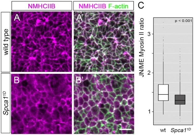
Myosin localization and phosphorylation in Spca11D mutants. (A-B′) Immunodetection of NMHCIIB and F-actin in E9.5 wild-type (A,A′) and Spca11D (B,B′) embryos. Scale bar: 5 µm. (C) Quantification of junctional (JN) and medial (ME) NMHCIIB in E9.5 wild-type (white) and Spca11D (gray) embryos (n>1000 cells from four embryos for each genotype). The line represents the median, the box indicates the distance between the first and third quartile (interquartile range; IQR), whiskers represent ±1.5 *IQR and points indicate outliers.
