Fig. 7.

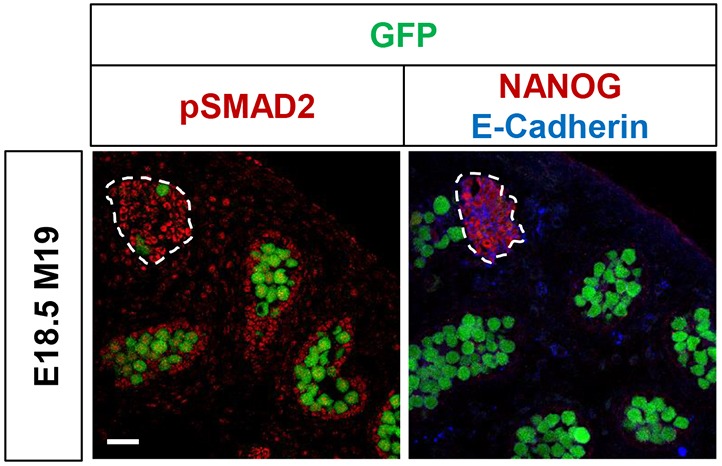
TGFβ signaling pathways are active in EC cells. Representative confocal microscopy images of serial sections from an Oct4ΔPE::GFP transgenic E18.5 M19 testis immunostained for phospho-SMAD2 (pSMAD2) or NANOG and E-cadherin. EC cell foci are outlined. Scale bar: 50 μm. See also Fig. S12.
