Wellcome Open Research logoLink to Wellcome Open Research
. 2019 Mar 27;4:5. Originally published 2019 Jan 17. [Version 2] doi: 10.12688/wellcomeopenres.15026.2

Case Report: Gastric Mucormycosis- a rare but important differential diagnosis of upper gastrointestinal bleeding in an area of Helicobacter pylori endemicity

Sudeep Adhikari 1,a, Ajaya Raj Gautam 1, Buddhi Paudyal 1, Keshav Raj Sigdel 1, Buddha Basnyat 2
PMCID: PMC6518441  PMID: 31131332

Version Changes

Revised. Amendments from Version 1

In this version of the manuscript, we have expanded the details of case presentation, including answers to the queries of the reviewers. In addition, we have corrected some grammar errors as suggested by the reviewers.

Abstract

A 57 years female from the hills of Nepal presented with upper gastrointestinal bleeding with gastric ulcer evident on endoscopy. Though initially treated with Helicobacter pylori ( H. pylori) eradication therapy alone, biopsy later on revealed both mucormycosis and H. pylori infection. She was then treated with antifungals liposomal amphotericin B followed by posaconazole which led to complete recovery. Mucormycosis is a rare but life-threatening fungal disease of immunocompromised host though our patient was immunocompetent. If recognized and treated at early stage, as in our patient, prognosis is good. A high index of suspicion is required for considering this disease in H. pylori endemic regions such as Nepal, and is crucial for early recognition and treatment.

Keywords: gastric mucormycosis, mucormycosis, helicobacter pylori, peptic ulcer disease, upper gastrointestinal disease

Introduction

Upper gastrointestinal bleeding is a common medical emergency which is usually treated with initial stabilization followed by upper gastrointestinal endoscopy. Peptic ulcer disease is a common finding in endoscopy which is usually associated with infection with Helicobacter pylori ( H. pylori) 1. In a developing country such as Nepal, H. pylori infection is so common 2 that treatment with two antibiotics and one proton pump inhibitor therapy, popularly called as ‘triple therapy’, is usually started empirically for eradication even before the presence of the infection is established. But there can be instances when some rare disease can be the cause of ulcers and bleeding, which if overlooked may lead to a fatal outcome without proper treatment. Here we present a case of upper gastrointestinal bleeding diagnosed as gastric mucormycosis.

Mucormycosis is a rare and often a life-threatening fungal disease, caused by a mold of the order Mucorales, characterized by vascular invasion by hyphae with thrombosis and necrosis 3. This is classically a fatal disease of immunocompromised state, with rhino-cerebral, pulmonary and cutaneous being the common types 4. Contrary to the classical description of this disease, our patient with gastric mucormycosis was an immunocompetent individual who recovered after antifungal treatment.

Case report

A 57 year old female from Sindhuli, Nepal, with no known medical comorbidities other than smoking, presented to the emergency department with two episodes of hematemesis over 24 hours. She had a 4-month history of dyspepsia, melena and fatigability. On physical exam, she was pale, blood pressure was 90/50 mm Hg with a heart rate of 130 beats per minute. Other examinations, including abdominal exam, were unremarkable. She was resuscitated with intravenous fluids and packed cell transfusion.

Laboratory parameters with normal ranges in parenthesis, are as follow:

Complete blood count before transfusion: white cell count 7.9 (4–10) × 10 9/L; neutrophils 70%; lymphocytes 26%; monocytes 4%; red blood cells 2.8 (4.2–5.4) × 10 12/L; haemoglobin 9 (12–15) g/dL; platelets 295 (150–400) × 10 9/L.

Biochemistry: random blood sugar 124 (65–110) mg/dL; urea 49 (17–45) mg/dL; creatinine 0.9 (0.8–1.3) mg/dL; sodium 140 (135–145) mmol/L and potassium 4 (3.5–5) mmol/L.

Hepatic panel: bilirubin total 1 (0.1–1.2) mg/dL and direct 0.6 (0–0.4) mg/dL; alanine transaminase 35 (5–30) units/L; aspartate transaminase 40 (5–30) units/L; alkaline phosphatase 98 (50–100) IU/L; albumin 3.5 (3.5–5) g/dL

After stabilization, she underwent upper gastrointestinal endoscopy the same day which revealed an ulcer (10 mm × 6 mm) in lesser curvature of the stomach without active bleeding. The provisional diagnosis was gastric ulcer due to H. pylori infection. The main differential diagnosis was gastric carcinoma, hence biopsy was taken from the ulcer.

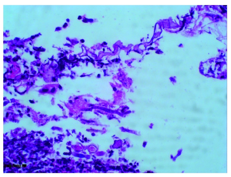
She was started on triple therapy regimen empirically for H. pylori eradication containing clarithromycin 500 mg, amoxicillin 1 gm and pantoprazole 40 mg twice daily for 14 days, and discharged. When she returned for follow-up, the histopathology showed fungi with broad ribbon like morphology, fruiting bodies consistent with mucor in the ulcer, as well as in granulation tissue but no necrosis or vascular thrombi ( Figure 1). It was also positive for H. pylori in Giemsa staining. She was diagnosed with gastric mucormycosis with H. pylori coinfection. But the confirmation of mucormycosis via culture or molecular tests could not be done because of unavailability of these tests in our facility. The abdominal imaging was not done because there was no evidence of invasive form of the disease in histopathology.

She was readmitted and started on liposomal amphotericin B, 5 mg/kg on the first day followed by 10 mg/kg for the next 13 days. She completed a total of 2 weeks of therapy without any complication. She had no dyspepsia, melena or hematemesis by then. She was then discharged with posaconazole 300 mg once daily, as an oral step-down therapy. However the blood level of posaconazole to ensure the therapeutic level was not tested due to its unavailability in resource- limited setup in Nepal.

Figure 1. Histopathology examination of ulcer showing fungi with broad ribbon like morphology and fruiting bodies consistent with mucor.

Figure 1.

On repeat follow up in 1 month, she was asymptomatic. Repeat endoscopy showed a healing ulcer in lesser curvature of stomach. Repeat biopsy showed absence of fungal hyphae. She took posaconazole for total 2 months and stopped the medicine due to financial constraints. She was doing well when she was last followed up 4 months later.

Discussion

Our patient presented with upper gastrointestinal bleed, and received H. pylori eradication treatment empirically after ulcer was evident on endoscopy. This is standard practice in Nepal as prevalence of H. pylori infection is very high in the general population 2, and it is also not always possible to perform tests for H. pylori detection as the tests are either unavailable or unaffordable. Fortunately, we took a gastric biopsy to try to rule out gastric malignancy. The biopsy serendipitously helped to clinch the proper diagnosis of gastric mucormycosis, which otherwise would not have been possible. Though our patient had coinfection with H. pylori, the diagnosis of mucormycosis became alarming because of its high fatality, and the need of specific antifungal treatment with amphotericin B.

Mucormycosis is a life threatening disease occurring in immunocompromised patients. The known risk factors for this disease include diabetes mellitus, particularly with ketoacidosis, treatment with glucocorticoids, hematologic malignancies, hematopoietic cell or solid organ transplantation, treatment with deferoxamine, iron overload, AIDS, injection drug use, trauma/burns, premature birth and malnutrition 5. But our patient had none of these risk factors, yet suffered from the disease.

The common sites of mucormycosis are the sinuses (39%), lungs (24%) and skin (19%). The gastrointestinal tract is involved in only 7% of patients, with the stomach being the most common site. Gastrointestinal mucormycosis is usually fatal (85% mortality) 4, 6. It can be classified into three forms- colonization of pre-existing ulcer, infiltration, and vascular invasion, with increasing fatality in this order 7. Our patient had early infiltrative form of the disease, without vascular invasion. Surgical debridement and antifungal therapy remain the mainstay of therapy, but we treated our patient with medical therapy alone. There are reports where early institution of antifungal therapy without the surgical intervention has led to the recovery in cases with non-invasive form of the disease like in our patient. Tathe et al. reported a case of gastric mucormycosis associated with gastric ulcer who recovered with antifungal treatment, and hypothesized that survival was due to detection and treatment at early nonfatal stage of the disease (colonization with early infiltration of pre-existing ulcer by the fungi rather than invasive ulcerative form of the disease which is almost always fatal) 8. Alfano et al. reported another case of gastric mucormycosis in a liver and kidney transplant recipient who recovered with antifungal therapy for total of 6 months 9. However our patient could not continue the medicine for more than 2 months due to financial constraints.

While transitioning the amphotericin to posaconazole, they should be overlapped until the therapeutic level of posaconazole is achieved in blood 10. But testing of blood level of the drug is not available in Nepal, hence the overlapping was not done. The resource limitation led to many pitfalls during management of our patient, yet she recovered from the infection as evidenced by clinical and histopathological remission. The survival of our patient may be attributed to it being the non-invasive form of disease, the absence of necrosis and thrombosis, and possibly also because of early diagnosis and prompt treatment. However this therapeutic strategy in early form of disease needs to be established as a standard of care by further studies.

Though validation by epidemiological studies is yet to be done, the estimated burden of mucormycosis in Nepal has been reported as 0.2 per 100000 per year 11, while in India it is estimated at 0.14 per 100000 12. A study in Nepal reported 3 cases of mucormycosis among 331 cases of benign sinonasal masses 13, but no account of the gastrointestinal form of disease has been documented in Nepal. The prevalence of mucormycosis appears to be low in Nepal, and we do not routinely consider its possibility in the differential diagnosis of peptic ulcer disease. Our case demonstrates the obvious usefulness of doing a proper gastric histopathological study, where available, even in areas of H. pylori endemicity.

Conclusion

Rare diseases like gastric mucormycosis may present as upper gastrointestinal bleeding, mimicking peptic ulcer disease. It may also co-exist with H. pylori infection. Mucormycosis is a life-threatening fungal disease of immunocompromised state, but it may rarely affect immunocompetent individuals too. Though usually fatal, recognition and treatment at its early stage may lead to recovery.

Consent

Written informed consent for publication of clinical details and clinical images was obtained from the patient herself.

Data availability

All data underlying the results are available as part of the article and no additional source data are required

Funding Statement

This work was supported by the Wellcome Trust [106680].

The funders had no role in study design, data collection and analysis, decision to publish, or preparation of the manuscript.

[version 2; peer review: 3 approved]

References

  • 1. Chen ZJ, Freeman ML: Management of upper gastrointestinal bleeding emergencies: evidence-based medicine and practical considerations. World J Emerg Med. 2011;2(1):5–12. [DOI] [PMC free article] [PubMed] [Google Scholar]
  • 2. Sherpa TW, Sherpa KT, Nixon G, et al. : The prevalence of Helicobacter pylori infection in Sherpa residents of the Upper Khumbu, an isolated community in Eastern Nepal. N Z Med J. 2012;125(1365):30–7. [PubMed] [Google Scholar]
  • 3. Hu J, Zhou X, Xu C, et al. : Fatal gastrointestinal mucormycosis in an immunocompetent patient: a case report and review of the literature. Int J Clin Exp Med. 2018;11(4):4301–4306. Reference Source [Google Scholar]
  • 4. Roden MM, Zaoutis TE, Buchanan WL, et al. : Epidemiology and outcome of zygomycosis: a review of 929 reported cases. Clin Infect Dis. 2005;41(5):634–653. 10.1086/432579 [DOI] [PubMed] [Google Scholar]
  • 5. Cox GM: Mucormycosis (zygomycosis). In: UpToDate, Kauffman CA, Thorner AR (Ed), UpToDate, Waltham, MA. (Accessed on October 27, 2018). Reference Source [Google Scholar]
  • 6. Agha FP, Lee HH, Boland CR, et al. : Mucormycoma of the colon: early diagnosis and successful management. AJR Am J Roentgenol. 1985;145(4):739–741. 10.2214/ajr.145.4.739 [DOI] [PubMed] [Google Scholar]
  • 7. Thomson SR, Bade PG, Taams M, et al. : Gastrointestinal mucormycosis. Br J Surg. 1991;78(8):952–954. 10.1002/bjs.1800780819 [DOI] [PubMed] [Google Scholar]
  • 8. Tathe SP, Dani AA, Chawhan SM, et al. : Gastric mucormycosis: Diagnosis by imprint cytology. Diagn Cytopathol. 2016;44(10):820–822. 10.1002/dc.23518 [DOI] [PubMed] [Google Scholar]
  • 9. Alfano G, Fontana F, Francesca D, et al. : Gastric Mucormycosis in a Liver and Kidney Transplant Recipient: Case Report and Concise Review of Literature. Transplant Proc. 2018;50(3):905–909. 10.1016/j.transproceed.2017.11.036 [DOI] [PubMed] [Google Scholar]
  • 10. Dioverti MV, Cawcutt KA, Abidi M, et al. : Gastrointestinal mucormycosis in immunocompromised hosts. Mycoses. 2015;58(12):714–8. 10.1111/myc.12419 [DOI] [PubMed] [Google Scholar]
  • 11. Khwakhali US, Denning DW: Burden of serious fungal infections in Nepal. Mycoses. 2015;58 Suppl 5:45–50. 10.1111/myc.12393 [DOI] [PubMed] [Google Scholar]
  • 12. Chakrabarti A, Sood P, Denning DW: Estimating fungal infection burden in India using computational models: mucormycosis burden as a case study. In, 23rd European Congress of Clinical Microbiology and Infectious Diseases (ECCMID, 23rd) Berlin 2013 Apr (p. P1044). Reference Source [Google Scholar]
  • 13. Nepal A, Chettri ST, Joshi RR, et al. : Benign sinonasal masses: a clinicopathological and radiological profile. Kathmandu Univ Med J (KUMJ). 2013;11(41):4–8. 10.3126/kumj.v11i1.11015 [DOI] [PubMed] [Google Scholar]
Wellcome Open Res. 2019 May 15. doi: 10.21956/wellcomeopenres.16593.r35145

Reviewer response for version 2

M Veronica Dioverti 1

Authors did a good job amending comments.

I have read this submission. I believe that I have an appropriate level of expertise to confirm that it is of an acceptable scientific standard.

Wellcome Open Res. 2019 May 13. doi: 10.21956/wellcomeopenres.16593.r35470

Reviewer response for version 2

Raju Pangeni 1

The authors have nicely presented an interesting case regarding an uncommon disease which is difficult to diagnose in a resource limited setting. Despite appropriate therapy, invasive mucormycosis itself is a deadly disease regardless of organ involved and requires surgical debridement in most cases.

In the present case, the tissue/vascular invasion couldn't be demonstrated and the patient was treated against H Pylori and Mucor simultaneously. Hence, it may not be able to label mucor as the etiological agent behind bleeding gastric ulcer. In my opinion, Mucor should be regarded as a co-infection.

However, this report makes a point that the endoscopists should consider biopsy among all patients with gastric ulcers.

I have read this submission. I believe that I have an appropriate level of expertise to confirm that it is of an acceptable scientific standard.

Wellcome Open Res. 2019 Apr 5. doi: 10.21956/wellcomeopenres.16593.r35144

Reviewer response for version 2

Gaetano Alfano 1

The manuscript is improved after the revision. You have provided an updated version of the article that fulfills my requests.

In conclusion, despite some important technical limitations in the diagnosis of Mucormycosis, the text can be useful in the differential diagnosis of gastric ulcers in an area of Helicobacter pylori endemicity like Nepal.

I have read this submission. I believe that I have an appropriate level of expertise to confirm that it is of an acceptable scientific standard.

Wellcome Open Res. 2019 Mar 20. doi: 10.21956/wellcomeopenres.16394.r34802

Reviewer response for version 1

M Veronica Dioverti 1

The authors presented a case of a 57 yo immunocompetent patient that presented with upper GI bleed, diagnosed with H pylori and latter with mucormycosis based on path. She was treated with antifungals alone. There is no mention of evaluation for surgical intervention, which is mainstay therapy for this invasive fungal infection.

Overall, grammar needs to be further reviewed and corrected (probably needs re-writting); for example: "A 57 years old, smoker female from Sindhuli, a hilly district of Nepal, with no known comorbidities, presented to the Emergency Department of Patan Hospital, Lagankhel, Lalitpur in April of 2018 with one day history of two episodes of bright red blood-mixed vomitus (around 500 ml per episode), but she had dyspepsia, malena and fatigability for 4 months duration. On assessment, she was pale with blood pressure of 90/50 mmHg and heart rate of 130 beats per minute" should read: A 57 year old female from Sindhuli, Nepal, no known medical comorbidities other than smoking, presented to the emergency department with two episodes of hematemesis over 24 hours. She had a 4-month history of dyspepsia, melena and fatigability. On physical exam, she was pale, blood pressure was 90/50 mmgHg with a heart rate of 130 beats per minute"

"Laboratory parameters with normal ranges in parenthesis, are as follow:"

Question #1 - Background history/case progression:

  • Histopathology: granulation tissue suggesting fungal invasion? Was there fungal invasion or no? This should be discernable on path, there should be clear necrosis and vascular invasion in order to make a diagnosis of invasive mucor.

  • Was surgical intervention entertained? This is a crucial component of mucormycosis management 

  • Was posaconazole level checked? This is important for treatment follow up

Question #2:

Physical exam/labs: only physical exam comments were that patient was "pale" and they provided blood pressure/heart rate. How about abdominal pain, guarding? Any other ominous signs/symptoms? Was imaging obtained? Oftentimes, mucor will invade nearby tissue. For laboratory, was there a culture sent? In terms of treatment follow up, did she tolerate high doses of amphotericin? Was a posaconazole level checked prior to stopping amphotericin?

Question #3:

Authors should extend discussion to include how were other patients managed? Typically, management includes surgery. Duration of antifungal therapy is typically longer that 1 month, indicating that perhaps this was not the correct diagnosis? How long were the other case reported treated for?

Question #4: 

More details need to be included as mentioned above

I have read this submission. I believe that I have an appropriate level of expertise to state that I do not consider it to be of an acceptable scientific standard, for reasons outlined above.

References

  • 1. Dioverti MV, Cawcutt KA, Abidi M, Sohail MR, Walker RC, Osmon DR: Gastrointestinal mucormycosis in immunocompromised hosts. Mycoses.2015;58(12) : 10.1111/myc.12419 714-8 10.1111/myc.12419 [DOI] [PubMed] [Google Scholar]
Wellcome Open Res. 2019 Mar 23.
Sudeep Adhikari 1

Thank you for your constructive feedback to our manuscript.

The grammar has been reviewed and corrected as suggested by the reviewer.

Question #1 - Background history/case progression:

  • Histopathology: granulation tissue suggesting fungal invasion? Was there fungal invasion or no? This should be discernable on path, there should be clear necrosis and vascular invasion in order to make a diagnosis of invasive mucor.

  • Was surgical intervention entertained? This is a crucial component of mucormycosis management 

  • Was posaconazole level checked? This is important for treatment follow up

Response to question 1

  • In the histopathology examination there was no evidence of fungal invasion, as necrosis and vascular invasion were not present. Changes have been made in the revised manuscript.

  • Surgical intervention was not done, as our patient had early form of the disease. Case reports where medical therapy alone was used for the management, have been cited in the revised manuscript. (1,2)

  • The need of overlapping amphotericin and posaconazole before stopping amphotericin till the therapeutic level of posaconazole is achieved has been discussed in the revised manuscript. (3) But the unavailability of the test of blood level of posaconazole in our setup hindered the use of the standard management practice in our case.

Question #2:

Physical exam/labs: only physical exam comments were that patient was "pale" and they provided blood pressure/heart rate. How about abdominal pain, guarding? Any other ominous signs/symptoms? Was imaging obtained? Oftentimes, mucor will invade nearby tissue. For laboratory, was there a culture sent? In terms of treatment follow up, did she tolerate high doses of amphotericin? Was a posaconazole level checked prior to stopping amphotericin?

Response to question 2

  • Other physical examinations were unremarkable, including the abdominal exam.

  • Abdominal imaging to exclude the invasion of nearby tissue was not done, as our patient had non-invasive form of the disease.

  • Confirmation of the infection via culture was not done, because this test is not readily available in our setup.

  • Our patient had no complication on using amphotericin B. The hospital stay was uneventful.

  • Posaconazole level testing is not available in our setup.

Question #3: 

Authors should extend discussion to include how were other patients managed? Typically, management includes surgery. Duration of antifungal therapy is typically longer that 1 month, indicating that perhaps this was not the correct diagnosis? How long were the other case reported treated for?

Response to question 3

  • Case reports of using medical therapy alone without the surgery, have been included in the discussion.

  • Our patient received posaconazole for 2 months only due to financial constraints. But she had clinical as well as histopathological remission from the disease. One case was treated for 6 months with antifungal therapy. (1)

Question #4: 

More details need to be included as mentioned above

Response to question 4

  • Details have been added as suggested.

References

  1.  Alfano G, Fontana F, Francesca D, Assirati G, Magistri P, Tarantino G, Ballarin R, Rossi G, Franceschini E, Codeluppi M, Guaraldi G, Mussini C, Di Benedetto F, Cappelli G: Gastric Mucormycosis in a Liver and Kidney Transplant Recipient: Case Report and Concise Review of Literature. Transplant Proc. 2018;  50 (3): 905-909

  2. Tathe SP, Dani AA, Chawhan SM, et al.: Gastric mucormycosis: Diagnosis by imprint cytology. Diagn Cytopathol. 2016;44(10):820–822. 27321416 10.1002/dc.23518

  3. Dioverti MV, Cawcutt KA, Abidi M, Sohail MR, Walker RC, Osmon DR: Gastrointestinal mucormycosis in immunocompromised hosts. Mycoses. 2015;  58 (12): 714-8  PubMed Abstract | Publisher Full Text 

Wellcome Open Res. 2019 Mar 13. doi: 10.21956/wellcomeopenres.16394.r34951

Reviewer response for version 1

Gaetano Alfano 1

Adhikari et al. describe an interesting case of gastric mucormycosis in an immunocompetent patient coming from an area where H. Pylori infection is endemic (Nepal). The case is singular because histological examination of the biopsy specimen obtained from the gastric ulcer showed a confection of Mucormycetes and H. Pyroli. Additionally, the case underlined the importance of histologic examination in the early diagnosis of Mucormycosis organisms.

Some further information should be provided in order to improve the quality of the manuscript:

  • The text lacks data regarding the clinical course of the patients after admission. In particular, the authors should describe the change in gastrointestinal symptomatology with the start of the specific therapy.

  • Please discuss if culture of gastric mucosal specimen or molecular analysis were performed to identify Mucormycetes

  • It is well-known that the management of Mucormycetes infection is based on the prompt institution of appropriate antifungal therapy and surgery. Studies report that surgical treatment has been associated with improved survival when combined with medical treatment (Roden et al., 2005 1; Turner, Soudry, Nayak, & Hwang, 2013 2).

Since the patient did not undergo surgical debridement of the gastric ulcer, the authors should discuss why surgical intervention was not performed.

On this issue, we published a similar case that was successfully treated with a conservative strategy. After the diagnosis of Mucormycosis, surgery was postponed because the institution of antifungal therapy controlled the disease and resolved the gastrointestinal symptoms progressively (Alfano et al., 2018 3). Although this therapeutic strategy cannot be generalized in absence of randomized controlled trial, antifungal therapy may be considered a first attempt in the early stage of a gastric mucormycosis of non-critically ill patients. Authors may underline that further studies are necessary to establish the best treatment of uncomplicated cases of gastric mucormycosis.

I have read this submission. I believe that I have an appropriate level of expertise to confirm that it is of an acceptable scientific standard, however I have significant reservations, as outlined above.

References

  • 1. Roden MM, Zaoutis TE, Buchanan WL, Knudsen TA, Sarkisova TA, Schaufele RL, Sein M, Sein T, Chiou CC, Chu JH, Kontoyiannis DP, Walsh TJ: Epidemiology and outcome of zygomycosis: a review of 929 reported cases. Clin Infect Dis.2005;41(5) : 10.1086/432579 634-53 10.1086/432579 [DOI] [PubMed] [Google Scholar]
  • 2. Turner JH, Soudry E, Nayak JV, Hwang PH: Survival outcomes in acute invasive fungal sinusitis: a systematic review and quantitative synthesis of published evidence. Laryngoscope.2013;123(5) : 10.1002/lary.23912 1112-8 10.1002/lary.23912 [DOI] [PubMed] [Google Scholar]
  • 3. Alfano G, Fontana F, Francesca D, Assirati G, Magistri P, Tarantino G, Ballarin R, Rossi G, Franceschini E, Codeluppi M, Guaraldi G, Mussini C, Di Benedetto F, Cappelli G: Gastric Mucormycosis in a Liver and Kidney Transplant Recipient: Case Report and Concise Review of Literature. Transplant Proc.2018;50(3) : 10.1016/j.transproceed.2017.11.036 905-909 10.1016/j.transproceed.2017.11.036 [DOI] [PubMed] [Google Scholar]
Wellcome Open Res. 2019 Mar 23.
Sudeep Adhikari 1

Thank you for your constructive feedback.

Question- The text lacks data regarding the clinical course of the patients after admission. In particular, the authors should describe the change in gastrointestinal symptomatology with the start of the specific therapy.

Response- Our patient's initial symptoms were resolved during the therapy with antifungal agents. This has been added in the mansucript.

Question- Please discuss if culture of gastric mucosal specimen or molecular analysis were performed to identify Mucormycetes

Response- Culture of gastric mucosal specimen or molecular analysis were not performed as these tests are not readily available in our setup.

Question- It is well-known that the management of Mucormycetes infection is based on the prompt institution of appropriate antifungal therapy and surgery. Studies report that surgical treatment has been associated with improved survival when combined with medical treatment (Roden et al., 2005 1; Turner, Soudry, Nayak, & Hwang, 2013 2).Since the patient did not undergo surgical debridement of the gastric ulcer, the authors should discuss why surgical intervention was not performed.

On this issue, we published a similar case that was successfully treated with a conservative strategy. After the diagnosis of Mucormycosis, surgery was postponed because the institution of antifungal therapy controlled the disease and resolved the gastrointestinal symptoms progressively (Alfano et al., 2018 3). Although this therapeutic strategy cannot be generalized in absence of randomized controlled trial, antifungal therapy may be considered a first attempt in the early stage of a gastric mucormycosis of non-critically ill patients. Authors may underline that further studies are necessary to establish the best treatment of uncomplicated cases of gastric mucormycosis.

Response- The surgical intervention is one important component of management. But we did not opt for the surgical management as our patient had early non-invasive form of the disease. Examples of cases where the early disease had been manged by antifungals alone have given in the revised manuscript.

Associated Data

    This section collects any data citations, data availability statements, or supplementary materials included in this article.

    Data Availability Statement

    All data underlying the results are available as part of the article and no additional source data are required


    Articles from Wellcome Open Research are provided here courtesy of The Wellcome Trust

    RESOURCES