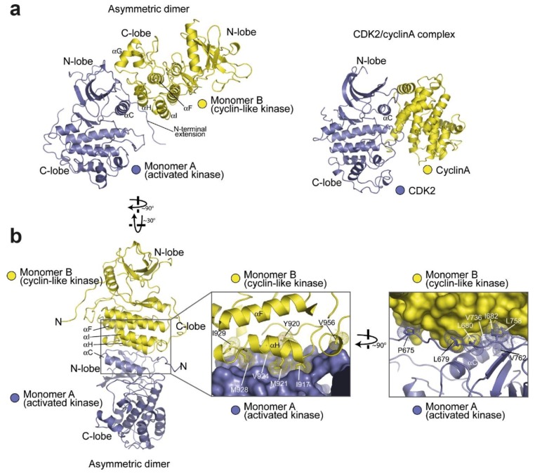
Figure 4.
Structure of the asymmetric crystallographic dimer of the EGFR kinase domain (taken, with permission, from Zhang et al. [36]). (a) Shows an overview of the dimer (PDB:2GS6) compared with the cyclin-dependent kinase-2 (CDK2)/cyclin A complex (PDB: 1HCL). (b) Shows detail of the dimer interface. The first zoomed-in area (left) shows a ribbon structure for Monomer B with its hydrophobic interface residues highlighted. The second zoomed-in area (right) shows the reverse, with the hydrophobic interface residues of monomer A highlighted.

